

HLA-E-restricted recognition of cytomegalovirus-derived peptides by human CD8+ cytolytic T lymphocytes
- PMID: 12960383
- PMCID: PMC196899
- DOI: 10.1073/pnas.1834449100
HLA-E-restricted recognition of cytomegalovirus-derived peptides by human CD8+ cytolytic T lymphocytes
Abstract
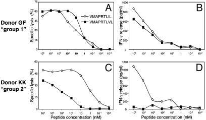
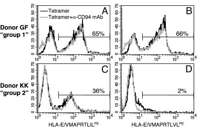
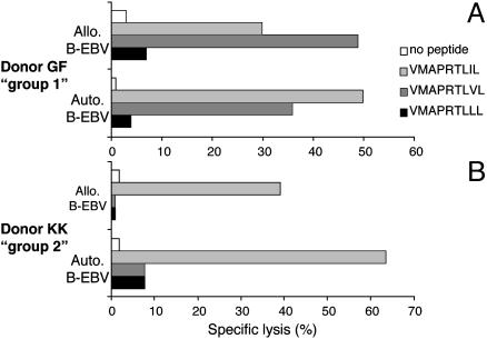
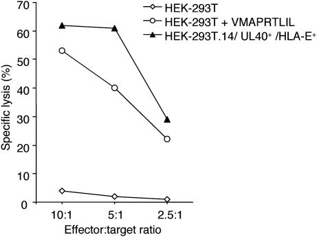
HLA-E-restricted T cell receptor alphabeta+ CD8+ cytolytic T lymphocytes (CTLs) exist as monoclonal expansions in the peripheral blood of some individuals. Here, we show that they recognize, with high avidity, peptides derived from the UL40 protein of different human cytomegalovirus (CMV) strains. Recognition results in the induction of cytotoxicity, IFN-gamma production and cell proliferation. Autologous cells pulsed with CMV-derived peptides become susceptible to lysis by HLA-E-restricted CTLs and induce their proliferation. The high avidity for CMV-derived peptides may explain how these cells are generated in vivo and suggest their possible role in the host defenses against CMV, a virus that evolved various mechanisms to down-regulate classical HLA class I molecules, thus escaping detection by conventional CTLs.
Figures
Similar articles
-
HLA-E-restricted recognition of human cytomegalovirus by a subset of cytolytic T lymphocytes.Hum Immunol. 2004 May;65(5):437-45. doi: 10.1016/j.humimm.2004.02.001. Hum Immunol. 2004. PMID: 15172443
-
Identification of HLA-E-specific alloreactive T lymphocytes: a cell subset that undergoes preferential expansion in mixed lymphocyte culture and displays a broad cytolytic activity against allogeneic cells.Proc Natl Acad Sci U S A. 2002 Aug 20;99(17):11328-33. doi: 10.1073/pnas.172369799. Epub 2002 Aug 7. Proc Natl Acad Sci U S A. 2002. PMID: 12167676 Free PMC article.
-
Soluble recombinant CMVpp65 spanning multiple HLA alleles for reconstitution of antiviral CD4+ and CD8+ T-cell responses after allogeneic stem cell transplantation.J Immunother. 2010 Jan;33(1):60-72. doi: 10.1097/CJI.0b013e3181b56dcc. J Immunother. 2010. PMID: 19952955
-
The emerging role of HLA-E-restricted CD8+ T lymphocytes in the adaptive immune response to pathogens and tumors.J Biomed Biotechnol. 2010;2010:907092. doi: 10.1155/2010/907092. Epub 2010 Jun 22. J Biomed Biotechnol. 2010. PMID: 20634877 Free PMC article. Review.
-
HLA-E: exploiting pathogen-host interactions for vaccine development.Clin Exp Immunol. 2019 May;196(2):167-177. doi: 10.1111/cei.13292. Epub 2019 Apr 9. Clin Exp Immunol. 2019. PMID: 30968409 Free PMC article. Review.
Cited by
-
HLA-E-restricted cross-recognition of allogeneic endothelial cells by CMV-associated CD8 T cells: a potential risk factor following transplantation.PLoS One. 2012;7(11):e50951. doi: 10.1371/journal.pone.0050951. Epub 2012 Nov 30. PLoS One. 2012. PMID: 23226431 Free PMC article.
-
Distinctive phenotype for HLA-E- versus HLA-A2-restricted memory CD8 αβT cells in the course of HCMV infection discloses features shared with NKG2C+CD57+NK and δ2-γδT cell subsets.Front Immunol. 2022 Dec 1;13:1063690. doi: 10.3389/fimmu.2022.1063690. eCollection 2022. Front Immunol. 2022. PMID: 36532017 Free PMC article.
-
A conserved energetic footprint underpins recognition of human leukocyte antigen-E by two distinct αβ T cell receptors.J Biol Chem. 2017 Dec 22;292(51):21149-21158. doi: 10.1074/jbc.M117.807719. Epub 2017 Sep 25. J Biol Chem. 2017. PMID: 28972140 Free PMC article.
-
Viral sequence determines HLA-E-restricted T cell recognition of hepatitis B surface antigen.Nat Commun. 2024 Nov 22;15(1):10126. doi: 10.1038/s41467-024-54378-9. Nat Commun. 2024. PMID: 39578466 Free PMC article.
-
The structure and stability of the monomorphic HLA-G are influenced by the nature of the bound peptide.J Mol Biol. 2010 Mar 26;397(2):467-80. doi: 10.1016/j.jmb.2010.01.052. Epub 2010 Feb 1. J Mol Biol. 2010. PMID: 20122941 Free PMC article.
References
-
- Rock, K. L. & Goldberg, A. L. (1999) Annu. Rev. Immunol. 17, 739–779. - PubMed
-
- Townsend, A. & Bodmer, H. (1989) Annu. Rev. Immunol. 7, 601–624. - PubMed
-
- Braud, V. M., Allan, D. S. & McMichael, A. J. (1999) Curr. Opin. Immunol. 11, 100–108. - PubMed
-
- Li, J., Goldstein, I., Glickman-Nir, E., Jiang, H. & Chess, L. (2001) J. Immunol. 167, 3800–3808. - PubMed
-
- Pietra, G., Romagnani, C., Falco, M., Vitale, M., Castriconi, R., Pende, D., Millo, E., Anfossi, S., Biassoni, R., Moretta, L. & Mingari, M. C. (2001) Eur. J. Immunol. 31, 3687–3693. - PubMed
Publication types
MeSH terms
Substances
LinkOut - more resources
Full Text Sources
Other Literature Sources
Molecular Biology Databases
Research Materials
