. 2003 Sep 16;100(19):11104-9.
doi: 10.1073/pnas.1831452100. Epub 2003 Sep 5.

Proteinase-activated receptor 1 activation induces epithelial apoptosis and increases intestinal permeability

Affiliations

Proteinase-activated receptor 1 activation induces epithelial apoptosis and increases intestinal permeability

Alex C Chin et al. Proc Natl Acad Sci U S A. .

Abstract

Proteinase-activated receptor 1 (PAR1)-mediated inflammation remains poorly understood. Here we characterize previously unrecognized effects of PAR1-induced apoptosis signaling, which contributes to epithelial barrier dysfunction. Incubation of epithelial cells with PAR1 agonists induced apoptosis and increased epithelial permeability in a caspase-3-dependent manner. Similarly, studies in vivo demonstrated that intracolonic infusion with PAR1 agonists increased colonic permeability in mice, and that this effect was abolished by pretreatment with a caspase-3 inhibitor. PAR1 agonists induced tight junctional zonula-occludens 1 disruption and apoptotic nuclear condensation. Investigation into signaling pathways showed that these effects were dependent on caspase-3, tyrosine kinase, and myosin light chain kinase. Conversely, the Src kinase inhibitor PP1 augmented zonula-occludens 1 injury and nuclear condensation induced by PAR1 agonists. These results support a role for proteinases and PARs in intestinal disease and provide new directions for possible therapeutic applications of PAR1 antagonists.


Figures
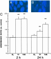
Fig. 1.
Fig. 1.
PAR1 agonists induce enterocyte apoptosis. Shown are representative Hoechst fluorescence micrographs of viable cells exhibiting uniform nuclear staining (A), and apoptotic cells exhibiting nuclear condensation (B). (C) Levels of apoptosis in epithelial monolayers incubated with 5% DMEM growth medium alone (-) or with 25 μM TFLLR-NH2 (TF), 5 units/ml thrombin (THR), or 25 μM RLLFT-NH2 (RL). Values at 30 min ELISA development were calculated as absorbance ratios versus control values arbitrarily set at 1.0 (-). Values are mean ± SEM; n = 18 per group; **, P < 0.01 compared with control.
Fig. 2.
Fig. 2.
PAR1-induced enterocyte apoptosis increases epithelial permeability. Transepithelial fluxes of FITC-dextran 3000 across epithelial monolayers pretreated with or without caspase-3 inhibitor Z-DEVD-FMK (C-3) before incubation with either apical or basolateral 5% DMEM, 2 μg/ml camptothecin (CAM), 25 μM RLLFT-NH2 (RL), 25 μM TFLLR-NH2 (TF), or 5 units/ml thrombin (THR) for2 h(A and B) or 24 h(C and D). Values are mean ± SEM; n = 9–16 per group; *, P < 0.05 compared with control.
Fig. 3.
Fig. 3.
Induction of apoptosis by activation of PAR1 increases colonic permeability. Blood levels of 51Cr-EDTA after intracolonic administration with saline vehicle, 200 μg of TFLLR-NH2,or200 μg of RLLFT-NH2 (A) or with saline vehicle, 200 μg of TFLLR-NH2, or with 200 μg of TFLLR-NH2 after pretreatment with caspase-3 inhibitor Z-DEVD-FMK (B). Values are mean ± SEM; n = 8 per group; *, P < 0.05 compared with control peptide; +, P < 0.05 compared with TFLLR-NH2 treatment.
Fig. 4.
Fig. 4.
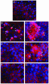
PAR1-induced enterocyte apoptosis disrupts tight junctional ZO-1 in a caspase-3-, tyrosine kinase-, and MLCK-dependent manner. Shown are representative micrographs illustrating ZO-1 and nuclear integrity in epithelial monolayers after 2 h. Preparations were coincubated with 5% DMEM growth medium (A), 25 μM TFLLR-NH2 (B), 5 units/ml thrombin (C), caspase-3 inhibitor Z-DEVD-FMK and 25 μM TFLLR-NH2 (D), Src inhibitor PP1 and 25 μM TFLLR-NH2 (E), tyrosine kinase inhibitor tyrphostin and 25 μM TFLLR-NH2 (F), or MLCK inhibitor ML-9 and 25 μM TFLLR-NH2 (G). Cellular changes include focal disruption of ZO-1 along the pericellular junctions (arrowhead), punctate ZO-1 redistribution (small arrows), diffuse intracellular ZO-1 relocalization (asterisk), and apoptotic nuclear condensation (large arrows). All micrographs were obtained at a magnification of ×400.
Fig. 5.
Fig. 5.
PAR1 agonists disrupt tight junctional ZO-1 via caspase-3, tyrosine kinases, and MLCK. Quantification of ZO-1 disruption in monolayers incubated with 5% DMEM growth medium (CON), 25 μM TFLLR-NH2 (TF), thrombin (THR), caspase-3 inhibitor Z-DEVD-FMK and 25 μM TFLLR-NH2 (TF+C3), Src inhibitor PP1 and 25 μM TFLLR-NH2 (TF+PP1), tyrosine kinase inhibitor tyrphostin and 25 μM TFLLR-NH2 (TF+TP), or MLCK inhibitor ML-9 and 25 μM TFLLR-NH2 (TF+ML9) for 2 h(A) or 24 h(B). Values are mean ± SEM; n = 8 per group; *, P < 0.05 compared with control; †, P < 0.05 compared with TFLLR-NH2-treated monolayers.
Fig. 6.
Fig. 6.
PAR1 agonists induce enterocyte apoptosis in a caspase-3-, tyrosine kinase-, and MLCK-dependent fashion. Quantification of apoptotic nuclear condensation in monolayers incubated with 5% DMEM growth medium (CON), 25 μM TFLLR-NH2 (TF), thrombin (THR), caspase-3 inhibitor Z-DEVD-FMK and 25 μM TFLLR-NH2 (TF+C3), Src inhibitor PP1 and 25 μM TFLLR-NH2 (TF+PP1), tyrosine kinase inhibitor tyrphostin and 25 μM TFLLR-NH2 (TF+TP), or MLCK inhibitor ML-9 and 25 μM TFLLR-NH2 (TF+ML9) for 2 h(A) or 24 h(B) is shown. Values are mean ± SEM; n = 8 per group; *, P < 0.05 compared with control; †, P < 0.05 compared with TFLLR-NH2-treated monolayers.

Similar articles

Cited by

References

    1. Dery, O., Corvera, C. U., Steinhoff, M. & Bunnett, N. W. (1998) Am. J. Physiol. 274, C1429-C1452. - PubMed
    1. Coughlin, S. R. (2000) Nature 407, 258-264. - PubMed
    1. Hollenberg, M. D. & Compton, S. J. (2002) Pharmacol. Rev. 54, 203-217. - PubMed
    1. Buresi, M. C., Schleihauf, E., Vergnolle, N., Buret, A., Wallace, J. L., Hollenberg, M. D. & MacNaughton, W. K. (2001) Am. J. Physiol. 281, G323-G332. - PubMed
    1. Buresi, M. C., Buret, A. G., Hollenberg, M. D. & MacNaughton, W. K. (2002) FASEB J. 16, 1515-1525. - PubMed

Publication types