. 2003 Sep 16;100(19):10728-33.
doi: 10.1073/pnas.1832185100. Epub 2003 Sep 5.

Ionizing radiation induces heritable disruption of epithelial cell interactions

Collaborators, Affiliations

Ionizing radiation induces heritable disruption of epithelial cell interactions

Catherine C Park et al. Proc Natl Acad Sci U S A. .

Abstract

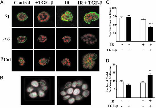
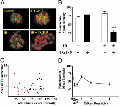
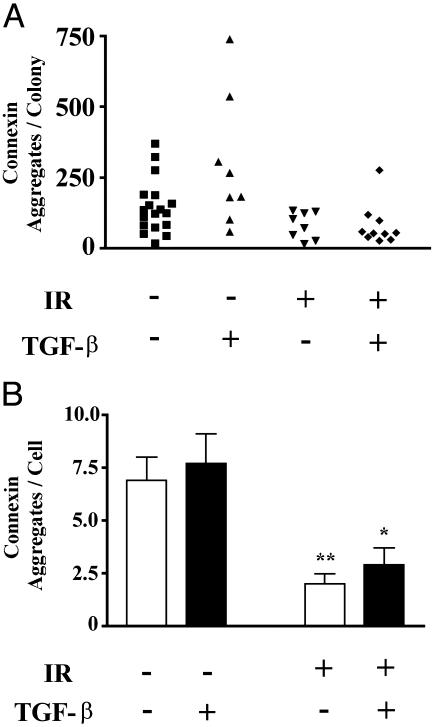
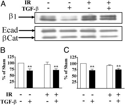
Ionizing radiation (IR) is a known human breast carcinogen. Although the mutagenic capacity of IR is widely acknowledged as the basis for its action as a carcinogen, we and others have shown that IR can also induce growth factors and extracellular matrix remodeling. As a consequence, we have proposed that an additional factor contributing to IR carcinogenesis is the potential disruption of critical constraints that are imposed by normal cell interactions. To test this hypothesis, we asked whether IR affected the ability of nonmalignant human mammary epithelial cells (HMEC) to undergo tissue-specific morphogenesis in culture by using confocal microscopy and imaging bioinformatics. We found that irradiated single HMEC gave rise to colonies exhibiting decreased localization of E-cadherin, beta-catenin, and connexin-43, proteins necessary for the establishment of polarity and communication. Severely compromised acinar organization was manifested by the majority of irradiated HMEC progeny as quantified by image analysis. Disrupted cell-cell communication, aberrant cell-extracellular matrix interactions, and loss of tissue-specific architecture observed in the daughters of irradiated HMEC are characteristic of neoplastic progression. These data point to a heritable, nonmutational mechanism whereby IR compromises cell polarity and multicellular organization.


Figures

Fig. 1.
Fig. 1.
Perturbed protein localization and acinar organization as a function of TGF-β, IR, and dual treatment. Colonies develop and organize in 3D rBM culture over the course of 10 days, during which time the cells are fed every other day. EGF, to stimulate proliferation, is removed at day 6, and the cells are harvested at day 10. (A) Representative images of colonies from control, TGF-β-, IR-, or dual-treated cultures. The image is representative of the mean intensity for each marker based on image analysis of 20 randomly chosen colonies. Immunostaining of β1 integrin, α6 integrin, and β-catenin was detected by using secondary antibodies labeled with Alexa Fluor 488 (green). Nuclei are counterstained with TO-PROR-3 iodide, shown in red. Note the loss of acinar organization in the irradiated colonies. (B) Acinar organization was measured by nuclear segmentation of the colony confocal midsection fit to an ellipse as shown for a control (Left) and dual-treated (Right) colony. (C) Acinar organization as a function of treatment group (n > 100 colonies per treatment). Acinar organization was significantly (P < 0.0001) decreased in colonies that arose from irradiated cells that were cultured in the presence of TGF-β.(D) The number of nuclei per colony midsection as a function of treatment group. The number of nuclei was significantly (P < 0.001) increased in colonies arising from irradiated cells treated with TGF-β.
Fig. 2.
Fig. 2.
E-cadherin immunoreactivity and localization are significantly reduced by IR and TGF-β.(A) Confocal images of E-cadherin immunoreactivity in midsections of colonies representative of the average response as measured by image analysis of 20 colonies are shown for each treatment group. E-cadherin (green) and nuclei (red) were detected as described in Fig. 1. (B) Quantified E-cadherin immunoreactivity as a function of treatment group. The mean intensity of E-cadherin immunofluorescence was significantly (P < 0.0001) reduced in TGF-β-treated, irradiated colonies. (C) Display of relative intensity versus colony area for sham (black circles) and dual-treated (red triangles) colonies. Comparison of the treated to control populations show that >75% of the treated colonies exhibit loss of E-cadherin, a frequency that cannot be explained by mutation rates. (D) Quantified E-cadherin immunoreactivity as a function of radiation exposure. The dose–response shows significant loss of E-cadherin immunoreactivity at doses that do not lead to any detectable loss of cell viability.
Fig. 3.
Fig. 3.
Gap junctions are decreased in irradiated colonies. (A) Connexin 43 was localized by immunostaining and randomly selected colonies were imaged by confocal microscopy. Data shown are representative of two independent experiments. The number of aggregates per colony are displayed for 8–18 colonies per treatment. (B) The average (±SE) number of connexin 43 foci per cell is displayed as a function of treatment group. Colonies arising from irradiated cells showed significantly (P < 0.05, two-tailed t test) fewer connexin foci than those from nonirradiated cells.
Fig. 4.
Fig. 4.
Protein levels as a function of IR and TGF-β. (A) Representative immunoblots of β1 integrin, E-cadherin, and β-catenin from total protein extracted from cultures. β1 integrin protein abundance increased in irradiated samples, regardless of TGF-β exposure. E-cadherin and β-catenin protein abundance were decreased in extracts from TGF-β-treated cultures, regardless of irradiation. Quantitation of E-cadherin (B) and β-catenin (C) protein abundance from three independent experiments normalized to β-actin are shown as mean and standard error. The protein levels in cell extracts were significantly (P < 0.01) reduced in TGF-β-treated cultures.

Similar articles

Cited by

References

    1. Mattsson, A., Ruden, B.-I., Wilking, N. & Rutqvist, L. E. (1993) J. Natl. Cancer Inst. 85, 1679–1685. - PubMed
    1. Mauch, P. (1995) Int. J. Radiat. Oncol. Biol. Phys. 33, 959–960. - PubMed
    1. Davis, F. G., Boice, J. D., Hrubec, Z. & Monson, R. R. (1989) Cancer Res. 49, 6130–6136. - PubMed
    1. Tokunaga, M., Land, C. E., Yamamoto, T., Asano, M., Tokuoka, S., Ezaki, H. & Nishimori, I. (1987) Radiat. Res. 112, 243–272. - PubMed
    1. Grosovsky, A. J. (1999) Proc. Natl. Acad. Sci. USA 96, 5346–5347. - PMC - PubMed

Publication types