. 2003 Dec;14(12):4971-83.
doi: 10.1091/mbc.e03-07-0463. Epub 2003 Sep 5.

Requirement for neo1p in retrograde transport from the Golgi complex to the endoplasmic reticulum

Affiliations

Requirement for neo1p in retrograde transport from the Golgi complex to the endoplasmic reticulum

Zhaolin Hua et al. Mol Biol Cell. 2003 Dec.

Abstract

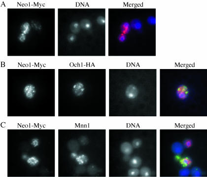
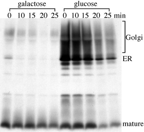
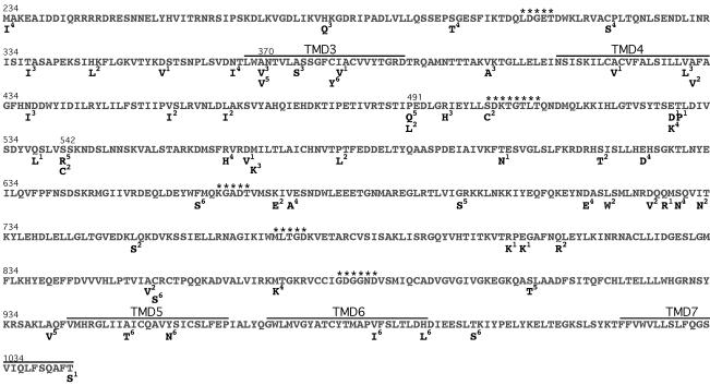
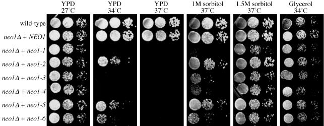
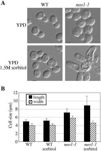
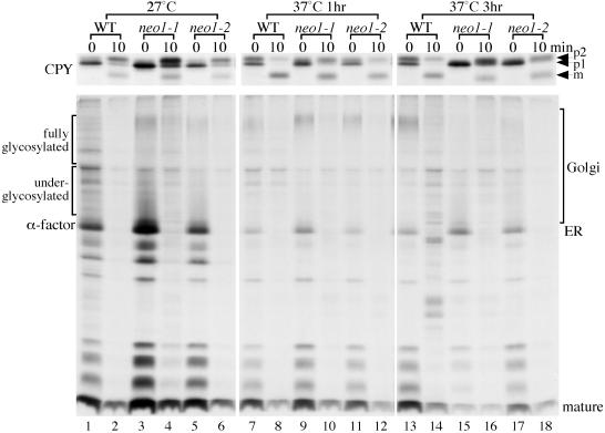
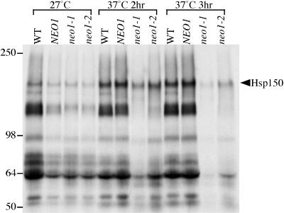
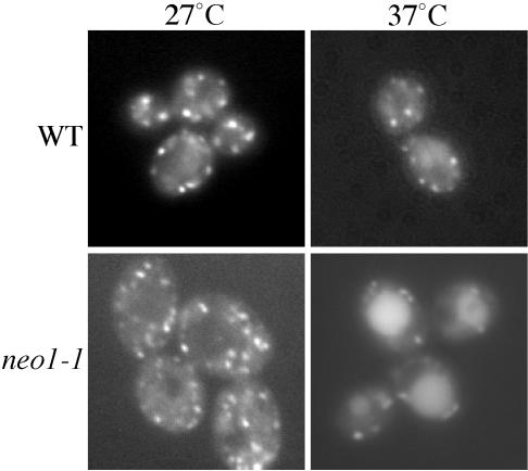
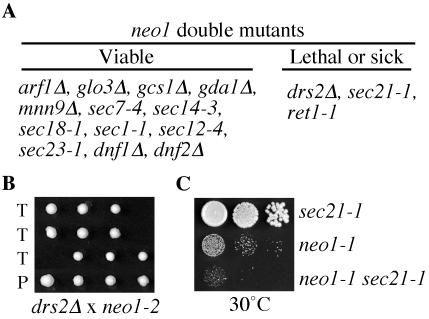
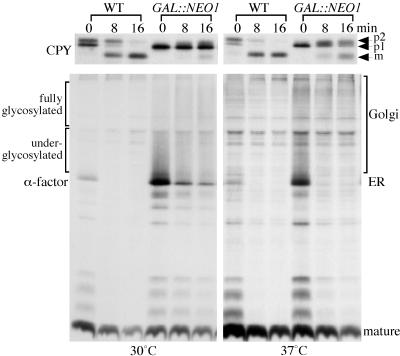
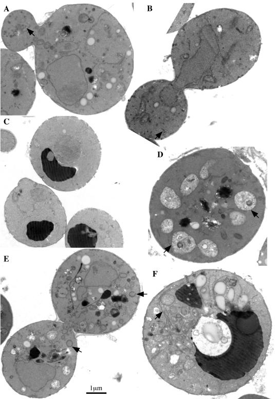
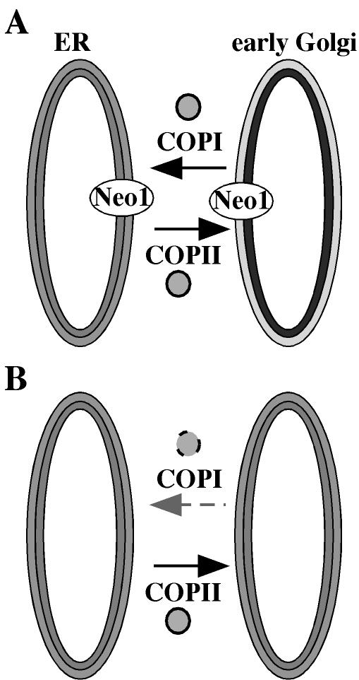
Neo1p from Saccharomyces cerevisiae is an essential P-type ATPase and potential aminophospholipid translocase (flippase) in the Drs2p family. We have previously implicated Drs2p in protein transport steps in the late secretory pathway requiring ADP-ribosylation factor (ARF) and clathrin. Here, we present evidence that epitope-tagged Neo1p localizes to the endoplasmic reticulum (ER) and Golgi complex and is required for a retrograde transport pathway between these organelles. Using conditional alleles of NEO1, we find that loss of Neo1p function causes cargo-specific defects in anterograde protein transport early in the secretory pathway and perturbs glycosylation in the Golgi complex. Rer1-GFP, a protein that cycles between the ER and Golgi complex in COPI and COPII vesicles, is mislocalized to the vacuole in neo1-ts at the nonpermissive temperature. These phenotypes suggest that the anterograde protein transport defect is a secondary consequence of a defect in a COPI-dependent retrograde pathway. We propose that loss of lipid asymmetry in the cis Golgi perturbs retrograde protein transport to the ER.


Figures

Figure 1.
Figure 1.
Neo1-Myc localizes to the ER and Golgi complex. (A) Strain ZHYNEO1-MYC was labeled with a mouse monoclonal anti-myc antibody. (B) The Neo1-Myc strain (ZHYNEO1-MYC) expressing HA-tagged OCH1 (from pOH-URA3) was labeled with rabbit anti-HA antibody and mouse anti-myc to detect Och1-HA and Neo1-Myc. (C) Strain ZHYNEO1-MYC overexpressing Mnn1p (pRS426 MNN1) was stained with affinity-purified polyclonal rabbit anti-Mnn1p and mouse anti-myc. DNA was stained with DAPI to identify the position of the nucleus in all samples. For the overlay images, Neo1-Myc localization is shown in red, Och1-HA or Mnn1p is in green, and DNA is in blue.
Figure 2.
Figure 2.
Neo1p depletion causes protein transport and glycosylation defects. ZHY9075E (neo1Δ GAL::NEO1) was grown in galactose medium and subcultured in media containing either galactose or glucose media for 24 h. Galactose induces high-level expression of Neo1p and glucose shuts off expression from the GAL::NEO1 construct. Cells were labeled with [35S]methionine/cysteine for 15 min and then chased for the times indicated. α-Factor was recovered from each sample by immunoprecipitation and subjected to SDS-PAGE.
Figure 3.
Figure 3.
Mutations present in the six neo1-ts alleles. The NEO1 wild-type sequence from amino acid 234-1043 subjected to mutagenesis is shown with corresponding residue numbers on top. Amino acids substitutions found in the six neo1 ts alleles are aligned underneath the corresponding wild-type residue. The superscript for each amino acid substitution indicates the neo1 allele number from which it is derived (neo1-1 to neo1-6). P-type ATPase conserved motifs are marked with asterisks. Predicted transmembrane domains three through seven are labeled as TMD3-7.
Figure 4.
Figure 4.
The growth defect of neo1-ts mutants is suppressed by osmotic support or nonfermentable carbon sources. Serial dilutions of the wild-type strain (BY4742) or neo1Δ carrying indicated alleles (expressing from plasmids) were spotted onto YPD plates, YPD supplemented with 1 or 1.5 M sorbitol, or YP with 3% glycerol as the carbon source. Plates were incubated at the indicated temperatures for two or three days before photographing.
Figure 5.
Figure 5.
neo1-ts cells are large. (A) Wild-type (BY4742) and neo1-1 (ZHY628-15B) strains were grown at 27°C in YPD media or YPD media supplemented with 1.5 M sorbitol and imaged. (B) Fifty cells of wild-type and neo1-1 strains grown at 27°C in YPD media or YPD sorbitol media were measured for length and width. A Student's t test indicated that the mutant cell sizes are significantly different from the wild-type (p < 0.0001).
Figure 6.
Figure 6.
neo1-ts exhibits Golgi glycosylation and protein transport defects at the nonpermissive temperature. Wild-type (BY4742) and two neo1-ts strains (ZHY628-15B and ZHY628-34A) were grown at 27°C, and then shifted to 37°C for 1 or 3 h. Cells were then labeled with [35S]methionine/cysteine for 6 min, and chased for 0 and 10 min. CPY and α-factor were immunoprecipitated from cells collected at each chase time and subjected to SDS-PAGE.
Figure 7.
Figure 7.
General secretion defect in neo1-ts. Wild-type (BY4742) and neo1Δ harboring either NEO1 (ZHY219RR), neo1-1 (ZHY628–15B), or neo1-2 (ZHY628–34A) were grown at 27°C, and part of each culture was shifted to 37°C for 2 or 3 h. Cells were then labeled with [35S]methionine/cysteine for 10 min and then chased for 30 min. Cells and media were separated by centrifugation, and the proteins in the media were precipitated with TCA and subjected to SDS-PAGE.
Figure 8.
Figure 8.
Rer1-GFP is mislocalized to the vacuole of neo1-1 cells. Wild-type (BY4742) and neo1-1 (ZHY628-15B) harboring an Rer1-GFP plasmid were grown at 27°C, and half of each culture was shifted to 37°C for 2 h. Fluorescent images were captured and representative cells are shown. Cells with large vacuoles were chosen to better distinguish vacuolar fluorescence from the Golgi signal. No ER localization was observed in any of the cells.
Figure 9.
Figure 9.
Genetic interactions between neo1-1 and drs2 or COPI mutations. (A) Summary of double mutant phenotypes. Strains harboring the alleles indicated were crossed with integrated neo1-1 or neo1-2 mutant, sporulated, and subjected to tetrad dissection. Double mutants harboring neo1-1 and the alleles listed in the viable column were recovered at the expected frequency and showed a ts growth profile comparable to one of the two parents. In contrast, most neo1-1 drs2Δ or neo1-2 drs2Δ double mutants were inviable, and the neo1-1 sec21-1 and neo1-1 ret1-1 strains were more temperature sensitive than either parent (Lethal or sick column). (B) Synthetic lethality between neo1-2 and drs2Δ. Dissected tetrads from the neo1-2/drs2Δ cross are shown and the missing colonies in rows labeled “T” (tetratype) were predicted to be double mutants. “P” is a parental ditype with two pairs of single mutant progeny. (C) Growth profile of neo1-1 sec21-1 compared with both parental strains at 30°C, a semipermissive temperature for neo1-1.
Figure 10.
Figure 10.
Neo1p depletion causes a more severe protein transport defect at lower temperature. Wild-type and neo1Δ GAL::NEO1 strains were grown in galactose and then shifted to glucose media for 24 h at 30°C. For labeling at 37°C, half of the cultures cells were shifted to 37°C for the last 2.5 h before labeling. Cells were then labeled with [35S]methionine/cysteine for 8 min and chased for 0, 8, and 16 min. CPY and α-factor was immunoprecipitated from cells collected at each chase time and subjected to SDS-PAGE.
Figure 11.
Figure 11.
neo1-ts cells accumulate abnormal membrane structures. (A and B) neo1-1 (ZHY628–15B) was grown at the permissive temperature of 27°C and processed for electron microscopy. (C) Wild-type cells (BY4742) grown at 37°C for 3 h. Wild-type cells grown at 27°C were indistinguishable and are not shown. (D-F) neo1-1 grown at 37°C for 3 h. Arrows indicate small vesicles in A and B, poorly stained fragmented vacuoles in D, abnormal Golgi membranes in E, and enlarged ER membrane in F.
Figure 12.
Figure 12.
Model for the role of Neo1p in retrograde transport. (A) In wild-type cells, Neo1p probably establishes phospholipid asymmetry in early Golgi membranes (exhibited as light and dark shades of the bilayer membrane), and COPI vesicles form normally. (B) When Neo1p is not functional, lipid asymmetry is lost and the efficiency of COPI vesicle formation is greatly reduced.

Similar articles

Cited by

References

    1. Auland, M.E., Roufogalis, B.D., Devaux, P.F., and Zachowski, A. (1994). Reconstitution of ATP-dependent aminophospholipid translocation in proteoliposomes. Proc. Natl. Acad. Sci. USA 91, 10938-10942. - PMC - PubMed
    1. Balasubramanian, K., and Schroit, A.J. (2003). Aminophospholipid asymmetry: a matter of life and death. Annu. Rev. Physiol. 65, 701-734. - PubMed
    1. Bankaitis, V.A., Malehorn, D.E., Emr, S.D., and Greene, R. (1989). The Saccharomyces cerevisiae SEC14 gene encodes a cytosolic factor that is required for transport of secretory proteins from the yeast Golgi complex. J. Cell Biol. 108, 1271-1281. - PMC - PubMed
    1. Barlowe, C. (2002). COPII-dependent transport from the endoplasmic reticulum. Curr. Opin. Cell Biol. 14, 417-422. - PubMed
    1. Barlowe, C., and Schekman, R. (1993). SEC12 encodes a guanine-nucleotide-exchange factor essential for transport vesicle budding from the ER. Nature 365, 347-349. - PubMed

Publication types

MeSH terms

Substances