. 2003 Nov;14(11):4592-604.
doi: 10.1091/mbc.e03-06-0384. Epub 2003 Sep 5.

Genetic and biochemical evaluation of the importance of Cdc6 in regulating mitotic exit

Affiliations

Genetic and biochemical evaluation of the importance of Cdc6 in regulating mitotic exit

Vincent Archambault et al. Mol Biol Cell. 2003 Nov.

Erratum in

  • Mol Biol Cell. 2005 Mar;16(3):1568

Abstract

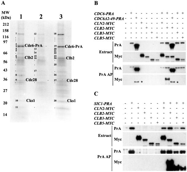
We evaluated the hypothesis that the N-terminal region of the replication control protein Cdc6 acts as an inhibitor of cyclin-dependent kinase (Cdk) activity, promoting mitotic exit. Cdc6 accumulation is restricted to the period from mid-cell cycle until the succeeding G1, due to proteolytic control that requires the Cdc6 N-terminal region. During late mitosis, Cdc6 is present at levels comparable with Sic1 and binds specifically to the mitotic cyclin Clb2. Moderate overexpression of Cdc6 promotes viability of CLB2Deltadb strains, which otherwise arrest at mitotic exit, and rescue is dependent on the N-terminal putative Cdk-inhibitory domain. These observations support the potential for Cdc6 to inhibit Clb2-Cdk, thus promoting mitotic exit. Consistent with this idea, we observed a cytokinesis defect in cdh1Delta sic1Delta cdc6Delta2-49 triple mutants. However, we were able to construct viable strains, in three different backgrounds, containing neither SIC1 nor the Cdc6 Cdk-inhibitory domain, in contradiction to previous work. We conclude, therefore, that although both Cdc6 and Sic1 have the potential to facilitate mitotic exit by inhibiting Clb2-Cdk, mitotic exit nevertheless does not require any identified stoichiometric inhibitor of Cdk activity.


Figures

Figure 1.
Figure 1.
Cdc6 associates with Clb2. (A) Cdc6 forms a complex with Clb2, Cdc28, and Cks1. Asynchronous CDC6-PRA cells (lane 1), α-factor–arrested CDC6-PRA bar1 cells (lane2) or glucose-arrested CDC6-PRA cdc20 GALL-CDC20 cells (lane 3) were submitted to a protein A affinity purification. Bands marked with either a dot or number were excised and analyzed by MALDI-mass spectrometry to identify proteins. Bands containing proteins identified with high confidence are numbered and the identifications are listed in the supplementary Table SI. Bands identified with a dot did not give a high-confidence identification. Proteins that seem to be specific to the Cdc6-PrA pullout are labeled. (B) Cdc6 associates with Clb2 and not with other cyclins and its N terminus is required for this association. CDC6-PRA or cdc6Δ2-49-PRA or CDC6-wt (untagged) cells also expressing CLN2-MYC, CLB2-MYC, CLB3-MYC, or CLB5-MYC were submitted to protein A affinity purification. Top, whole cell soluble extracts probed by Western blot for protein A and Myc. Bottom, purified proteins probed by Western blot for protein A and Myc. In the anti-MYC Western blot, the bands indicated by the asterisk (*) corresponded to the protein A tag from Cdc6Δ2-49-PrA that was recognized by the anti-MYC antibody due to its high abundance. (C) Sic1 associates with Clb2, Clb3, and Clb5 but not Cln2 (methods as in B).
Figure 2.
Figure 2.
Cdc6 temporally overlaps and associates with Clb2 in mitotic cells. (A) Timing of expression. CDC6-PRA (or cdc6Δ2-49-PRA, or SIC1-PRA) cdc20 GALL-CDC20 cells were grown in YPG, arrested in YPD for 3 h and released in YPG. Top, budding index (squares, CDC6-PRA cells; diamonds, cdc6Δ2-49-PRA cells; triangles, SIC1-PRA cells). Bottom, Western blot analysis of the cultures probing for protein A and Clb2. In cdc6Δ2-49-PRA cells, a breakdown fragment of Cdc6Δ2-49-PrA migrated at a molecular weight identical to Clb2, possibly accounting for the low signal observed for unbudded cells in the Clb2 blot because the protein A could be detected by the Clb2 antibody. (B) Cdc6 associates with Clb2 in mitotic cells. Right, a time course of protein A affinity purification was performed on CDC6-PRA cdc20 GALL-CDC20 cells grown in YPG, arrested in YPD for 3 h, and released in YPG. Samples were taken every 10 min after release for affinity purification. Left, controls for the affinity purification; all asynchronous cultures. Lane a, CDC6-PRA cells; lane b, CLB2-MYC cells; lane c, CDC6-PRA cells mixed with CLB2-MYC cells before cell breakage (control for postlysis binding); lane d, CDC6-PRA cdc20 GALL-CDC20 cells; and lane e, CDC6-PRA CLB2-MYC cdc20 GALL-CDC20 cells. Top, budding index; middle, whole cell soluble extracts probed by Western blot for protein A and Myc; bottom, purified proteins probed by Western blot for protein A and Myc.
Figure 3.
Figure 3.
Mutants expressing stabilized forms of Clb2 are rescued by CDC6 in an N terminus-dependent manner. CLB2Δdb GALSIC1(1 ×) or CLB2Δdb,ken GAL-SIC1 (2 ×) cells were transformed with plasmids expressing CDC6, cdc6Δ2-49, cdc6-3P-, and cdc6Δ8-17. Galactose-grown cultures were serially diluted (10-fold dilutions) on galactose- or glucose-containing rich medium and incubated for 2 d at 30°C.
Figure 4.
Figure 4.
cdc6Δ2-49 sic1 mutants are viable. (A) In the W303 background, a deletion of coding sequence for amino acids 2–49 was introduced in the chromosome with no associated marker, as described in the text and Figure S3. Left, sic1::HIS3 cdc6Δ2-49 pURA3-SIC1 strains were constructed by tetrad analysis, and loss of the pSIC1-URA3 plasmid selected on 5-FOA. Strains with and without the plasmid were tested for the CDC6 N-terminal deletion and for the sic1::HIS3 mutation by PCR, and for growth rate by serial dilution at 30 and 37°. Right, segregants from a cross with cdh1::LEU2, sic1::HIS3, and cdc6Δ2-49 segregating were tested by PCR. The second PCR analysis tests specifically for the presence of the coding sequence for amino acids 2-49. The strains were tested for growth rate as in the left panel. (B) In the S288C background (Figure 4B), we made a strain that was cdc6Δ::kanMX sic1Δ::kanMX pURA3-CDC6, and a control cdc6Δ::kanMX SIC1 pURA3-CDC6 strain (complete coding sequence deletions). All kanMX disruptions were confirmed by PCR. We transformed these strains with lowcopy (CEN) plasmids encoding wild-type and the three mutant CDC6 genes, expressed from the CDC6 promoter (Weinreich et al., 2001). All transformants, except for the vector controls could readily lose the URA3/CDC6 plasmid. (C) BF264-15D-congenic background: We backcrossed cdc6Δ2-49 from W303 (see A) into the BF264-15D congenic strain background (see text). Isogenic pairs of sic1Δ pURA3-SIC1 segregants (cdc6Δ2-49 or CDC6) from the fifth backcross were selected for pURA3-SIC1 plasmid loss by FOA and tested for growth rate by serial dilution at 30 and 37°. PCR characterization of the strains was performed as in A.
Figure 5.
Figure 5.
Removal of the Cdc6 N terminus reduces viability of sic1Δ cdh1Δ cells. Galactose-grown cultures were serially diluted (10-fold dilutions) on galactose- or glucose-containing rich medium and incubated for 3 d at 30°C. All strains contained the TUB1-GFP allele (used in Figure 6).
Figure 6.
Figure 6.
Removal of the Cdc6 N terminus confers a cytokinesis defect in sic1Δ cdh1Δ cells. (A) Cells with the indicated genotypes (the same strains as in Figure 5) were cultured in galactose-containing medium (left) and then transferred to glucose-containing medium for 4 h (right). Differential interference contrast (DIC) pictures, Tub1-GFP fluorescence pictures, and FACS profiles are shown for each condition. Brackets in the lower DIC picture indicate the four-cell body objects that were resistant to sonication (see text). (B) Cells with the indicated genotypes (wild-type TUB1) were cultured in galactose-containing medium and then transferred to glucose-containing medium. FACS profiles were taken after 2.5 and 5 h. Cells taken at 2.5 h were fixed and immunofluorescence was used to visualize spindles in DAPI-stained cells; long spindles with separated nuclei (anaphase spindles) were counted.
Figure 7.
Figure 7.
CDC6 rescues sic1Δ cdh1Δ cells. A cdh1Δ sic1Δ strain carrying a pSIC1-URA3 plasmid was transformed with LEU2 plasmids, either vector (RS415, low-copy, or RS425, high-copy), RS415 containing CDH1 as a positive control, or RS425 carrying wt and mutant CDC6. (A) Transformants were patched on -leu and then replica-plated to -ura (indicating retention of the pSIC1-URA3 plasmid) or FOA (indicating ability to lose this plasmid). Note that one RS425-CDC6 transformant spontaneously lost the pSIC1-URA3 plasmid. (B) Serial dilutions of transformants on -leu or FOA.
Figure 8.
Figure 8.
CDH1, SWI5, and CDC6 interact genetically. (A) cdh1Δ swi5Δ mutants are inviable and can be rescued by SIC1 and by CDC6. Rescue by CDC6 depends on both its N terminus and its DNA-replication activity. cdh1Δ swi5Δ GAL-SIC1 cells were transformed with plasmids expressing SIC1, CDC6, cdc6Δ2-49, cdc6-3P-, cdc6Δ8-17, and cdc6-K114E. Galactose-grown cultures were serially diluted (10-fold dilutions) on galactose-or glucose-containing rich medium and incubated for 2 d at 30°C. (B) Cells with the indicated genotype were grown in galactose-containing medium and transferred to glucose-containing medium for 3 h. FACS profiles are shown, with the percentage of binucleated cells in the top right corner.
Figure 9.
Figure 9.
Genetic interaction of cdc6Δ2-49 and sic1Δ with the mitotic exit network. (A) Interaction with cdc14-1. A W303-congenic cdc14-1 strain was constructed by repeated backcrossing and tetrad analysis, and a cdc6Δ2-49/+ sic1Δ/+ cdc14-1/+ GAL-SIC1::TRP1/trp1 diploid was constructed. Haploid segregants of the indicated genotype (all GAL-SIC1 cdc14-1) were tested for growth by serial dilution and incubation at the indicated temperature, on galactose (GAL-SIC1 on) or glucose (GAL-SIC1 off). (B) Interaction with cdc15-2. Methods as in A. (C) The indicated plasmids were introduced into a cdc14-1 strain and replica-plated to plates that were incubated at the indicated temperatures.

Similar articles

Cited by

References

    1. Aitchison, J.D., Blobel, G., and Rout, M.P. (1996). Kap104p: a karyopherin involved in the nuclear transport of messenger RNA binding proteins. Science 274, 624-627. - PubMed
    1. Bardin, A.J., and Amon, A. (2001). Men and sin: what's the difference? Nat. Rev. Mol. Cell Biol. 2, 815-826. - PubMed
    1. Basco, R.D., Segal, M.D., and Reed, S.I. (1995). Negative regulation of G1 and G2 by S-phase cyclins of Saccharomyces cerevisiae. Mol. Cell Biol. 15, 5030-5042. - PMC - PubMed
    1. Beavis, R.C., and Chait, B.T. (1996). Matrix-assisted laser desorption ionization mass-spectrometry of proteins. Methods Enzymol. 270, 519-551. - PubMed
    1. Bell, S.P., and Dutta, A. (2002). DNA replication in eukaryotic cells. Annu. Rev. Biochem. 71, 333-374. - PubMed

Publication types

MeSH terms

Substances

LinkOut - more resources