. 2003 Dec;14(12):4813-25.
doi: 10.1091/mbc.e03-05-0308. Epub 2003 Sep 5.

Integration of cell attachment, cytoskeletal localization, and signaling by integrin-linked kinase (ILK), CH-ILKBP, and the tumor suppressor PTEN

Affiliations

Integration of cell attachment, cytoskeletal localization, and signaling by integrin-linked kinase (ILK), CH-ILKBP, and the tumor suppressor PTEN

Sarah Attwell et al. Mol Biol Cell. 2003 Dec.

Abstract

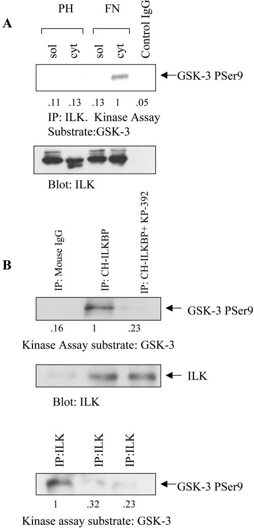
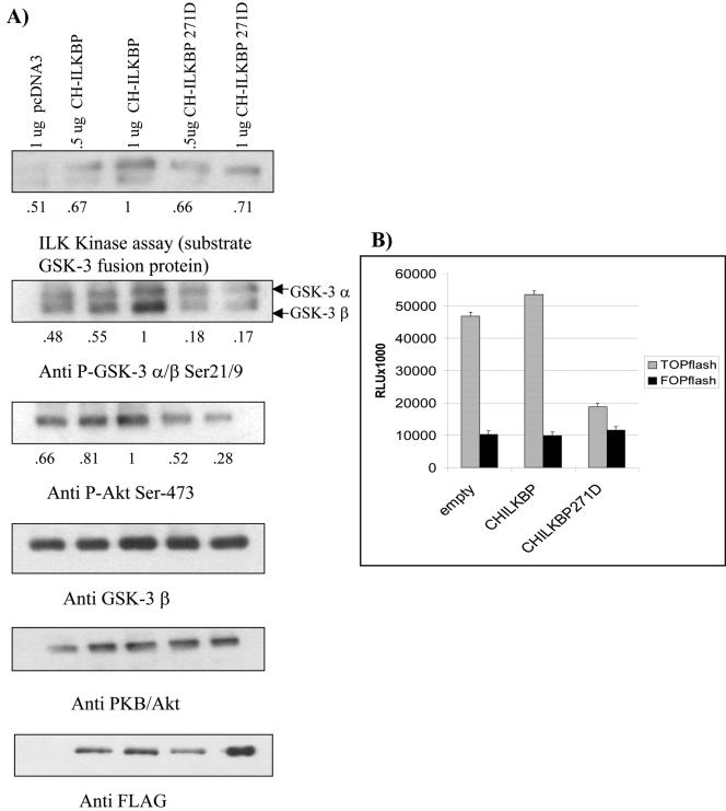
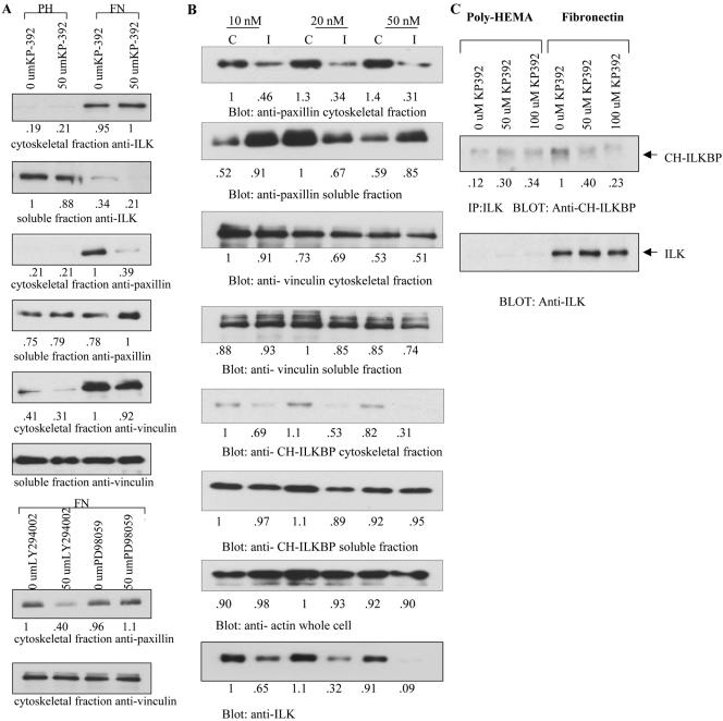
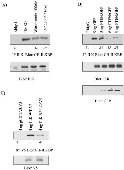
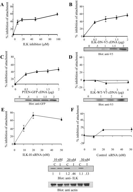
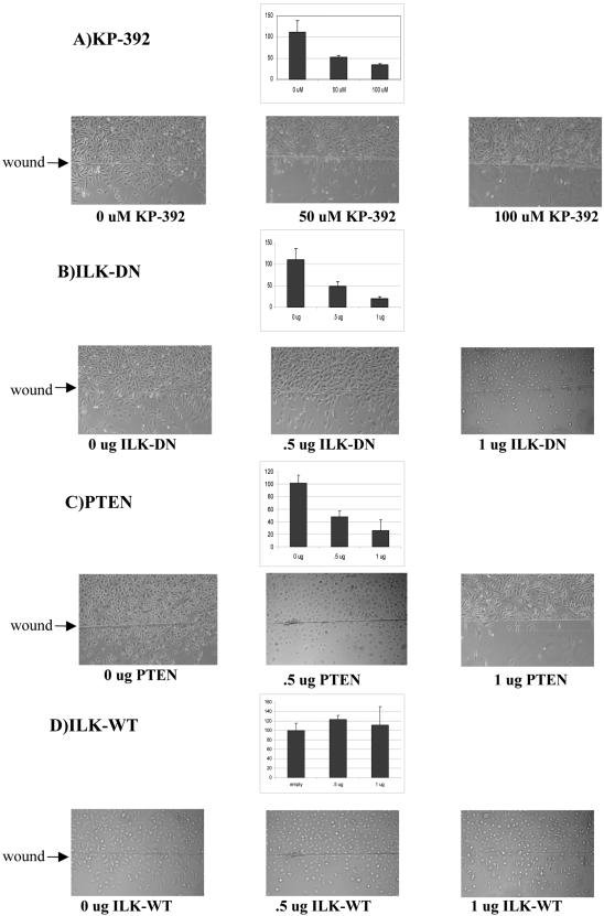
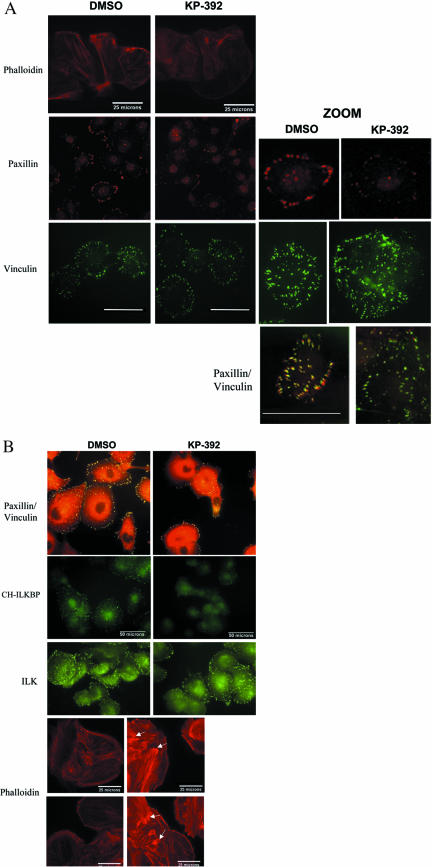
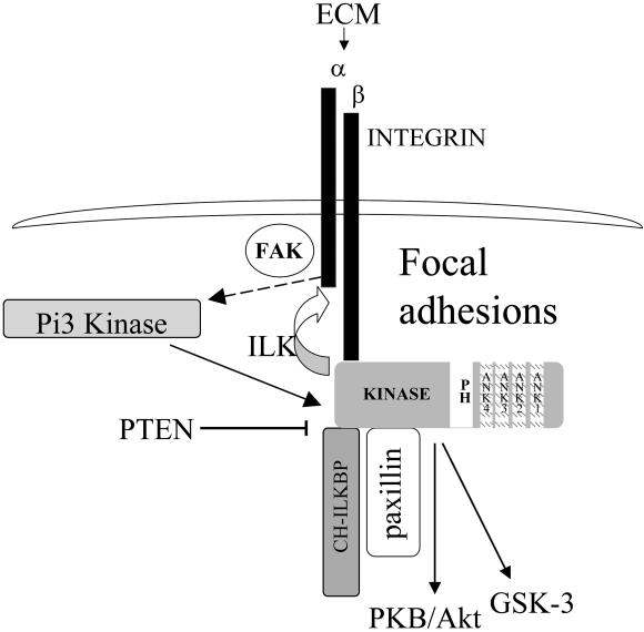
Cell attachment and the assembly of cytoskeletal and signaling complexes downstream of integrins are intimately linked and coordinated. Although many intracellular proteins have been implicated in these processes, a new paradigm is emerging from biochemical and genetic studies that implicates integrin-linked kinase (ILK) and its interacting proteins, such as CH-ILKBP (alpha-parvin), paxillin, and PINCH in coupling integrins to the actin cytoskeleton and signaling complexes. Genetic studies in Drosophila, Caenorhabditis elegans, and mice point to an essential role of ILK as an adaptor protein in mediating integrin-dependent cell attachment and cytoskeletal organization. Here we demonstrate, using several different approaches, that inhibiting ILK kinase activity, or expression, results in the inhibition of cell attachment, cell migration, F-actin organization, and the specific cytoskeletal localization of CH-ILKBP and paxillin in human cells. We also demonstrate that the kinase activity of ILK is elevated in the cytoskeletal fraction and that the interaction of CH-ILKBP with ILK within the cytoskeleton stimulates ILK activity and downstream signaling to PKB/Akt and GSK-3. Interestingly, the interaction of CH-ILKBP with ILK is regulated by the Pi3 kinase pathway, because inhibition of Pi3 kinase activity by pharmacological inhibitors, or by the tumor suppressor PTEN, inhibits this interaction as well as cell attachment and signaling. These data demonstrate that the kinase and adaptor properties of ILK function together, in a Pi3 kinase-dependent manner, to regulate integrin-mediated cell attachment and signal transduction.


Figures

Figure 5.
Figure 5.
Active ILK is bound to CH-ILKBP, and is localized to the cytoskeletal fraction when cells are plated on FN. (A) Serum-starved PC3 cells were plated on the substrates poly-HEMA (PH) or fibronectin (FN) for 1 h, and the soluble and cytoskeletal fractions were separated. ILK (or control mouse IgG) was immunoprecipitated from the cytoskeletal fraction, and kinase assays were performed using GSK-3 fusion protein as a substrate. Samples were then Western blotted with antiphospho GSK-3α/β Ser 21/9, and anti-ILK. (B) PC3 cells lysates were immunoprecipitated with anti-mouse IgG, or with anti CH-ILKBP, and kinase assays were performed using GSK-3 fusion protein as a substrate. The blot was stripped and reprobed with anti-ILK, to show immunoprecipitation. The leftover lysates (cleared with anti mouse IgG or anti–CH-ILKBP) were then immunoprecipitated with anti-ILK antibody, and the ILK kinase assay was then performed on the GSK-3 fusion protein substrate. Results are representative of three independent trials.
Figure 6.
Figure 6.
CH-ILKBP, but not an ILK-binding mutant (CH-ILKBP F271D) stimulates ILK signaling in DU145 prostate cancer cells. (A) Cells were transfected with empty vector, CH-ILKBP, or CH-ILKBP F271D. After 48 h, cells were serum-starved overnight, then refed for 1 h. Samples were Western blotted with anti-phospho GSK-3α/β Ser 21/9, anti-phospho PKB/Akt Ser 473, anti-GSK-3 β, anti-PKB/Akt, and anti-FLAG. Kinase assay was performed using GSK-3 fusion protein as a substrate, and then Western blotting with anti-phospho GSK-3α/β Ser 21/9. (B) HEK-293 cells were transfected with empty vector, CH-ILKBP, or CH-ILKBP F271D, as well as either the TOP or FOP FLASH reporter constructs, and pRenilla. After 48 h, a dual luciferase reporter assay was performed. Results are representative of three independent trials.
Figure 3.
Figure 3.
Inhibition of ILK activity by KP-392 disrupts the localization of paxillin to the Triton-insoluble fraction, and disrupts ILK:CH-ILKBP binding. (A) Inhibition by KP-392. Du145 cells were plated on either poly-HEMA (PH) or fibronectin (FN)-coated plates, and treated with either DMSO or 50 μM KP-392 for 16 h. Soluble and cytoskeletal fractions were then separated, and Western blots performed, with 20 μg of lysate per sample, as described in MATERIALS AND METHODS. (B) PC3 cells were transiently transfected with ILK-H siRNA specific to the PH-like domain of ILK (I), or with control siRNA (C), at concentrations of either 10, 25, or 50 nM. After 72 h, cells were then treated as in A. (C) Du145 cells were plated on poly-HEMA or FN and treated with increasing amounts of KP-392. The cytoskeletal fraction was isolated, and this fraction was immunoprecipitated with anti-ILK antibody. Western blots were then performed as described in MATERIALS AND METHODS. All results are representative of 3 independent trials.
Figure 7.
Figure 7.
THE ILK:CH-ILKBP interaction is Pi3 kinase dependent. (A) Chemical inhibitors disrupt the ILK:CH-ILKBP interaction. PC3 cells were treated with either DMSO, wortmannin, or LY294002 for 3 h. Cells were then lysed with NP-40, and immunoprecipitated with anti-ILK. Samples were then Western blotted with anti-CH-ILKBP, and stripped and reprobed with anti-ILK. (B) PTEN reintroduction disrupts the ILK:CH-ILKBP interaction. PC3 cells were transfected with increasing amounts of PTEN. After 48 h, cells were treated as in A, and samples were also stripped and reprobed with anti-GFP as a transfection control. (C) The ILK:CH-ILKBP interaction is reduced when the proposed PiP3 binding domain of ILK is mutated. PC3 cells were transfected with pCDNA3:V5, ILK-WT:V5, or ILK R211A:V5. After 48 h, cells were lysed with NP-40 lysis buffer, immunoprecipitated with anti-V5 antibody, and then treated as in A. Results are representative of three independent trials.
Figure 1.
Figure 1.
Inhibition of ILK activity decreases cell attachment. PC3 cells were treated with increasing amounts of inhibitor (A) for 24 h, were transiently transfected with ILK-DN:V5 (B), PTEN:GFP (C), or ILK-WT:V5 (D) and left to recover for 48 h, or were transfected with ILK-H siRNA specific to the PH-like domain of ILK (I) (E), or control siRNA (C) (F), and left to recover for 72 h. Attachment assay was performed as described in methods. Each sample was repeated in triplicate, as indicated by error bars, and the experiment was repeated three times. Data are plotted as % increase in the inhibition of attachment vs. control.
Figure 2.
Figure 2.
Inhibition of ILK activity disrupts cell migration. (A) A wound was introduced to PC3 cells as described in MATERIALS AND METHODS, and cells were then treated with increasing amounts of KP-392 for 24 h. Migrated cells were photographed and counted in five separate fields. Alternatively, cells were transfected with PTEN:GFP (B), ILK-DN:V5 (C), or ILK-WT:V5 (D). Wounding assay was then performed 24 h posttransfection. Results are representative of three independent trials.
Figure 4.
Figure 4.
Inhibition of ILK activity with KP-392 disrupts the localization of paxillin and CH-ILKBP, but not ILK and vinculin, to focal adhesion plaques. Du145 cells were plated on FN-coated coverslips, with DMSO or 50 μM KP-392, for 16 h. Cells were then either solubilized with a Triton wash (A), or fixed as whole cells (B). Samples were then stained with the appropriate antibodies. (A) Solubilized staining. Rhodamine; bar, 25 μm. Paxillin (rhodamine) and vinculin (FITC), bar represents 50 μm. Paxillin and vinculin zoom, Bar, 39 μm. (B) Whole cell staining. Paxillin (rhodamine) and vinculin (FITC) merge. CH-ILKBP (FITC), ILK (FITC), and phalloidin. Paxillin and CH-ILKBP are specifically dissociated from focal adhesion plaques upon inhibition of ILK activity. Results are representative of three independent trials. Bar, 50 μm.
Figure 8.
Figure 8.
Summary of ILK recruitment and activity at focal adhesions. On integrin engagement with the extracellular matrix (ECM), Pi3 kinase is activated, through FAK, and clustering and coactivation of growth factor receptor tyrosine kinases. ILK is then activated through Pi3 kinase, allowing binding with CH-ILKBP and paxillin, and recruitment to focal adhesion plaques. At the focal adhesion plaques, ILK activity is crucial for maintaining upstream signaling to β1 integrins, and downstream signaling to PKB/Akt and GSK-3.

References

    1. Attwell, S., Roskelley, C., and Dedhar, S. (2000). The integrin-liked kinase (ILK) suppresses anoikis. Oncogene 19, 3811–3815. - PubMed
    1. Brakebusch, C., Bouvard, D., Stanchi, F., Sakai, T., and Fassler, R. (2002). Integrins in invasive growth. J. Clin. Invest. 109, 999–1006. - PMC - PubMed
    1. Calderwood, D.A., Shattil, S.J., and Ginsberg, M.H. (2000). Integrins and actin filaments: reciprocal regulation of cell adhesion and signaling. J. Biol. Chem. 275, 22607–22610. - PubMed
    1. Carrieras, F., Rigot, V., Cruet, S., Andre, F., Gauduchon, P., and Marvaldi, J. (1999). Migration properties of the human ovarian adenocarcinoma cell line IGROV 1, Importance of αvβ3 integrins and vitronectin. Int. J. Cancer. 80, 285–94. - PubMed
    1. Cruet-Hennequart, S., Maubant, S., Luis, J., Gauduchon, P., Staedel, C., and Dedhar, S. (2003). αv integrins regulate cell proliferation through integrin-linked kinase (ILK) in ovarian cancer cells. Oncogene 22, 1688–1702. - PubMed

Publication types

MeSH terms