. 2003 Dec;14(12):4885-95.
doi: 10.1091/mbc.e03-05-0295. Epub 2003 Sep 5.

The simultaneous production of phosphatidic acid and diacylglycerol is essential for the translocation of protein kinase Cepsilon to the plasma membrane in RBL-2H3 cells

Affiliations

The simultaneous production of phosphatidic acid and diacylglycerol is essential for the translocation of protein kinase Cepsilon to the plasma membrane in RBL-2H3 cells

Maria Jose Lopez-Andreo et al. Mol Biol Cell. 2003 Dec.

Abstract

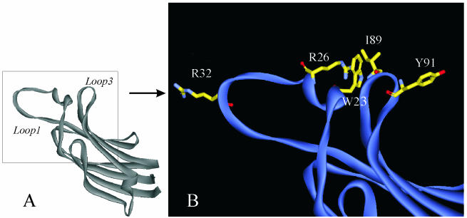
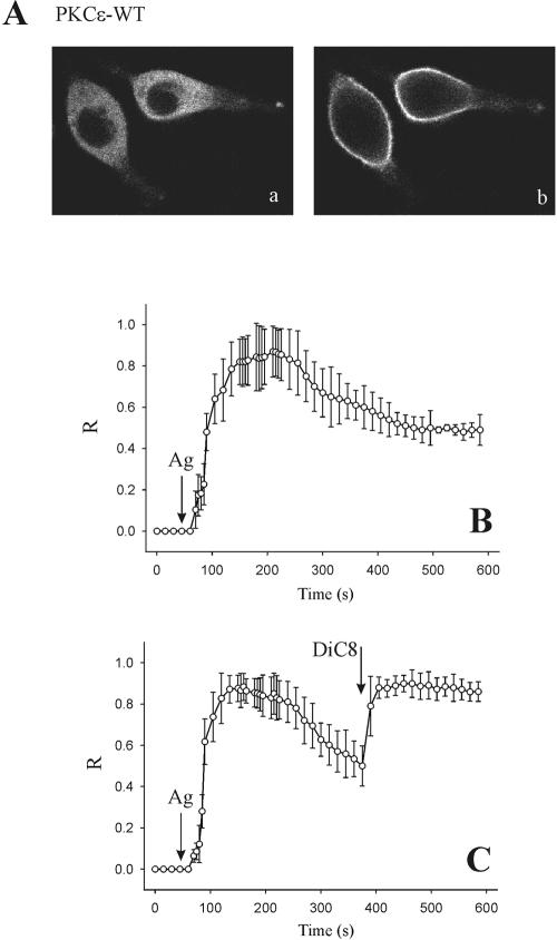
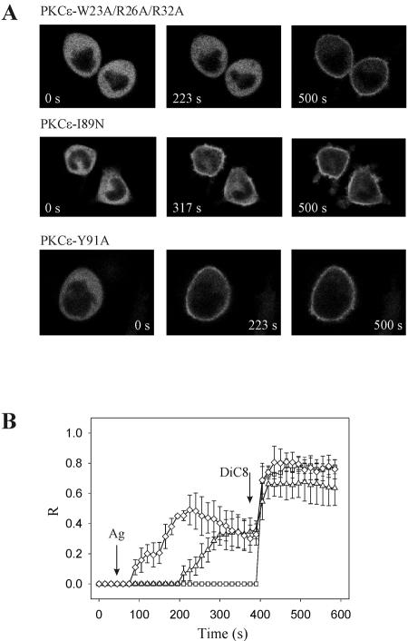
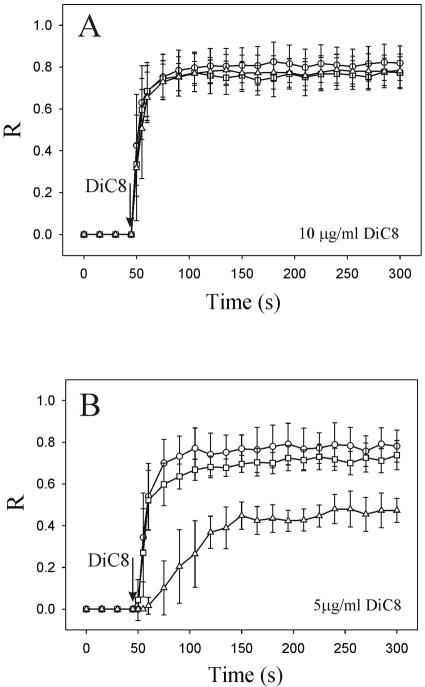
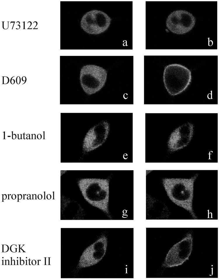
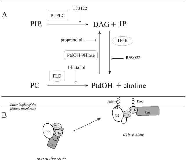
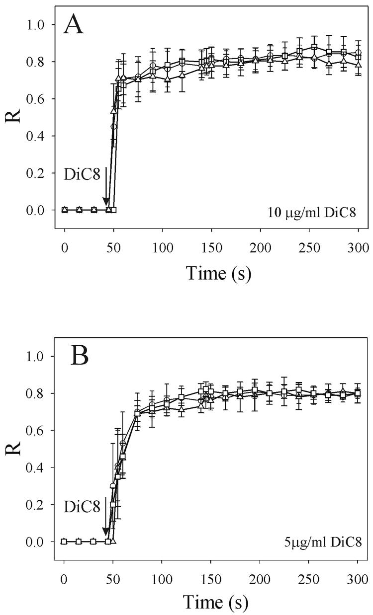
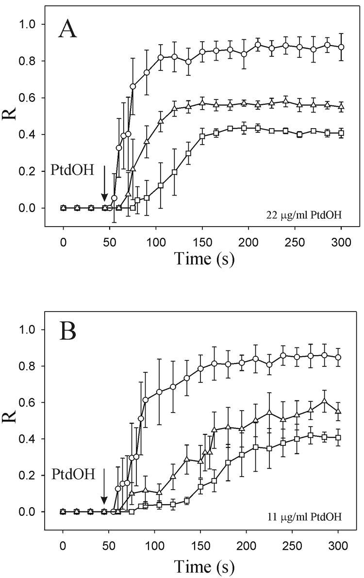
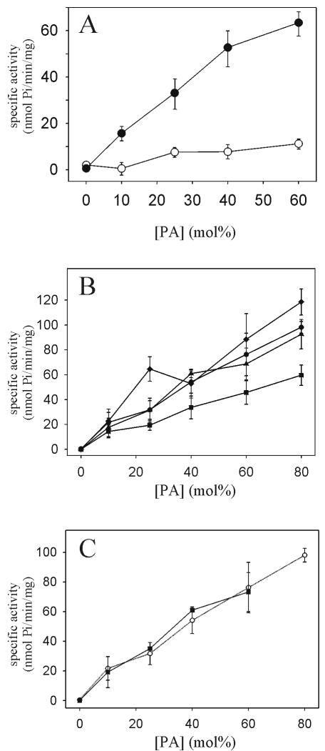
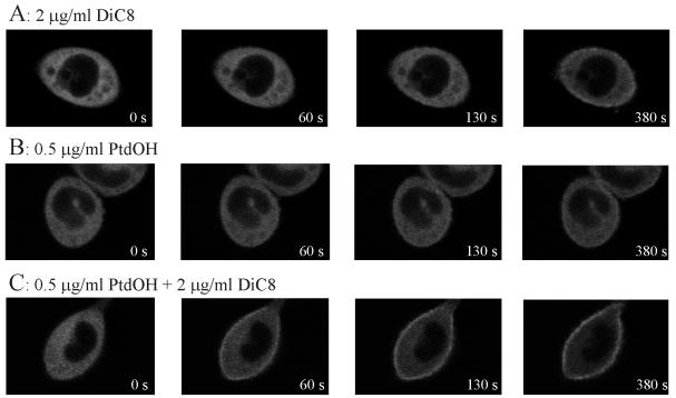
To evaluate the role of the C2 domain in protein kinase Cepsilon (PKCepsilon) localization and activation after stimulation of the IgE receptor in RBL-2H3 cells, we used a series of mutants located in the phospholipid binding region of the enzyme. The results obtained suggest that the interaction of the C2 domain with the phospholipids in the plasma membrane is essential for anchoring the enzyme in this cellular compartment. Furthermore, the use of specific inhibitors of the different pathways that generate both diacylglycerol and phosphatidic acid has shown that the phosphatidic acid generated via phospholipase D (PLD)-dependent pathway, in addition to the diacylglycerol generated via phosphoinosite-phospholipase C (PLC), are involved in the localization of PKCepsilon in the plasma membrane. Direct stimulation of RBL-2H3 cells with very low concentrations of permeable phosphatidic acid and diacylglycerol exerted a synergistic effect on the plasma membrane localization of PKCepsilon. Moreover, the in vitro kinase assays showed that both phosphatidic acid and diacylglycerol are essential for enzyme activation. Together, these results demonstrate that phosphatidic acid is an important and essential activator of PKCepsilon through the C2 domain and locate this isoenzyme in a new scenario where it acts as a downstream target of PLD.


Figures

Figure 1.
Figure 1.
(A) Overall structure of the C2 domain of PKCε. The central structural feature of this C2 domain is an eight-stranded antiparallel β-sandwich of type II topology. Loop 1 corresponds to the connection between β1 and β2 strands and, loop 3 corresponds to the connection between β5 and β6 strands. The square box is amplified in B. (B) Represents the top region of the C2 domain, corresponding to loops 1, 2, and 3. The side chains of the mutated amino acids are shown in yellow. The three mutants generated in this work are: PKCε-W23A/R26A/R32A in loop 1, PKCε-I89N and PKCε-Y91A in loop 3.
Figure 2.
Figure 2.
(A) Receptor-induced translocation of PKCε to the plasma membrane. The FcεRI of RBL-2H3 cells were sensitized by the addition of 0.5 μg/ml anti-DNP IgE for 16 h. Confocal images were obtained in a time lapse experiment, during which a series of 120 images were recorded at 5-s intervals. Receptor cross-linking was performed by addition of 4 μg/ml DNP-HSA after 45 s of recording. The images shown were recorded at 0 (a) and 200 s (b). (B) Average data of the time course of plasma membrane translocation of PKCε after stimulation of the receptor by antigen. R, relative plasma membrane translocation calculated as explained in MATERIALS AND METHODS. (C) Average data of the time course of plasma membrane translocation of PKCε upon antigen and DiC8 stimulation, DiC8 was added after 370 s of recording.
Figure 3.
Figure 3.
(A) Confocal fluorescence images of RBL-2H3 cells expressing the different phospholipid-binding mutants, PKCε-W23A/R26A/R32A, PKCε-I89N, and PKCε-Y91A at different points of the time-lapse experiment. Antigen was added after 45 s and DiC8 after 395 s of recording. (B) Average of the time courses of plasma membrane localization of PKCε-W23A/R26A/R32A (□), PKCε-I89N (▵) and PKCε-Y91A (⋄) under the conditions stated above.
Figure 4.
Figure 4.
Average of the time courses of plasma membrane localization when RBL-2H3 cells were transfected with wild-type PKCε (○), PKCε-W23A/R26A/R32A (□) and PKCε-I89N (▵) and stimulated with 10 μg/ml DiC8 (A) and 5 μg/ml DiC8 (B). R, relative plasma membrane translocation.
Figure 5.
Figure 5.
The cultures were preincubated at 37°C with the appropriate concentrations of inhibitors for 10 min before the addition of 4 μg/ml DNP-HSA. a, c, e, g, and i represent the cells before antigen stimulation. b, d, f, h, and j represent the same cells 200 s after antigen addition. The inhibitors were used at 30 μM U73122 (a and b), 30 μM D-609 (c and d), 50 mM 1-butanol (e and f), 100 μM propranolol (g and h), and 20 μM DGK inhibitor II (i and j). The cells shown are representative of four independent assays.
Figure 10.
Figure 10.
Mechanism of activation of PKCε in RBL-2H3 cells upon activation of the IgE receptor. (A) Scheme showing the lipid intermediate generated upon receptor stimulation. In a first step, phosphatidylinositol-4,5-bisphosphate is hydrolyzed to produce DAG and inositol triphosphate by PI-PLC; simultaneously PC is hydrolyzed by PLD to produce PtdOH and choline. In a second step, DAG and PtdOH generated are transformed to PtdOH and DAG, respectively, by the action of DGK and PtdOH-phosphohydrolase. The inhibitors of each reaction are marked with a bar arrow. (B) Together, the results obtained in this work suggest that when the IgE receptor is activated, the enzyme translocates to the plasma membrane when PtdOH and DAG are generated simultaneously in the plasma membrane, thus leading to the catalytic activation of the enzyme (active state). It has been shown the C1b domain as DAG receptor assuming the model proposed by Szallasi et al. (1996).
Figure 6.
Figure 6.
Average of the time courses of plasma membrane localization when RBL-2H3 cells were transfected with wild-type PKCε and stimulated with 10 μg/ml DiC8 (A) and 5 μg/ml DiC8 (B) in the absence (○) and in the presence of 50 mM 1-butanol (□) or 100 μM propranolol (▵). R, relative plasma membrane translocation.
Figure 7.
Figure 7.
PtdOH-dependent plasma membrane localization of wild-type PKCε (○), PKCε-W23A/R26A/R32A (□) and PKCε-I89N (▵). Averages of the time courses of localization when the cells transfected with the different constructs were stimulated with 22 μg/ml PtdOH (A) and 11 μg/ml PtdOH (B).
Figure 8.
Figure 8.
(A) Specific activity of PKCε by using large unilamellar vesicles containing POPC/POPA (99-X: X, with X being the molar fraction of POPA in each case) (○) and POPC/DOG/POPA (95-X:5:X, with X being the molar fraction of POPA in each case) (•). (B) PtdOH-dependent activation of wild-type PKCε (•), PKCε-W23A/R26A/R32A (▪), PKCε-I89N (▴), and PKCε-Y91A (♦) in large unilamellar vesicles. PKC activity was measured using POPC/DOG/POPA large unilamellar vesicles (95-X:5:X, with X being the molar fraction of POPA in each case). (C) Specific activity of PKCε by using large unilamellar vesicles containing POPC/POPA (99-X: X, with X being the molar fraction of POPA in each case) (○) (these results are the same that in part B of this figure, and have been represented here to facilitate the comparison of the data) and POPC/POPS/DOG/POPA (95-X:20:5:X, with X being the molar fraction of POPA in each case) (▪). The total lipid concentration was 0.2 mM and Histone II-SS was used as a substrate. Error bars indicate the SEM for triplicate determinations.
Figure 9.
Figure 9.
RBL-2H3 cells were stimulated with 2 μg/ml DiC8 (A), 0.5 μg/ml PtdOH (B), or a combination of the two activators together, 2 μg/ml DiC8 + 0.5 μg/ml PtdOH (C). Time series were collected each 5 s during 10 min. These cells are representative of three independent experiments.

Similar articles

Cited by

References

    1. Akita, Y., et al. (1994). Overproduction of a Ca2+-independent protein kinase C isozyme, nPKC epsilon, increases the secretion of prolactin from thyrotropin-releasing hormone-stimulated rat pituitary GH4C1 cells. J. Biol. Chem. 269, 4653–4660. - PubMed
    1. Andresen, B.T., Rizzo, M.A., Shome, K., and Romero, G. (2002). The role of phosphatidic acid in the regulaton of the Ras/MEK/ERK signalling cascade. FEBS Lett. 531, 65–68. - PubMed
    1. Barker, S.A., Caldwell, K.K., Pfeiffer, J.R., and Wilson, B.S. (1998). Wortmannin-sensitive phosphorylation, translocation and activation of PLCγ1 but not PLCγ2, in antigen-stimulated RBL-2H3 mast cells. Mol. Biol. Cell 9, 483–496. - PMC - PubMed
    1. Besson, A., Wilson, T.L., and Yong, V.W. (2002). The anchoring protein RACK1 links protein kinase C epsilon to integrin beta chains. J. Biol. Chem. 277, 22073–22084. - PubMed
    1. Bleasdale, J.E., Thakur, N.R., Gremban, R.S., Bundy, G.L., Fitzpatrick, F.A., Smith, R.J., and Bunting, S. (1990). Selective inhibition of receptor-coupled phospholipase C-dependent processes in human platelets and polymorphonuclear neutrophils. J. Pharmacol. Exp. Ther. 255, 756–768. - PubMed

Publication types

MeSH terms

LinkOut - more resources