. 2003 Nov;14(11):4654-66.
doi: 10.1091/mbc.e03-03-0187. Epub 2003 Sep 5.

Myosin IIA drives neurite retraction

Affiliations

Myosin IIA drives neurite retraction

Steven R Wylie et al. Mol Biol Cell. 2003 Nov.

Abstract

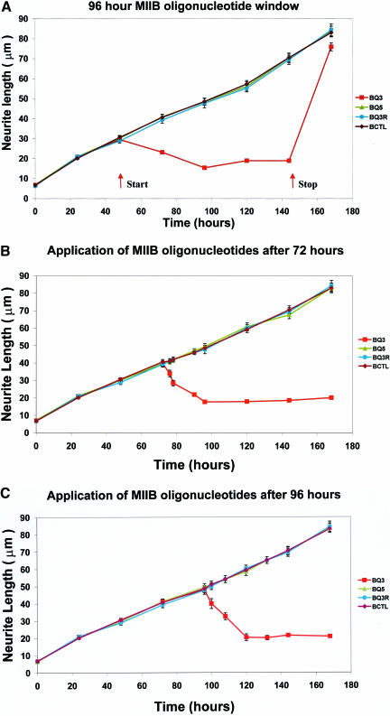
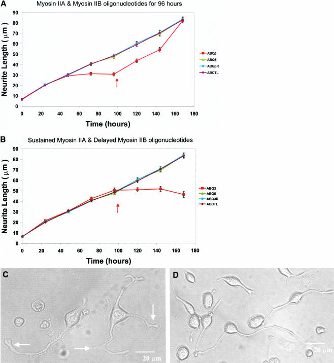
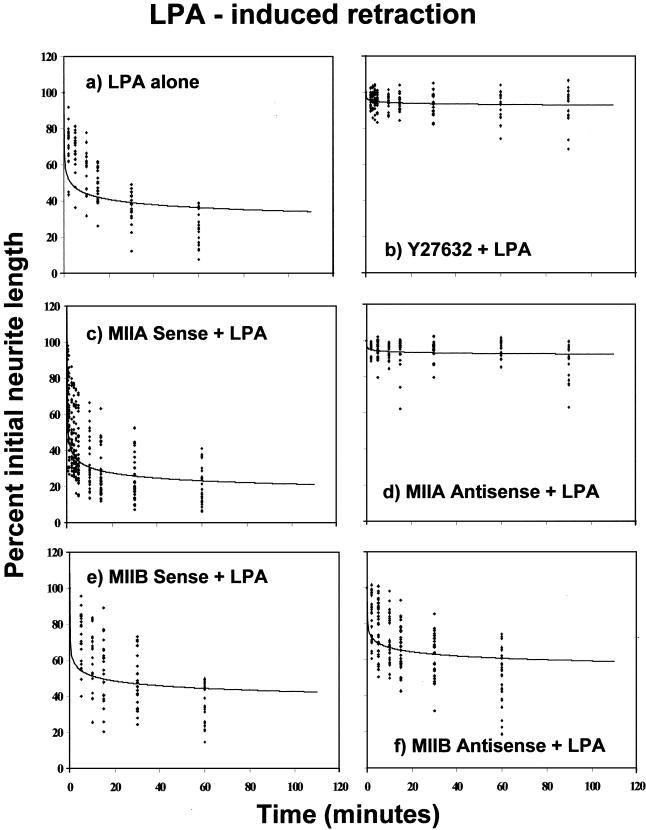
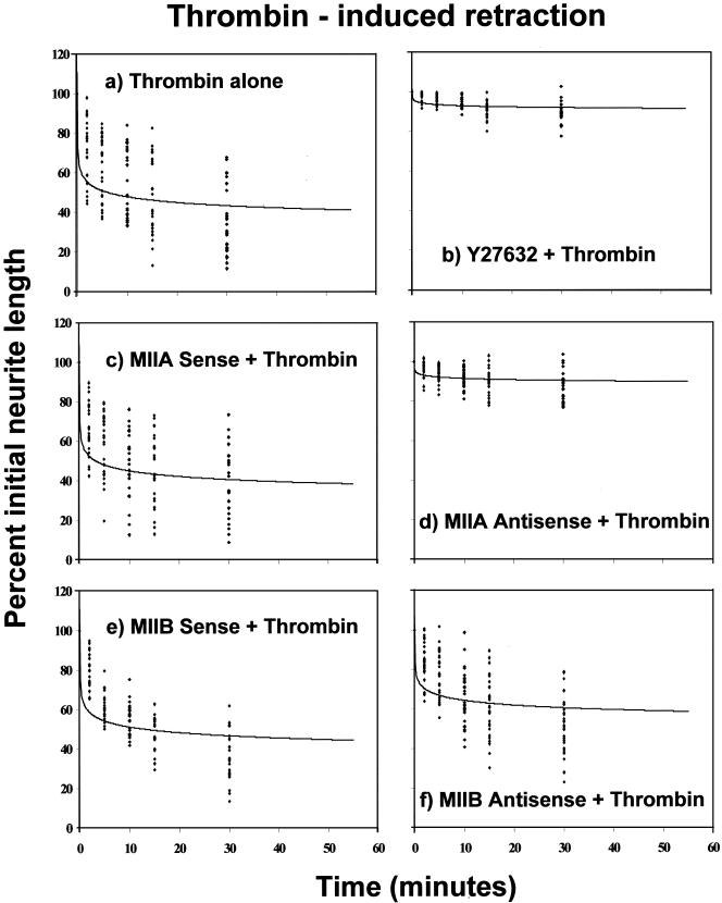
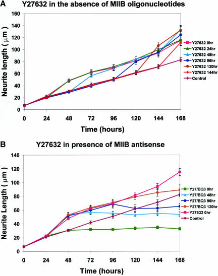
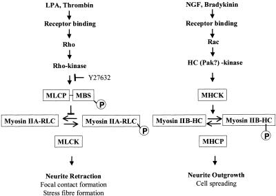
Neuritic extension is the resultant of two vectorial processes: outgrowth and retraction. Whereas myosin IIB is required for neurite outgrowth, retraction is driven by a motor whose identity has remained unknown until now. Preformed neurites in mouse Neuro-2A neuroblastoma cells undergo immediate retraction when exposed to isoform-specific antisense oligonucleotides that suppress myosin IIB expression, ruling out myosin IIB as the retraction motor. When cells were preincubated with antisense oligonucleotides targeting myosin IIA, simultaneous or subsequent addition of myosin IIB antisense oligonucleotides did not elicit neurite retraction, both outgrowth and retraction being curtailed. Even during simultaneous application of antisense oligonucleotides against both myosin isoforms, lamellipodial spreading continued despite the complete inhibition of neurite extension, indicating an uncoupling of lamellipodial dynamics from movement of the neurite. Significantly, lysophosphatidate- or thrombin-induced neurite retraction was blocked not only by the Rho-kinase inhibitor Y27632 but also by antisense oligonucleotides targeting myosin IIA. Control oligonucleotides or antisense oligonucleotides targeting myosin IIB had no effect. In contrast, Y27632 did not inhibit outgrowth, a myosin IIB-dependent process. We conclude that the conventional myosin motor, myosin IIA, drives neurite retraction.


Figures

Figure 1.
Figure 1.
Application of myosin IIB oligonucleotide treatment at various times after initiation of neurite outgrowth. These experiments examine the effect of myosin IIB antisense treatment windows on neurite outgrowth at different times of onset after plating: 48 h (a), 72 h (b), and 96 h (c). (a) Application of a 96-h duration myosin IIB oligonucleotide treatment window, beginning at 48 h; oligonucleotides were removed and replaced by oligonucleotide-free media at 144 h. Arrows indicate the start and termination times of the oligonucleotide treatment and illustrate the reversibility of the process by showing that outgrowth can be restored. Measurements are plotted as mean lengths and SEMs for at least 100 neurites per data point (range 163-847 neurites). (b and c) Measurement of the rate of neurite retraction after induction by myosin IIB antisense oligonucleotide treatment at various times after initiation of neurite outgrowth. To measure the initial rate of retraction, myosin IIB antisense oligonucleotides were applied 72 (b) or 96 (c) hours after initiation of neurite outgrowth and measurements of neurite length were followed over time intervals of 6-8 h during the first 24 h after oligonucleotide application. As in a, measurements are plotted as mean lengths and SEMs for at least 100 neurites per data point. Ranges were 308-537 neurites (72 h) and 240-421 neurites (96 h). Symbols for sense (BQ5), antisense (BQ3), and scrambled (BQ3R) treated cells, or untreated (BCTL) control cells, are shown in the side panels. Note the progressive retraction of neuritic process length, regardless of initial neurite length, at the start of each treatment window. Also note that retraction stalls at a minimum value of ∼15-20 μm, below which it does not proceed further.
Figure 2.
Figure 2.
Combined application of oligonucleotides targeting both myosin IIA and myosin IIB to cultured Neuro-2A cells. These experiments examine the effect of a combined treatment of myosin IIA and myosin IIB oligonucleotides on neurite outgrowth. (a) Myosin IIA and myosin IIB oligonucleotides were provided together for 96 h from the time of plating. (b) Myosin IIA oligonucleotides alone were provided for 96 h from the time of plating and then myosin IIB and myosin IIA oligonucleotides were provided simultaneously during the subsequent 96 h. Mean lengths and SEMs are plotted using at least 100 neurites per individual data point (range 100-453 neurites). Arrows indicate either initiation of the recovery phase through replenishment with oligonucleotide-free media (a) or the addition of myosin IIB oligonucleotide treatment (b). Symbols denoting the use of combined myosin IIA and myosin IIB sense (ABQ5), antisense (ABQ3), and scrambled (ABQ3R) treated cells, or untreated (ABCTL) control cells, are defined in the side panels. Note that simultaneous exposure to double antisense oligonucleotides from time zero to 96 h (a) suppresses neurite outgrowth to a similar extent to that observed with IIB antisense alone (Wylie et al., 1998); however, the recovery phase after cessation of treatment is prolonged. When cells are first exposed to myosin IIA antisense for 96 h before myosin IIB antisense exposure (b), both neurite outgrowth and retraction are attenuated. DIC images of Neuro-2A cells treated for 96 h with antisense oligonucleotides targeting both myosin IIA and myosin IIB (c) are shown alongside untreated control cells observed in parallel wells (d). Note that although both neurite outgrowth and retraction are inhibited (a and b) in the cells shown in c, lamellipodial outgrowth continues to occur giving rise to aberrant, extended growth cones. This emphasizes that local lamellipodial dynamics are mechanistically different from the overall mass dynamics of neurite extension. Arrows (c) indicate abnormal club-like lamellipodia. Bars, 20 μm.
Figure 3.
Figure 3.
Brief treatment of Neuro-2A cells with antisense oligonucleotides targeting myosin IIA does not bring about extensive disruption of the actin cytoskeleton. Neuro-2A cells were treated for either 48 or 96 h with sense or antisense oligonucleotides targeting myosin IIA, as indicated. Disruption to the actin cytoskeleton, observed at 96 h simultaneous with the ablation of myosin IIA and paxillin expression, was not discernible after 48 h treatment with myosin IIA antisense oligonucleotides. Images obtained by confocal microscopy. Filamentous actin detected by rhodamine phalloidin staining. Myosin IIA detected by indirect immunofluorescence (Alexa-Fluor 633). Paxillin detected by direct immunofluorescence (fluorescein isothiocyanate). Bar, 20 μm.
Figure 4.
Figure 4.
Induction of Neuro-2A cell neurite retraction by LPA, after pretreatment with the Rho-kinase inhibitor Y27632 or with oligonucleotides targeting either myosin IIA or myosin IIB. Neuro-2A cells, cultured for 72-120 h in serum-free medium to induce significant neurite outgrowth, were treated with a pulse of LPA (1 μM) either alone (a), or preceded by 30-min incubation with Y27632 (25 μM) (b), 48-h incubation with sense (c) or antisense (d) oligonucleotides directed against myosin IIA sequence, or 18-h incubation with sense (e) or antisense (f) oligonucleotides directed against myosin IIB sequence. In the case of the myosin IIB oligonucleotides treatments, exposure was kept to the minimum necessary to ensure an effect, so that myosin IIB antisense-induced retraction did not eliminate neurites before LPA treatment. At each time point, taken over a 60-min period after LPA addition, the lengths of the same 20 neurites were measured and plotted. For illustrative purposes, data points were fitted using a polynomial equation of the form y = Ax-B; curve-fitting to a polynomial provided a better fit by eye than single- or multiexponential fits. Values for A and B, respectively, were 56.451, 0.1094 (a); 95.847, 0.0074 (b); 44.130, 0.1593 (c); 95.431, 0.0072 (d); 61.688, 0.0818 (e); and 75.235, 0.0529 (f).
Figure 5.
Figure 5.
Induction of Neuro-2A cell neurite retraction by thrombin addition, after pretreatment with the Rho-kinase inhibitor Y27632, or with oligonucleotides targeting either myosin IIA or myosin IIB. Neuro-2A cells, cultured for 72-120 h in serum-free medium to induce significant neurite outgrowth, were treated with a pulse of thrombin (5 NIH units) either alone (a) or preceded by 30-min incubation with Y27632 (25 μM) (b); 48-h incubation with sense (c) or antisense (d) oligonucleotides directed against myosin IIA sequence; or 18-h incubation with sense (e) or antisense (f) oligonucleotides directed against myosin IIB sequence. Comments provided in the legend for the LPA experiments are also pertinent here. Data points were fitted using a polynomial equation of the form y = Ax-B. Values for A and B, respectively, were 58.830, 0.0917 (a); 94.776, 0.0096 (b); 55.926, 0.0962 (c); 93.731, 0.0113 (d); 61.957, 0.0847 (e); and 73.032, 0.0558 (f).
Figure 6.
Figure 6.
Y27632 neither inhibits neurite outgrowth from Neuro-2A cells nor antagonizes attenuation of outgrowth by antisense oligonucleotides that target myosin IIB. (a) Neuro-2A cells were treated with Y27632 at various times (0, 24, 48, 96, 120, and 144 h) after plating. Note that no inhibition of outgrowth is observed irrespective of the time of Y27632 administration; rather, a small but significant increase in the rate of outgrowth is seen during times subsequent to Y27632 application. (b) Neuro-2A cells were treated with Y27632 continuously from the time of plating. At various times (0, 48, 96, and 120 h), antisense oligonucleotides targeting myosin IIB were administered and continuously applied. Note that attenuation of neurite outgrowth occurred subsequent to application of the myosin IIB oligonucleotides, and this followed a familiar time course (Figure 2) even in the continued presence of Y27632. Mean neurite lengths and SEMs are plotted for at least 100 neurites per data point (ranges 100-165 neurites in a and 100-218 neurites in b). Symbols used to illustrate the various treatments are defined in the side panels.
Figure 7.
Figure 7.
Scheme to illustrate the separate functions and regulation of conventional nonsarcomeric myosin IIA and myosin IIB. We propose separate pathways to regulate the distinctive functions of nonsarcomeric, conventional myosin isoforms, in particular with regard to the dynamic process of neurite extension. We have shown (this study) that myosin IIA is the motor involved in neurite retraction, a process that can be triggered by a number of effectors, including LPA (Tigyi and Miledi, 1992; Jalink et al., 1993, 1994; Tigyi et al., 1996b) and thrombin (Jalink and Moolenaar, 1992; Suidan et al., 1992; Jalink et al., 1994). A direct pathway relating cause and effect is shown (LHS) and involves Rho activation, which, in turn, activates Rho-kinase (Matsui et al., 1996; Ridley, 1996; Bishop and Hall, 2000). When the myosin binding subunit (MBS) of myosin light chain phosphatase (MLCP) is phosphorylated by activated Rho-kinase (Kimura et al., 1996; Ridley, 1996), dephosphorylation of the regulatory light chain (RLC) of myosin is inhibited, allowing myosin IIA to maintain a level of activity determined by the level of phosphorylation at Ser19 on the RLC (Bresnick, 1999). The activity of Rho-kinase is inhibited by Y27632 (Uehata et al., 1997; Davies et al., 2000), leading to activation of MLCP, dephosphorylation of myosin IIA and a decline in cross-bridge cycling, preventing retraction. Interestingly, in vitro studies have demonstrated that the heavy chain of myosin IIA can act as a substrate for protein kinase C (Murakami et al., 1995) and metastasis-associated protein (Mts 1) (Murakami et al., 2000) so dual, or even triple, regulation at the level of the myosin molecule may also be possible. Myosin IIA is also known to be involved in focal contact and stress fiber formation (Chrzanowska-Wodnicka and Burridge, 1996; Wei and Adelstein, 2000; Wylie and Chantler, 2001), processes that can be also be inhibited by Y27632 (Uehata et al., 1997). It is likely that there are other intermediates in the pathway shown as indicated by a requirement for two different tyrosine kinases (Aoki et al., 1999). We have shown previously that myosin IIB is involved in neurite outgrowth (Wylie et al., 1998), a process that can be initiated in many neuronal cells by nerve growth factor (Gundersen, 1985) or bradykinin (van Leeuwen et al., 1999) (although not required to stimulate outgrowth in the case of Neuro-2A cells). A proposed chain of command is shown (RHS) in which agonist binding activates Rac, leading ultimately to activation of a myosin heavy chain kinase (MHCK) (van Leeuwen et al., 1999). Phosphorylation of the myosin heavy chain has been shown to correlate with cell spreading (van Leeuwen et al., 1999). We propose that myosin IIB is also activated by MHCK in Neuro-2A cells, leading to neurite outgrowth. Casein kinase II and protein kinase C have been implicated as possible candidates for MHCK by in vitro experiments (Murakami et al., 1998). PAK-kinase may also be involved (Sanders et al., 1999; van Leeuwen et al., 1999) and has been demonstrated to phosphorylate the RLC (Chew et al., 1998) in vitro. Once again, the results suggest that some form of dual regulation at the level of the myosin molecule may occur. Putative cross talk between the pathways has not been included in the interest of clarity but may involve reciprocal actions of cAMP (Hirose et al., 1998), PAK (van Leeuwen et al., 1999), and further actions of the small GTPase protein family (Ridley, 1996; Sander et al., 1999; Bishop and Hall, 2000; Yuan et al., 2003).

Similar articles

Cited by

References

    1. Albertinazzi, C., Gilardelli, D., Paris, S., Longhi, R., and de Curtis, I. (1998). Overexpression of a neural-specific Rho family GTPase, cRac1B, selectively induces enhanced neuritogenesis and neurite branching in primary neurons. J. Cell Biol. 142, 815-825. - PMC - PubMed
    1. Amano, M., Chihara, K., Nakamura, N., Fukata, Y., Yano, T., Shibata, M., Ikebe, M., and Kaibuchi, K. (1998). Myosin II activation promotes neurite retraction during the action of Rho and Rho-kinase. Genes Cells 3, 177-188. - PubMed
    1. Aoki, J., Katoh, H., Yasui, H., Yamaguchi, Y., Nakamura, K., Hasegawa, H., Ichikawa, A., and Negishi, M. (1999). Signal transduction pathway regulating prostaglandin EP3 receptor-induced neurite retraction: requirement for two different tyrosine kinases. Biochem. J. 340, 365-369. - PMC - PubMed
    1. Berg, J.S., and Cheney, R.E. (2002). Myosin X is an unconventional myosin that undergoes intrafilopodial motility. Nat. Cell Biol. 4, 246-250. - PubMed
    1. Berg, J.S., Powell, B.C., and Cheney, R.E. (2001). A millennial myosin census. Mol. Biol. Cell 12, 780-794. - PMC - PubMed

Publication types

MeSH terms

LinkOut - more resources