. 2003 Dec;14(12):4997-5010.
doi: 10.1091/mbc.e03-01-0041. Epub 2003 Sep 5.

Intersectin regulates fission and internalization of caveolae in endothelial cells

Affiliations

Intersectin regulates fission and internalization of caveolae in endothelial cells

Sanda A Predescu et al. Mol Biol Cell. 2003 Dec.

Abstract

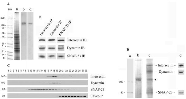
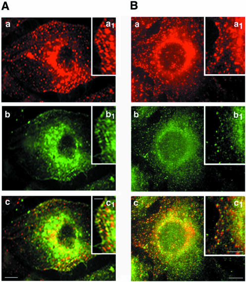
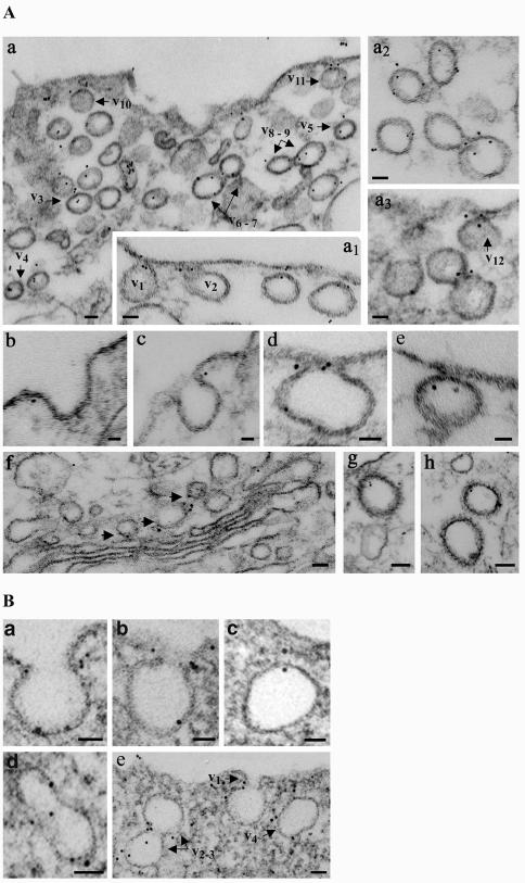
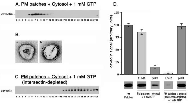
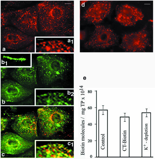
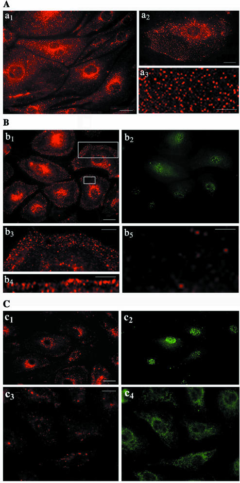
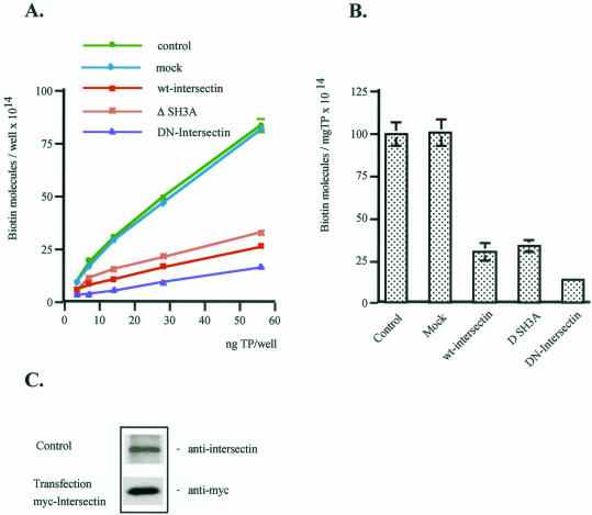
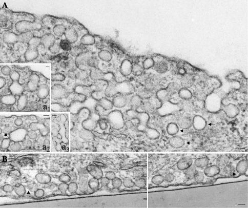
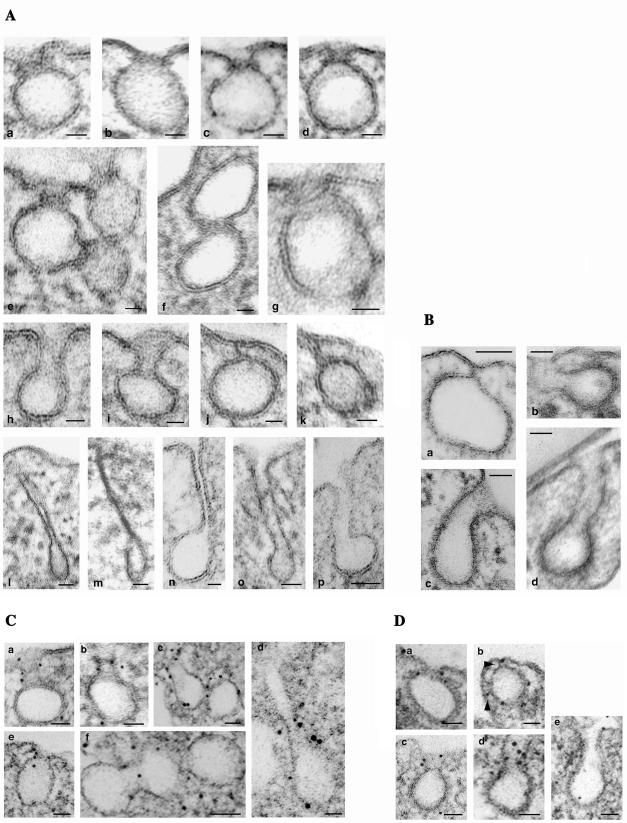
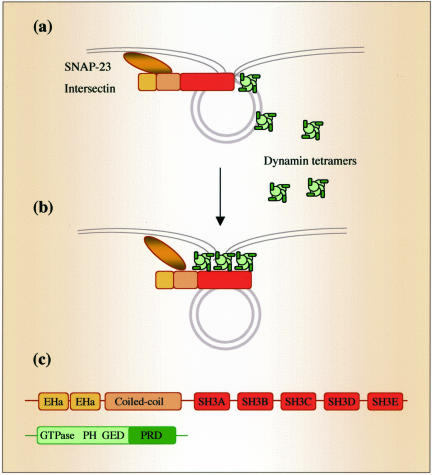
Intersectin, a multiple Eps15 homology and Src homology 3 (SH3) domain-containing protein, is a component of the endocytic machinery in neurons and nonneuronal cells. However, its role in endocytosis via caveolae in endothelial cells (ECs) is unclear. We demonstrate herein by coimmunoprecipitation, velocity sedimentation on glycerol gradients, and cross-linking that intersectin is present in ECs in a membrane-associated protein complex containing dynamin and SNAP-23. Electron microscopy (EM) immunogold labeling studies indicated that intersectin associated preferentially with the caveolar necks, and it remained associated with caveolae after their fission from the plasmalemma. A cell-free system depleted of intersectin failed to support caveolae fission from the plasma membrane. A biotin assay used to quantify caveolae internalization and extensive EM morphological analysis of ECs overexpressing wt-intersectin indicated a wide range of morphological changes (i.e., large caveolae clusters marginated at cell periphery and pleiomorphic caveolar necks) as well as impaired caveolae internalization. Biochemical evaluation of caveolae-mediated uptake by ELISA showed a 68.4% inhibition by reference to control. We also showed that intersectin interaction with dynamin was important in regulating the fission and internalization of caveolae. Taken together, the results indicate the crucial role of intersectin in the mechanism of caveolae fission in endothelial cells.


Figures

Figure 1.
Figure 1.
Intersectin is present in ECs in a membrane-associated protein complex containing dynamin and SNAP-23. (A) Western blotting of cultured ECs (b) and rat lung (c) detergent extracts using affinity-purified anti-intersectin pAb shows a band of immunoreactivity of ∼140 kDa. (a) Coomassie staining of total EC lysate. MW, protein standards. (B) Immunoprecipitation analysis applied on cultured EC lysates using the specific anti-intersectin, anti-dynamin, and anti-SNAP-23 antibodies indicates the presence of the same endothelial proteins in the immunoprecipitate. (C) Total protein (500 μg) from EC lysate was subjected to 10–35% glycerol gradient centrifugation, at 4°C, 42,000 rpm, overnight, in a SW55 Beckman rotor. Fractions (28 fractions of 180 μl each) were collected. Aliquots (20 μl) from each fraction were subjected to SDS-PAGE, followed by Western blotting using each indicated antibody. The similar sedimentation pattern indicates that intersectin is a protein component of a complex containing dynamin and SNAP-23 in ECs, whereas caveolin-1 is not part of this complex. An ECL detection system was used in all cases to visualize the bound antibodies. All results above are representative of four separate experiments. (D) The cross-linked products generated by DST in intact cultured ECs were immunoprecipitated with anti-dynamin mAb and analyzed by 6% SDS-PAGE in 8 M urea. Commassie R-250 staining indicated the presence of an aggregate >250 kDa (b) only in the membrane fraction. Second-dimensional analysis of the cross-linked product on 10% SDS-PAGE followed by silver staining suggests the presence of intersectin, dynamin, and SNAP-23 complex. The identity of the 70-kDa protein (*) is unknown. (c) Immunoblotting analysis with anti-intersectin, anti-dynamin, and anti–SNAP-23 pAbs confirmed the identity of the protein bands (d). MW standards (a) apply only to lane b.
Figure 2.
Figure 2.
Subcellular distribution of intersectin by double immunofluorescence in cultured ECs. Intersectin displays a punctate staining pattern both at the plasma membrane and throughout the cytoplasm in cultured ECs permeabilized by methanol fixation (A and B, red staining, a and a1). (A) Caveolin immunostaining in cultured ECs (b). The merged image (c) indicated extensive colocalization of the signals for intersectin and caveolin (panel c and inset c1), both intracellular and at the plasma membrane. (B) Anti-dynamin mAb revealed a similar punctate pattern that overlapped a predominant diffuse pattern (b). The merged image (c) indicates marked colocalization of intersectin (red) and dynamin (green) both at the plasma membrane and cytoplasm of an EC. Insets a1 and b1 show in detail the punctate intersectin and dynamin staining at the plasma membrane. The majority of the reactive small vesicles at the plasma membrane contain both proteins (c1). Results are representative of three experiments. Bars, (a–c) 10 μm; (insets) 5 μm.
Figure 3.
Figure 3.
Localization of intersectin (A) and dynamin (B) in cultured ECs by EM immunogold labeling using DNP-conjugated rabbit IgG and protein A gold. (A) Gold particles (5 nm) are found at the plasma membrane associated with caveolae profiles open to the apical front (a1, v1 and v2) and on apparently free caveolae in the cytosol (a, v3–v5). Note the gold particles associated with caveolae just released from the plasma membrane (a, v10 and v11; a3, v12; and e). Gold particles are often found at the constriction region between two adjoining caveolae (a, v6 and v7, v8 and v9; a2; and a3). Note the preferential association of intersectin with the caveolae neck region (b–d). Gold particles are also found associated with Golgi-derived vesicles (f, arrowheads) and CCVs (g and h). Bars, (a, a1, a2, a3, f, g, and h) 50 nm, (b, c, d, and e) 20 nm. (B) Gold particles (6 nm) are preferentially associated with the neck region of caveolae open to the EC surface (a, b, and e, v1). Frequently, more than two gold particles labeled a caveolar profile or its neck. Gold particles are also frequently found at the constriction region between two adjoining caveolae (d and e, v1 and v2) and associated with caveolae apparently free in the cytosol (c and e, v3). Bars, (a–c) 20 nm; (d and e) 50 nm.
Figure 4.
Figure 4.
Intersectin is required for caveolae fission. (A) Low-density caveolin-enriched membranes (fractions. 5–11) were obtained by density centrifugation on sucrose gradients when rat lung EC plasma membrane patches bearing attached caveolae were incubated with cytosol, 1 mM GTP, and the ATP regenerating system. (B) EM negative staining applied on aliquots from fraction 7 of sucrose gradients revealed a homogeneous population of caveolae (a), which by EM immunocytochemistry were heavily labeled by 10 nm gold conjugated anti-caveolin pAb (b). (C) When both cytosol and plasma membranes sheets were depleted in intersectin, caveolin was not detected by Western blotting in the light fractions of sucrose gradients. The GTP-induced caveolae fission did not occur. (D) The signal detected for caveolin by immunoblotting of aliquots containing equal amounts of total protein from PM patches (starting material), fractions 5–10 of sucrose gradients, and repelleted silica-coated membranes, were quantified by scanning densitometry and plotted as a percentage of signal detected in the starting material. All data are representative for four different experiments.
Figure 5.
Figure 5.
Internalization of biotinylated cell surface proteins in ECs by caveolae. Contribution of caveolae to the internalization of biotinylated cell surface proteins was evaluated using a double internalization assay and CT-FITC as a marker for the caveolar uptake. Cell surface proteins were biotinylated and then internalized for 30 min, at 37°C. Biotinylated proteins still on the cell surface after 30 min were reduced with glutathione, and the uptake of biotinylated surface proteins was analyzed morphologically by fluorescence microscopy using neutrAvidin-Texas Red (a). CT-FITC bound on the cell surface at 4°C (inset b.1) was internalized for 30 min at 37°C (b). Virtually similar staining pattern indicates the dominant contribution of caveolae to the internalization process. The merged image (c) indicates the degree of colocalization of the two probes. Inset c.1 shows in detail that majority of small vesicles contain both probes. Results are representative of three experiments. Bars, (a–c) 10 μm; (insets) 4 μm. (d) K+ depletion used to inhibit internalization by CCVs did not affect caveolae internalization. (e) The number of biotin molecules in the final supernates of ECs lysates when biotinylated cell surface proteins or CT-biotin were used as probes. A similar number of biotin molecules are found in the lysates of ECs K+-depleted. Results are the averages ± SD of four different experiments.
Figure 6.
Figure 6.
Expression of wt-intersectin inhibits caveolae internalization. (A) In control cells subjected to biotinylation of cell surface proteins and internalization assay, fluorescent staining with neutrAvidin Texas Red indicates a strong punctate pattern throughout the cytosol a1 (see inset a3 for a higher magnification) with accumulation in the perinuclear area. Inset a2 shows an enlarged EC subjected to the internalization assay. Note the relatively limited labeling at the cell surface because of the internalization. (B) ECs transiently transfected with myc-tagged wt-intersectin were selected based on their resistance to blasticidin and subjected to the internalization assay; anti-myc mAb followed by anti-mouse IgG FITC were used to visualize the transfected cells (b2). NeutrAvidin Texas Red revealed a generalized prominent fine punctate cell surface staining (b1 and b4). A strong punctate, belt-like staining was often seen at the plasma membrane (b3) below which was a region of low density of puncta (b5). Panels b3 and b5 are enlarged versions of the boxed regions in b1. (C) Effects of ΔSH3A and DN-intersectin on biotinylated cell surface protein internalization. ECs expressing ΔSH3A (c1) or DN-intersectin (c3) were subjected to the biotin internalization assay. NeutrAvidin Texas Red showed a limited staining in both cases. Anti-myc mAb followed by anti-mouse IgG FITC were used to visualize the transfected cells (c2, c4). Bars, (a1,a2,b1,c1, and c3)10 μm, (a3 and b3)10 μm, and (b4 and b5) 7.5 μm.
Figure 7.
Figure 7.
Effects of wt-intersectin overexpression on caveolae-mediated uptake. (A) The number of biotin molecules present in the EC lysates containing the internalized biotinylated cell surface proteins in control, mock, wt-intersectin ΔSH3A, or DN-intersectin transfected cells was determined by ELISA in 4–5 experiments. Ordinate: number of biotin molecules per well; abscissa: ng total protein (TP) per well. (B) Degree of inhibition of caveolae-mediated uptake in transfected cells compared with control. Bars, ±SD. (C) Cultured ECs were transiently transfected with the DNA construct encoding myc tagged wt-intersectin. After protein expression, cells were lysed and processed along with an aliquot of untransfected ECs lysate (control) for Western blots with anti-intersectin pAb or anti-myc mAb, respectively.
Figure 8.
Figure 8.
Electron micrographs of ECs overexpressing wt-intersectin. Micrographs show partial views of cultured ECs overexpressing wt-intersectin. The cells maintain a large population of caveolae open to the apical (A) or basolateral (B) fronts of the cells or apparently free in the cytosol (arrows). The majority of caveolae profiles are part of large clusters of interconnected caveolae, “grape-like” structures or with unusual shapes, accumulated at the cell periphery. Insets in A illustrate additional “grape-like” (a1, a2) or unusually shaped (a3), caveolae clusters. Note the caveolar profiles displaying staining-dense rings (arrowheads). Bars, 50 nm.
Figure 9
Figure 9
(facing page). Gallery of representative caveolae profiles (A) and CCPs (B) and dynamin immunoreactivity (C and D) in ECs overexpressing wt-intersectin. Micrographs in A show highly magnified staining-dense rings surrounding the necks of caveolae open to the cell surface (a–d), constricted region between two adjoining caveolae (e and f), caveolae with elongated necks (h and i), caveolae attached to the plasmalemma and unable to move into the cytosol (j and k), and caveolae with extremely long necks (l–p). The micrograph in panel g shows in a favorable section an almost complete ring encircling a caveolae neck. A caveolae neck of ∼70 nm length (the vesicle diameter) was considered as elongated neck. The figures shown in these panels were not seen in normal cells. Bars, (a–k and n) 20 nm, (l, m, o, and p) 50 nm. (B) CCV attached to the plasmalemma, just before release into the cytosol (a) and CCPs displaying elongated (b and c) or long necks (d). Bars, 50 nm. (C) Representative electron micrographs show dynamin immunoreactivity at the level of elongated caveolar necks (a–c), constricted region between adjacent caveolae (c and f), and staining-dense ring encircling a caveolae neck (e). A long caveolae neck immunoreactive to dynamin antibody is shown in d. Bars, (a, b, d, and e) 25 nm; (c and f) 35 nm. (D) Dynamin immunoreactivity is associated with a staining-dense ring surrounding the neck of a deep pit (a), elongated pits (c–e), or a CCP displaying two necks (d, arrowheads). Bars, 50 nm.
Figure 10.
Figure 10.
Role of intersectin-dynamin interaction in caveolae dynamics. In this model, the intersectin-dynamin interaction plays a central role in caveolae fission. Both intersectin and dynamin are preferentially associated with the caveolae neck (a). Intersectin binds and clusters dynamin in the proximity of the plasma membrane generating a high local dynamin concentration required for ring formation around caveolar necks. Dynamin bound to intersectin is functionally impaired and fission is restrained (b). Domain structures of intersectin and dynamin (c). Intersectin binds the PRD domain of dynamin via a subset of its SH3 domains (SH3A, SH3C, and SH3E).

Similar articles

Cited by

References

    1. Adams, A., Thorn, J.M., Yamabhai, M., Kay, B.K., and O'Brian, J.P. (2000). Intersectin, an adaptor protein involved in clathrin-mediated endocytosis activates mitogenic signaling pathways. J. Biol. Chem. 275, 27414-27420. - PubMed
    1. Anderson, E., Hellman, L., Gullberg, U., and Olsson, I. (1998). The role of the propeptide for processing and sorting of human myeloperoxidase. J. Biol. Chem. 273, 4747-4753. - PubMed
    1. Apodaca, G., Cardone, M.H., Whiteheart, S.W., DasGupta, B.R., and Mostov, K.E. (1996). Reconstitution of transcytosis in SLO-permeabilized MDCK cells: existence of an NSF-dependent fusion mechanism with the apical surface of MDCK cells. EMBO J. 15(7), 1471-1481. - PMC - PubMed
    1. Boyles, J., L'Hernault, N., Laks, H., and Palade, G.E. (1981). Evidence for a vesicular shuttle in heart capillaries. J. Cell Biol. 91, 418a.
    1. Bretscher, M.S., and Lutter, R. (1988). A new method for detecting endocytosed proteins. EMBO J. 7, 4087-4092. - PMC - PubMed

Publication types

MeSH terms

LinkOut - more resources