. 2003 Nov;14(11):4448-57.
doi: 10.1091/mbc.e02-11-0758. Epub 2003 Sep 5.

Recycling of Raft-associated prohormone sorting receptor carboxypeptidase E requires interaction with ARF6

Affiliations

Recycling of Raft-associated prohormone sorting receptor carboxypeptidase E requires interaction with ARF6

Irina Arnaoutova et al. Mol Biol Cell. 2003 Nov.

Abstract

Little is known about the molecular mechanism of recycling of intracellular receptors and lipid raft-associated proteins. Here, we have investigated the recycling pathway and internalization mechanism of a transmembrane, lipid raft-associated intracellular prohormone sorting receptor, carboxypeptidase E (CPE). CPE is found in the trans-Golgi network (TGN) and secretory granules of (neuro)endocrine cells. An extracellular domain of the IL2 receptor alpha-subunit (Tac) fused to the transmembrane domain and cytoplasmic tail of CPE (Tac-CPE25) was used as a marker to track recycling of CPE. We show in (neuro)endocrine cells, that upon stimulated secretory granule exocytosis, raft-associated Tac-CPE25 was rapidly internalized from the plasma membrane in a clathrin-independent manner into early endosomes and then transported through the endocytic recycling compartment to the TGN. A yeast two-hybrid screen and in vitro binding assay identified the CPE cytoplasmic tail sequence S472ETLNF477 as an interactor with active small GTPase ADP-ribosylation factor (ARF) 6, but not ARF1. Expression of a dominant negative, inactive ARF6 mutant blocked this recycling. Mutation of residues S472 or E473 to A in the cytoplasmic tail of CPE obliterated its binding to ARF6, and internalization from the plasma membrane of Tac-CPE25 mutated at S472 or E473 was significantly reduced. Thus, CPE recycles back to the TGN by a novel mechanism requiring ARF6 interaction and activity.


Figures
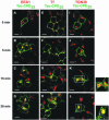
Figure 1.
Figure 1.
Tac-CPE25 traffics from the PM through the endocytic recycling compartment to the TGN. Neuro2A cells expressing Tac-CPE25 were labeled for 30 min at 4°C with anti-Tac monoclonal antibodies alone (A–D and I–L) or with both anti-Tac antibodies and fluorescent-labeled Tf (red) (E–H). Cells were chased at 37°C for the indicated times. The cells were then fixed in 4% paraformaldehyde, permeabilized, and incubated with anti-EEA1 (A–D) or anti-TGN38 (I–L) antibodies followed by appropriate fluorescent-labeled secondary antibodies (both red). To follow anti-Tac antibody uptake, cells were probed with Alexa 488-conjugated goat anti-mouse (E–H and I–L) or rabbit anti-mouse (A–D) secondary antibodies (green). Single confocal microscope sections are shown. Colocalization on the merged images looks yellow. High resolution analysis of the TGN area (boxed K and L) are shown to the right of the panel confirming colocalization of Tac-CPE25 with TGN38 at 30 min. Bars, 10 μM. Note that Tac-CPE25 was found in early endosomes after 5 min, in the endocytic recycling compartment, labeled with Tf, after 15 min, and in TGN after 30 min of internalizaition.
Figure 2.
Figure 2.
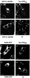
Eps15 mutant and dynamin-2 mutant K44A do not block internalization of Tac-CPE25. Neuro2A cells cotransfected with GFP-EΔ95/295 and Tac-CPE25 (A–D) or with K44A and Tac-CPE25 (E–H) were incubated with anti-Tac ab (A, B, E, and F) or with Tf-Alexa 594 (C, D, G, and H) for 30 min at 4°C. Cells were then chased for 15 min at 37°C in Tf- or ab-free media, fixed, permeabilized, and probed with Alexa 568-conjugated anti-mouse antibody to follow internalization of Tac-CPE25. Single confocal microscope sections are shown. Arrows point to the cells where mutants were expressed and Tf was not internalized. Of 100 cells transfected with Eps15 mutant (GFP-EΔ95/295) or dominant negative mutant of Dynamin 2 (K44A-GFP) counted, >85% of them showed internalization of Tac-CPE25 but not transferrin. Bars, 10 μM.
Figure 3.
Figure 3.
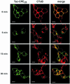
Tac-CPE25 and the lipid raft marker, cholera toxin B subunit, internalize and reach the TGN by the same endocytic pathway. Neuro2A cells expressing Tac-CPE25 were labeled for 30 min at 4°C with anti-Tac monoclonal ab and CTxB-Alexa 594 (red), and then chased for the indicated times at 37°C. Cells were fixed, permeabilized, and labeled with goat anti-mouse secondary antibody-Alexa 488 (green) to visualize anti-Tac antibody uptake. Single confocal microscope sections are shown. Colocalization on the merged images looks yellow. Bars, 10 μM.
Figure 4.
Figure 4.
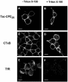
Tac-CPE25 remains in lipid rafts on the cell surface after exocytosis. Neuro2A cells expressing Tac-CPE25 were stimulated to exocytose and then were incubated at 4°C with anti-Tac ab (A and B) or CTxB-Alexa Fluor 594 (C and D). Before fixation, cells were treated for 30 min at 4°C with 1% Triton X-100. After fixation cells were stained with anti-TfR ab (E and F). Bars, 10 μM.
Figure 5.
Figure 5.
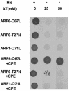
A yeast two-hybrid screen using ARF6-Q67L as bait and a fetal brain library identified a clone containing the last 90 amino acids of CPE. ARF6-Q67L, ARF6-T27N, and ARF1-Q71L were cloned into bait vector pAS2–1. Yeast strain CG-1945 was cotransformed with prey plasmid pACT2 alone (top three rows) or the pACT2 library clone containing the C-terminal 90 amino acids of CPE (bottom three rows). Transformants were spotted onto plates lacking leucine and tryptophan with (+) or without (-) histidine (His) in the presence of 25 or 50 mM 3-amino-1,2,4-triazole (3-AT). Vigorous growth was observed for cells coexpressing the ARF6-Q67L bait plasmid and the CPE clone, even at the highest concentration of 3-AT tested (fourth row). Slight growth was observed for cells coexpressing ARF6-T27N and CPE, but only at the lower concentration of 25 mM 3-AT (fifth row). ARF1-Q71L showed no interaction with CPE. No growth was observed at either concentration of 3-AT for cells expressing ARF6-Q67L and the pACT2 vector alone (top row).
Figure 6.
Figure 6.
The cytoplasmic tail of CPE interacts with ARF6. In vitro binding assay of fusion proteins shows interaction of six amino acids of CPE C-terminus with active form of ARF6. (A) Input (10%) of fusion proteins was analyzed by SDS-PAGE followed by staining with Silver Stain kit. (B) Fusion proteins, bound to S-agarose beads were analyzed by Western blot. Nitrocellulose membrane was probed with anti-GST ab (top) or with Ponceau S (bottom).
Figure 7.
Figure 7.
Inactive ARF6 blocks internalization of Tac-CPE25. (A) Neuro2A cells cotransfected with wtARF6 and Tac-CPE25 (wtARF6) or with ARF6-T27N and Tac-CPE25 (ARF6-T27N) constructs after stimulation were labeled with anti-Tac antibody for 30 min at 4°C and chased for 5, 10, and 15 min at 37°C in antibody-free media. Cells were fixed, permeabilized, and incubated with rabbit anti-ARF6 antiserum followed by Alexa 568 goat anti-rabbit and Alexa 488 goat anti-mouse secondary antibodies to detect expressed ARF6 and anti-Tac antibody uptake, respectively. Images were obtained from cells, expressing both Tac-CPE25 and wtARF6 (or ARF6-T27N) proteins. Single confocal microscopy sections show the Tac-CPE25 immunostaining only. Arrow points to a cell, where ARF6-T27N was not expressed and therefore internalization of Tac-CPE25 was not inhibited. Note that cells expressing wtARF6 demonstrate PM-bound and internalized anti-Tac antibody staining, whereas cells expressing negative mutant of ARF (ARF6-T27N) show only PM-bound anti-Tac antibodies. Bars, 10 μM. (B) Neuro2A cells cotransfected with wtARF6 and Tac-CPE25 (wtARF6) or with ARF6-T27N and Tac-CPE25 (ARF6-T27N) constructs were labeled with anti-Tac antibody for 30 min at 4°C and chased for 5, 10, and 15 min at 37°C in antibody-free media, as in A. To remove anti-Tac antibodies remaining at the PM, the cells were washed with Na-acetate buffer (pH 3.0) at 4°C before fixation. To detect ARF6 and internalized anti-Tac antibodies, fixed cells were labeled with anti-ARF6 rabbit antiserum followed by incubation with appropriate fluorescent-conjugated secondary antibodies. Images were taken from cells, expressing both Tac-CPE25 and wtARF6 (or ARF6-T27N) proteins. Single confocal microscopy sections show the Tac-CPE25 immunostaining only. Bars, 10 μM.
Figure 8.
Figure 8.
Mutation of the CPE cytoplasmic tail prevents its interaction with active form of ARF6. In vitro binding assay of fusion proteins shows two mutations that prevents interaction of CPE C terminus with ARF6-Q67L. Fusion proteins, bound to S-agarose beads were analyzed by Western blot. Nitrocellulose membrane was probed with anti-ARF6 ab (top) or with S-protein-HRP conjugate (bottom).
Figure 9.
Figure 9.
CPE mutants unable to bind active ARF6 are not internalized. Neuro2A cells expressing (A), Tac-CPES472A (B), and Tac-CPEE473A (C) were stimulated to exocytose, incubated at 4°C with anti-Tac ab and then chased for 15 min at 37°C. Cells were fixed, permeabilized, and labeled with goat anti-mouse secondary antibody-Alexa 488 (green) to visualize anti-Tac antibody uptake. Single confocal microscope sections are shown. Arrows point to cells that were counted as having internalized anti-Tac ab (Table 2), albeit the internalization of mutants (B and C) was much less than in wild-type Tac-CPE25 (A). Arrowheads show cells that were counted as having no internalized ab (Table 2). Bars, 10 mM.

Similar articles

Cited by

References

    1. Bamezai, A., Goldmacher, V.S., and Rock, K.L. (1992). Internalization of glycosyl-phosphatidylinositol (GPI)-anchored lymphocyte proteins. II. GPI-anchored and transmembrane molecules internalize through distinct pathways. Eur. J. Immunol. 22, 15-21. - PubMed
    1. Benmerah, A., Bayrou, M., Cerf-Bensussan, N., and Dautry-Varsat, A. (1999). Inhibition of clathrin-coated pit assembly by an Eps15 mutant. J. Cell Sci. 112, 1303-1311. - PubMed
    1. Bose, A., Cherniack, A.D., Langille, S.E., Nicoloro, S.M., Buxton, J.M., Park, J.G., Chawla, A., and Czech, M.P. (2001). G(alpha)11 signaling through ARF6 regulates F-actin mobilization and GLUT4 glucose transporter translocation to the plasma membrane. Mol. Cell Biol. 21, 5262-5275. - PMC - PubMed
    1. Brown, F.D., Rozelle, A.L., Yin, H.L., Balla, T., and Donaldson, J.G. (2001). Phosphatidylinositol 4, 5-bisphosphate and Arf6-regulated membrane traffic. J. Cell Biol. 154, 1007-1017. - PMC - PubMed
    1. Chatterjee, S., Smith, E.R., Hanada, K., Stevens, V.L., and Mayor, S. (2001). GPI anchoring leads to sphingolipid-dependent retention of endocytosed proteins in the recycling endosomal compartment. EMBO J. 20, 1583-1592. - PMC - PubMed