. 2003 Nov;14(11):4605-17.
doi: 10.1091/mbc.e02-11-0739. Epub 2003 Sep 5.

Rapid microtubule-dependent induction of neurite-like extensions in NIH 3T3 fibroblasts by inhibition of ROCK and Cbl

Affiliations

Rapid microtubule-dependent induction of neurite-like extensions in NIH 3T3 fibroblasts by inhibition of ROCK and Cbl

Robin M Scaife et al. Mol Biol Cell. 2003 Nov.

Abstract

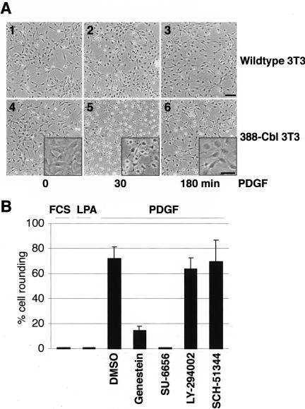
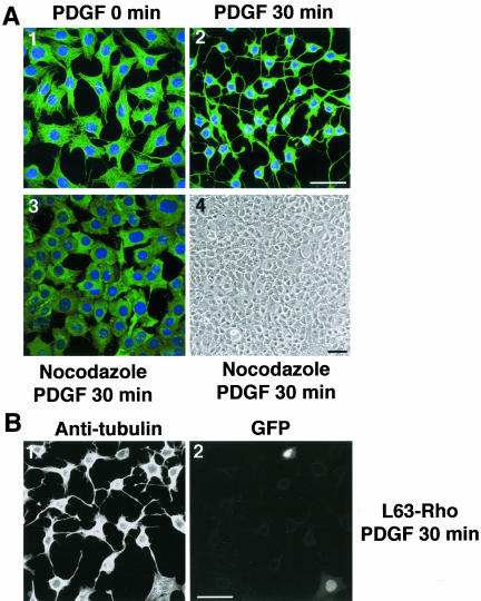
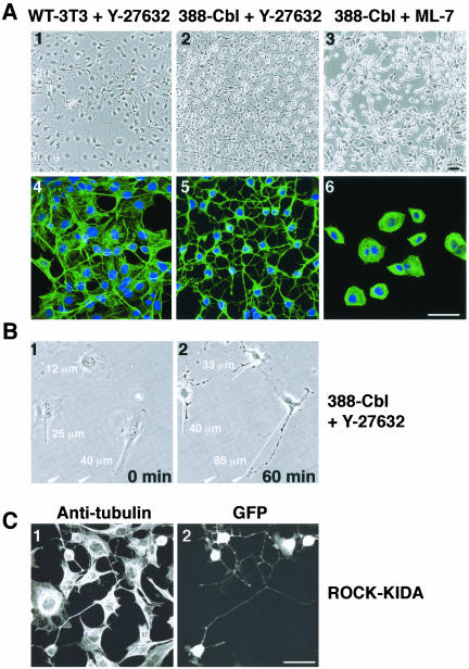
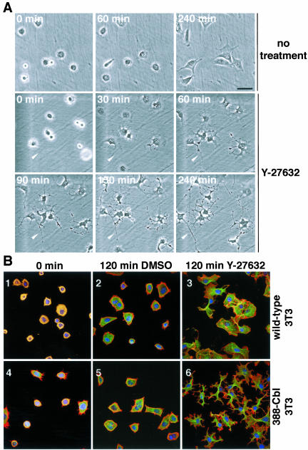
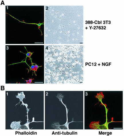
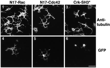
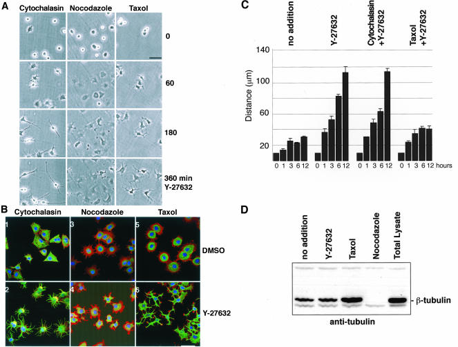
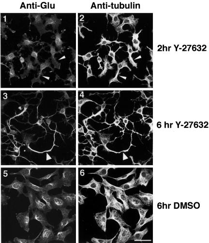
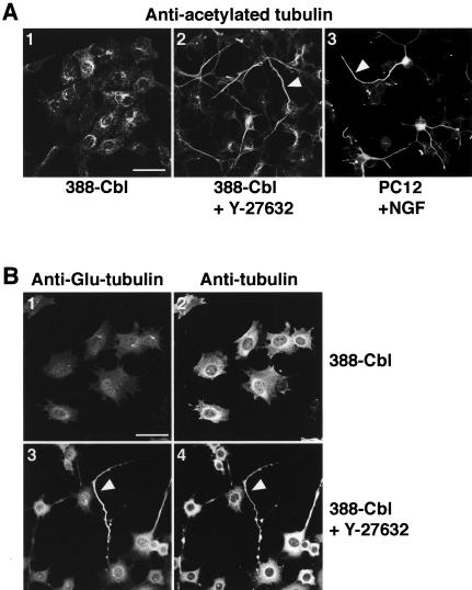
A number of key cellular functions, such as morphological differentiation and cell motility, are closely associated with changes in cytoskeletal dynamics. Many of the principal signaling components involved in actin cytoskeletal dynamics have been identified, and these have been shown to be critically involved in cell motility. In contrast, signaling to microtubules remains relatively uncharacterized, and the importance of signaling pathways in modulation of microtubule dynamics has so far not been established clearly. We report here that the Rho-effector ROCK and the multiadaptor proto-oncoprotein Cbl can profoundly affect the microtubule cytoskeleton. Simultaneous inhibition of these two signaling molecules induces a dramatic rearrangement of the microtubule cytoskeleton into microtubule bundles. The formation of these microtubule bundles, which does not involve signaling by Rac, Cdc42, Crk, phosphatidylinositol 3-kinase, and Abl, is sufficient to induce distinct neurite-like extensions in NIH 3T3 fibroblasts, even in the absence of microfilaments. This novel microtubule-dependent function that promotes neurite-like extensions is not dependent on net changes in microtubule polymerization or stabilization, but rather involves selective elongation and reorganization of microtubules into long bundles.


Figures

Figure 1.
Figure 1.
Involvement of Cbl in RTK-activation–dependent changes in cell morphology. (A) Serum-starved wild-type (1–3) and 388-Cbl–expressing (4– 6) NIH 3T3 cells were either left untreated (1 and 4) or were treated with PDGF for 30 min (2 and 5) or 180 min (3 and 6). Phase contrast images of the cells were collected at the indicated time points. The panel insets represent higher magnification views. Bar, 100 μm. (B) After a 30-min incubation with either no addition, DMSO, genestein, SU 6656, LY 294002, or SCH-51344. Serum-starved 388-Cbl–expressing NIH 3T3 cells were treated for a further 30 min with FCS, LPA, or PDGF, as indicated. Phase contrast images of the cells were collected at this time point, and the percentage of cells that had undergone extensive cell rounding was determined.
Figure 2.
Figure 2.
RTK-activation–dependent changes in cell morphology involves microtubule reorganization and signaling by Rho. (A) Serum-starved 388-Cbl–expressing NIH 3T3 cells were left untreated (1) or were either treated with PDGF (30 min) (2) or with nocodazole (20 min) followed by PDGF (30 min) (3 and 4). Cells were then either observed directly by phase contrast microscopy (4) or were stained with anti-tubulin antibody and Hoescht and imaged by confocal immunofluorescence microscopy (1–3). Bars, 50 μm. (B) 388-Cbl–expressing NIH 3T3 cells were cotransfected with L63RhoA and NLS-GFP cDNA. After serum starvation, the cells were treated with PDGF for 30 min and anti-tubulin (1) and GFP fluorescence (2) images were captured by confocal microscopy. Bar, 50 μm.
Figure 3.
Figure 3.
ROCK inactivation induces changes in cell morphology identical to RTK-induced changes. (A) Phase contrast images (1–3) and confocal immunofluorescence of anti-tubulin antibody and Hoescht staining (4–6) of serum-starved wild-type (1 and 4) and 388-Cbl–expressing NIH 3T3 cells (2, 3, 5, and 6) were taken after a 30-min treatment with Y-27632 (1, 2, 4, and 5) or ML-7 (3 and 6). Bar, 50 μm. (B) Phase contrast images were taken of a group of 388-Cbl–expressing cells before (0 min) and after (60 min) of treatment with Y-27632. The distances represented by the white lines are indicated numerically, and two positional markers are indicated by the arrowheads. (C) After transient cotransfection of 388-Cbl–expressing cells with ROCK-KIDA and NLSGFP–expressing plasmids, anti-tubulin antibody (1) and GFP staining (2) were imaged by confocal immunofluorescence microscopy. Bar, 50 μm.
Figure 4.
Figure 4.
Inhibition of ROCK induces polarity and microtubule-rich extensions in freshly plated 388-Cbl–expressing NIH 3T3 fibroblasts. (A) Phase contrast microscopy images of freshly plated 388-Cbl–expressing cells were collected during incubation in the absence (1–3) or presence of Y-27632 (4–9). The position of a cytoplasmic extension has been indicated by the arrowhead. Bar, 50 μm. (B) Confocal immunofluorescence microscopy images of TRITC-phalloidin (red), anti-tubulin antibody (green), and Hoescht (blue) staining of wild-type (1–3) and 388-Cbl–expressing NIH 3T3 cells (4– 6) were collected 30 min after replating (1 and 4), or after an additional 120 min of incubation in the absence (2 and 5) or presence of Y-27632 (3 and 6). Bar, 50 μm.
Figure 5.
Figure 5.
Inhibition of ROCK induces polarity and extensions in freshly plated 388-Cbl–expressing NIH 3T3 fibroblasts. (A) Confocal immunofluorescence microscopy images of TRITC-phalloidin (red), anti-tubulin antibody (green), and Hoescht (blue) staining (1 and 3), and phase contrast images (2 and 4) were collected 20 h after replating of 388-Cbl–expressing NIH 3T3 cells in the presence of Y-27632 (1 and 2), or 3 d after plating of PC12 cells in the presence of NGF (3 and 4). Bar, 50 μm. (B) Confocal immunofluorescence microscopy images of TRITC-phalloidin (1), anti-tubulin antibody (2) of 388-Cbl–expressing NIH 3T3 cells were collected 6 h after replating in the presence of Y-27632. A merged image of the TRITC-phalloidin and anti-tubulin signals is shown in 3. Bar, 12.5 μm.
Figure 6.
Figure 6.
Induction of extensions is independent of signaling by Rac, Cdc42, and Crk. After transfection with GFP-N17Rac1 (1 and 4), GFP-N17Cdc42 (2 and 5) or cotransfection with a Crk SH3 domain mutant (Crk-SH3*) and NLS-GFP (3 and 6), 388-Cbl–expressing cells were replated for 4 h in the presence of Y-27632. Anti-tubulin antibody (1–3) and GFP staining (4– 6) was then determined by confocal immunofluorescence microscopy. Bar, 50 μm.
Figure 7.
Figure 7.
Requirement of microtubules, but not microfilaments, for the formation of extensions. (A) After replating of 388-Cbl–expressing cells for 30 min in the presence of cytochalasin D, nocodazole, or taxol, phase contrast images were collected at 0, 60, 180, and 360 min of addition of Y-27632 to the culture media. Bar, 50 μm. (B) 388-Cbl–expressing cells were replated for 30 min in the presence of cytochalasin D (1 and 2), nocodazole (3 and 4), or taxol (5 and 6). Confocal immunofluorescence microscopy images of phalloidin (red), anti-tubulin antibody (green), and Hoescht (blue) staining of 388-Cbl were then collected after a 4-h incubation with DMSO (1, 3, and 5) or Y-27632 (2, 4, and 6). Bar, 50 μm. (C) 388-Cbl–expressing cells were replated in the absence (no addition) or presence of Y-27632, Y-27632 plus cytochalasin D, and Y-27632 plus taxol. Median maximal distances from the cell boundary to the nucleus were obtained from separate triplicate anti-tubulin immunofluorescence images as described in MATERIALS AND METHODS. Average values and standard deviations are indicated for samples taken at 0, 1, 3, 6, and 12 h of adhesion to the culture substrate after replating. (D) 388-Cbl–expressing cells were replated for 6 h either with no drug treatment (lanes 1 and 5), Y-27632 (lane 2), taxol (lane 3), or nocodazole (for the final 60 min, lane 4), and microtubule cytoskeleton fractions (lanes 1–4) or total cell lysates (lane 5) were probed with anti-tubulin antibody after SDS-PAGE and Western blot.
Figure 8.
Figure 8.
Microtubule bundles formed by ROCK inhibition are enriched in detyrosinated tubulin. Confocal immunofluorescence microscopy images of anti-Glu-tubulin (1, 3, and 5) and anti-β-tubulin (2, 4, and 6) staining of 388-Cbl–expressing cells were collected after 2 h (1 and 2) and after 6 h (3–6) of replating in the presence (3 and 4) and absence (5 and 6) of Y-27632. Non-Glu– and Glu-tubulin–containing microtubule bundles are indicated by narrow and wide arrowheads, respectively. Bar, 50 μm.
Figure 9.
Figure 9.
Accumulation of acetylated tubulin and nocodazole-resistant microtubules in Y-27632–induced neurite-like extensions. (A) Confocal immunofluorescence microscopy images of anti-acetylated-tubulin staining of either 388-Cbl–expressing cells replated for 20 h in the absence (1) or presence of Y-27632 (2) or PC12 cells exposed to NGF for 72 h (3). Acetylated tubulin containing microtubule bundles are indicated by the arrowheads. Bar, 50 μm. (B) 388-Cbl–expressing cells were replated for 20 h in the absence (1 and 2) or presence of Y-27632 (3 and 4). Confocal immunofluorescence microscopy images of anti-Glu-tubulin (1 and 3) and anti-β-tubulin (2 and 4) staining were then collected after exposure of the cells to 1 μM nocodazole for 30 min. Glu-tubulin–rich nocodazole-resistant microtubule bundles are indicated by the arrowheads. Bar, 50 μm.

Similar articles

Cited by

References

    1. Andoniou, C.E., Thien, C.B.F., and Langdon, W.Y. (1994). Tumour induction by activated abl involves tyrosine phosphorylation of the product of the cbl oncogene. EMBO J. 13, 4515-4523. - PMC - PubMed
    1. Aspenstrom, P. (1999). Effectors for the Rho GTPases. Curr. Opin. Cell Biol. 11, 95-102. - PubMed
    1. Bachmaier, K., Krawczyk, C., Kozieradzki, I., Kong, Y., Sasaki, T., Olivierados-santos, A., Mariathasan, S.D.B., Wakeham, A., Itle, A., et al. (2000) Negative regulation of lymphocyte activation and autoimmunity by the molecular adaptor Cbl-b. Nature 403, 211-216. - PubMed
    1. Bito, H., Furuyashiki, T., Ishihara, H., Shibasaki, Y., Ohashi, K., Mizuno, K., Maekewa, M., Ishizaki, T., and Narumiya, S. (2000). A critical role for a Rho-associated kinase, p160ROCK in determination of axon outgrowth in mammalian CNS neurons. Neuron 26, 431-441. - PubMed
    1. Chiang, Y.J., Kole, H., Brown, K., Naramura, M., Fukuhara, S., Hu, R.J., Kyung, J., Gutkind, J.S., Shevach, E., and Gu, H. (2000). Cbl-b regulates the CD28 dependence of T-cell activation. Nature 403, 216-220. - PubMed

Publication types

MeSH terms

LinkOut - more resources