. 2003 Sep 19;332(3):529-36.
doi: 10.1016/s0022-2836(03)00925-2.

Allosteric switching by mutually exclusive folding of protein domains

Affiliations

Allosteric switching by mutually exclusive folding of protein domains

Tracy L Radley et al. J Mol Biol. .

Abstract

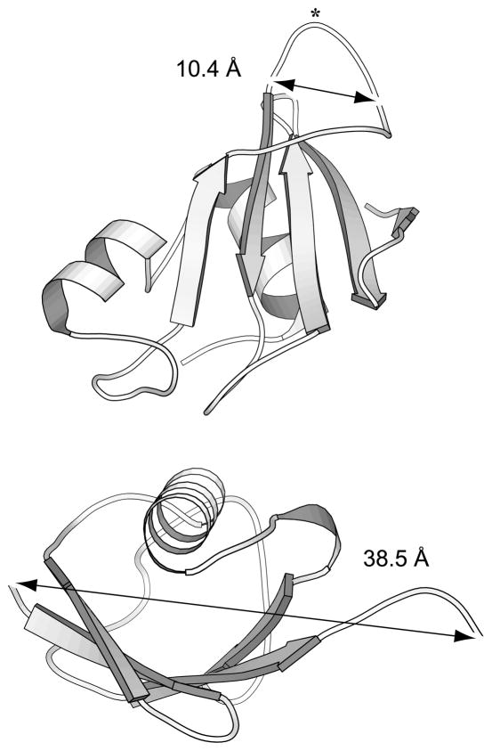
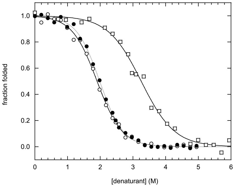
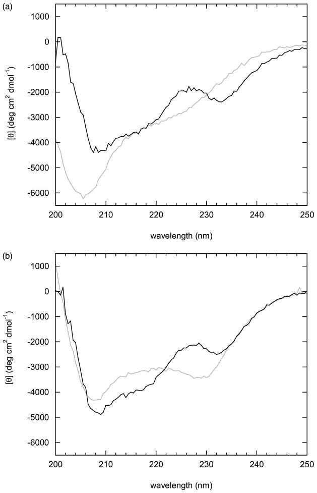
Many proteins are built from structurally and functionally distinct domains. A major goal is to understand how conformational change transmits information between domains in order to achieve biological activity. A two-domain, bi-functional fusion protein has been designed so that the mechanical stress imposed by the folded structure of one subunit causes the other subunit to unfold, and vice versa. The construct consists of ubiquitin inserted into a surface loop of barnase. The distance between the amino and carboxyl ends of ubiquitin is much greater than the distance between the termini of the barnase loop. This topological constraint causes the two domains to engage in a thermodynamic tug-of-war in which only one can exist in its folded state at any given time. This conformational equilibrium, which is cooperative, reversible, and controllable by ligand binding, serves as a model for the coupled binding and folding mechanism widely used to mediate protein-protein interactions and cellular signaling processes. The position of the equilibrium can be adjusted by temperature or ligand binding and is monitored in vivo by cell death. This design forms the basis for a new class of cytotoxic proteins that can be activated by cell-specific effector molecules, and can thus target particular cell types for destruction.


Figures

Figure 1
Figure 1
Ribbon diagrams of Bn (top) and Ub (bottom), showing Cα–Cα distances between Pro64 and Thr70 of Bn and between N and C termini of Ub. The BU gene was constructed by inserting the Ub gene between the Lys66 and Ser67 codons of Bn. Gly-Thr and Gly-Gly-Ser linker sequences were added to the N and C termini of Ub, respectively. The BU expression plasmid pETMT was created by inserting the BU gene into the pET25b(+) plasmid (Novagen). In order to maintain plasmid stability, it was necessary to introduce the Bs gene (together with its natural promoter from B. amyloli-quefaciens; a gift from Y. Bai, National Institutes of Health) into the same plasmid. BU was expressed in E. coli by co-transforming BL21(DE3) cells with a second, kanamycin-resistant plasmid containing the Bs gene under control of the phage T7 promoter. Cells were grown on Luria–Bertani medium to A600 = 1.0 and induced with 0.4 mM IPTG. Cells were centrifuged three hours later and lysed in 50 mM Tris (pH 7.5), 0.1 mM EDTA. Then 8 M urea was added to dissociate bound Bs, which was subsequently removed by passing the solution through a DE52 column (Whatman). The flow-through was loaded onto a HiTrap heparin column (Pharmacia), washed with 10 mM potassium phosphate (pH 7.5), 6 M urea and eluted with a 0–0.5 M NaCl gradient. BU eluted as one of a series of poorly resolved peaks; Western blot analysis using an anti-Ub antibody revealed that the contaminants were chiefly truncated BU proteins in which the Ub was partially proteolyzed. These products presumably arise because BU is expressed in E. coli as the BU–Bs complex, in which the Ub domain is unfolded and susceptible to the action of protease. We were able to purify BU to >95% homogeneity by choosing fractions conservatively; however, this reduced yields to 1–2 mg per liter of starting culture. Finally, urea was removed by extensive dialysis against double-distilled water and the protein was lyophilized.
Figure 2
Figure 2
(a) Temperature-induced conformational change of BU demonstrated by CD. All experiments were performed with 1 μM BU in 10 mM potassium phosphate (pH 7.5), 0.1 M NaCl. Circles and squares indicate 5 ºC and 50 ºC, respectively; other scans were recorded at 5 deg. C increments between these two limits. Data were collected using an Aviv model 202 CD spectropolarimeter. (b) Conversion from the Bn to the Ub form of BU monitored by ellipticity at 230 nm (open circles, continuous line) and Bn enzymatic activity (filled triangles, broken line). Lines are for illustrative purpose only. A 1 ml solution of 50 μM GpUp (Sigma) was allowed to come to thermal equilibrium at the indicated temperature, whereupon 15–25 μl of BU was added (final concentration 2 μM). Initial velocities were recorded by monitoring change in absorbance at 275 nm on a Varian Cary-100 spectrophotometer. Activity is reported as the average initial velocity from three measurements, and is normalized to the highest value observed.
Figure 3
Figure 3
Denaturation of the Bn domain (10 ºC, circles) and the Ub domain (50 ºC, squares) induced by urea and GdnHCl, respectively. Data were collected by CD at 230 nm (open symbols, black lines) or by Trp fluorescence at 320 nm (filled symbols, grey line), using a Jobin-Yvon/SPEX Fluoromax-3. Lines represent best fits to the linear extrapolation equation, and reported errors are the standard deviations obtained from three samples prepared on different days. Samples were prepared by adding equal volumes of BU to buffer alone and to buffer containing urea (Sigma) or GdnHCl (ICN Biochemicals). The solutions were then mixed in various ratios using a Hamilton Microlab 540B dispenser to yield the final indicated concentrations of denaturant. Denaturant concentrations were determined by measuring refractive index.
Figure 4
Figure 4
Thermodynamics of BU, Bn and Ub folding. (a) Temperature dependence of the free energies of free Bn (pH 5.5, continuous line) and free Ub (pH 4.0, broken line) folding. The following parameters were used to generate the curves: Bn, tm = 55.1 ºC, ΔHm = 119.6 kcal mol−1, ΔCp = 1.39 kcal mol−1 K−1; Ub, tm = 90.0 ºC, ΔHm = 73.7 kcal mol−1, ΔCp = 0.79 kcal mol−1 K−1. ΔCp was assumed to be independent of temperature. (b) Coupled folding/unfolding of the Bn domain (continuous line) and the Ub domain (broken line) of BU, calculated from equations (2) and (3) (see the text for details). Open circles are the experimental CD data points from Figure 2(b).
Figure 5
Figure 5
Bs binding induces folding of the Bn domain and unfolding of the Ub domain. CD spectra were recorded at (a) 40 ºC, (b) 15 ºC. Gray and black lines represent 10 μM BU in the absence and in the presence of 12.5 μM Bs, respectively. Black lines were generated by subtracting the CD spectra of free Bs.

References

    1. Dyson HJ, Wright PE. Coupling of folding and binding for unstructured proteins. Curr Opin Struct Biol. 2002;12:54–60. - PubMed
    1. Luque I, Leavitt SA, Friere E. The linkage between protein folding and functional cooperativity: two sides of the same coin. Annu Rev Biophys Biomol Struct. 2002;31:235–256. - PubMed
    1. Botuyan MV, Koth CM, Mer G, Chakrabartty A, Conaway JW, Conaway RC, et al. Binding of elongin A or a von Hippel-Lindau peptide stabilizes the structure of yeast elongin C. Proc Natl Acad Sci USA. 1999;96:9033–9038. - PMC - PubMed
    1. Botuyan MV, Mer G, Yi GS, Koth CM, Case DA, Edwards AM, et al. Solution structure and dynamics of yeast elongin C in complex with a von-Hippel-Lindau peptide. J Mol Biol. 2001;312:177–186. - PubMed
    1. Kim AS, Kakalis LT, Abdul-Manan N, Liu GA, Rosen MK. Autoinhibition and activation mechanisms of the Wiskott–Aldrich syndrome protein. Nature. 2000;404:151–158. - PubMed

Publication types

MeSH terms