. 2003 Sep 15;162(6):1135-47.
doi: 10.1083/jcb.200207056. Epub 2003 Sep 8.

Myostatin negatively regulates satellite cell activation and self-renewal

Affiliations

Myostatin negatively regulates satellite cell activation and self-renewal

Seumas McCroskery et al. J Cell Biol. .

Abstract

Satellite cells are quiescent muscle stem cells that promote postnatal muscle growth and repair. Here we show that myostatin, a TGF-beta member, signals satellite cell quiescence and also negatively regulates satellite cell self-renewal. BrdU labeling in vivo revealed that, among the Myostatin-deficient satellite cells, higher numbers of satellite cells are activated as compared with wild type. In contrast, addition of Myostatin to myofiber explant cultures inhibits satellite cell activation. Cell cycle analysis confirms that Myostatin up-regulated p21, a Cdk inhibitor, and decreased the levels and activity of Cdk2 protein in satellite cells. Hence, Myostatin negatively regulates the G1 to S progression and thus maintains the quiescent status of satellite cells. Immunohistochemical analysis with CD34 antibodies indicates that there is an increased number of satellite cells per unit length of freshly isolated Mstn-/- muscle fibers. Determination of proliferation rate suggests that this elevation in satellite cell number could be due to increased self-renewal and delayed expression of the differentiation gene (myogenin) in Mstn-/- adult myoblasts. Taken together, these results suggest that Myostatin is a potent negative regulator of satellite cell activation and thus signals the quiescence of satellite cells.


Figures
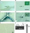
Figure 1.
Figure 1.
Myostatin is expressed in satellite cells. M. tibialis anterior was serially sectioned and immunostained with antibodies specific for (A) Myostatin and (B) Pax7. The myonuclei were stained with DAPI. The same muscle was used for in situ and probed for Myostatin (C) and Pax7 transcripts (D). Arrows indicate the stained satellite cells, and DAPI-stained myonuclei are also shown in the insets (B and D). (E) Micrograph showing in situ hybridization performed with myostatin antisense probe on muscle sections of myostatin-null mouse. (F, i) Agarose gel electrophoresis of RT-PCR products derived from satellite cell total RNA. Primers specific for 3′ region of myostatin amplify the expected 515-bp product in a combined RT-PCR reaction (Mstn). Amplicons are not detected in the absence of template (Negative control). Pax7 was amplified with primers designed to produce a 571-bp product (Pax7), and CD34 splice variants were also PCR amplified from the same RT reaction (CD34 splice variants). 1-kb plus DNA ladder is shown. (F, ii) Western blot showing the presence of the full-length 52-kD Myostatin protein in satellite cell protein extract. Bars, 10 μm.
Figure 2.
Figure 2.
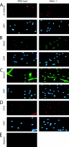
Immunocytochemistry of isolated satellite cells. Adult myoblasts were cultured from the hind leg muscles of either wild-type or Mstn−/−mice, fixed, and immunostained for M-cadherin (A), MyoD (B), Desmin (C), and CD34 (D). DAPI staining of the nuclei in the corresponding fields is also shown; panel E shows the background immunofluorescence when anti–mouse secondary antibody was used in the absence of primary antibody. Similar background was observed for all other secondary antibody negative controls. Greater than 95% of the isolated cultured cells were myogenic.
Figure 3.
Figure 3.
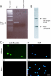
Myostatin is expressed in adult myoblasts derived from satellite cells. (A) Agarose gel electrophoresis of myostatin ORF amplified in a combined RT-PCR reaction using total RNA from adult myoblasts (lane 2). Amplicons are not detected in the absence of template (negative control) (lane 3). 1-kb plus DNA ladder is shown in lane 1. (B) Western analysis from proteins isolated from adult myoblasts cultured for 48 h showing both the full-length (52 kD) and LAP (38 kD) peptides of Myostatin. (C) Representative immunofluorescence showing the presence of Myostatin in wild-type adult myoblasts (i) and absence in myostatin-null (Mstn−/−) myoblasts (iii). DAPI staining of the nuclei is shown in the corresponding fields (ii and iv).
Figure 4.
Figure 4.
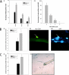
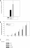
Lack of Myostatin increases the activation of satellite cells on myofibers. (A, i) In vivo quantification of activated satellite cells in the muscle of wild-type and myostatin-null mice. Activated satellite cells were labeled with BrdU in wild-type or myostatin-null (Mstn −/−) mice of 4 wk, 8 wk, or 6 mo and were isolated using Percoll gradient. 5,000–10,000 satellite cells were immunostained for BrdU, and percentages of nuclei that were positive for BrdU labeling are shown. **, P < 0.01 (as compared with wild type). At least a total of 1,000 cells were counted in each of three replicates. The data provided are an average of three animals each. (A, ii) Myostatin inhibits the migration of satellite cells from fibers. Single muscle fibers (n = 32) were isolated from the muscle and incubated in media conducive to the migration of myogenic precursor cells. The addition of Myostatin in increasing concentrations decreases the percentage of fibers with migrated satellite cells (**, P < 0.01). (B, i) The number of satellite cells (CD34 positive) per 100 myonuclei in wild-type and myostatin-null (Mstn−/−) myofibers is shown. **, P < 0.01 (as compared with wild type). More than 1,000 nuclei were counted in each of three replicates. The data presented are an average of three animals each. (B, ii) Micrograph showing typical CD34-immunostained satellite cell and (iii) myonuclei were visualized by counterstaining with DAPI. (C, i) An increased number of satellite cells migrate from myostatin-null fibers. Quantitative analysis demonstrates that an increased number of myogenic progenitor cells migrates from single myofibers isolated from myostatin-null mice as compared with wild-type myofibers (*, P < 0.05). (C, ii) An example of satellite cells surrounding an isolated myofiber after 72 h.
Figure 5.
Figure 5.
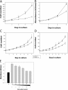
myostatin-null adult myoblasts proliferate faster than the wild-type myoblasts. Adult myoblasts were isolated from hind limbs of C57BL/10 (Mstn+/+) or myostatin-null mice (Mstn−/−) and seeded at a low number. Proliferation rate determined for the myoblasts isolated from E17 (A) and 4-wk-old muscle (B). In addition, the proliferation rate determined for m. tibialis anterior (C) and m. soleus (D) muscle fiber–specific adult myoblasts is also shown. Experiments were done in triplicate. Data shown are an average of three animals. *, P < 0.05; **, P < 0.01 (as compared with wild type). To determine the direct effect of Myostatin on satellite cell proliferation, adult myoblasts were isolated from wild-type and myostatin-null mice. Exogenous Myostatin was added to the proliferating myostatin-null myoblast cultures. As the concentration increased, the enhanced proliferative potential of the myostatin-null myoblast decreased to that of the wild type (E).
Figure 6.
Figure 6.
Lack of Myostatin results in deregulated S phase entry of adult myoblasts. (A) Freshly isolated adult myoblasts were stained with propidium iodide and analyzed on a flow cytometer. 10,000 cells of each genotype were analyzed on FACS® and were distributed into the phases of the cell cycle based on the DNA content. Percentage of myostatin-null (Mstn−/−) and wild-type (Mstn+/+) myoblasts in S phase are shown in panel A. (B) Isolated myoblasts were synchronized to G1 phase, and once released into S phase, they were BrdU labeled and the percentage of BrdU-positive cells was counted at various time points. myostatin-null cells progress into the S phase more rapidly as compared with wild-type myoblasts. **, P < 0.01. At least a total of 1,000 cells were counted in each of three replicates. Data presented are an average of three animals. (C) Western blot showing the levels of p21 and Cdk2 protein in primary myoblasts cultured with (+) or without (−) Myostatin for 24 h. p21 protein was detected using anti-p21 antibodies, and Cdk2 protein was detected using Cdk2 antibody. Tubulin protein levels are included to show equal loading.
Figure 7.
Figure 7.
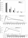
Delayed cell cycle withdrawal and differentiation of myostatin-null adult myoblasts. Isolated myoblasts from C57BL/10 (Mstn+/+) or myostatin-null mice (Mstn−/−) were switched to DM, and the cells were pulsed with BrdU before fixing at 0-, 6-, 12-, 24-, or 48-h time points. Percentage of cells that incorporate BrdU are shown for every time point in panel A. *, P < 0.05; **, P < 0.01 (as compared with wild type). Over 1,000 cells were counted in each of three replicates. The data presented are an average of three animals (A). Freshly isolated myoblasts from C57BL/10 (WT) or myostatin-null mice (KO) were switched to DM, and proteins were extracted at indicated time points. Time 0 indicates freshly isolated quiescent satellite cells. Western analysis (n = 3) with MyoD (B) and Myogenin (C) antibodies on protein extracts from differentiating myoblasts is shown. Maximum expression was termed 100%, and relative expression at various time points was plotted. Anti–α-tubulin Western indicates equal loading.
Figure 8.
Figure 8.
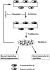
A model for the role of Myostatin in postnatal muscle growth. Quiescent satellite cells on muscle fibers are activated in response to muscle injury to give rise to myoblasts. Proliferating myoblasts can either fuse with the existing fiber or differentiate into a nascent myotube. A portion of proliferating myoblasts, however, can revert to become quiescent satellite cells, thus resulting in self-renewal. As Myostatin is a negative regulator of cell cycle progression, high levels of Myostatin in satellite cells block the activation to maintain quiescence.

Similar articles

Cited by

References

    1. Allen, R.E., C.J. Temm-Grove, S.M. Sheehan, and G. Rice. 1997. Skeletal muscle satellite cell cultures. Methods Cell Biol. 52:155–176. - PubMed
    1. Beauchamp, J.R., L. Heslop, D.S. Yu, S. Tajbakhsh, R.G. Kelly, A. Wernig, M.E. Buckingham, T.A. Partridge, and P.S. Zammit. 2000. Expression of CD34 and Myf5 defines the majority of quiescent adult skeletal muscle satellite cells. J. Cell Biol. 151:1221–1234. - PMC - PubMed
    1. Bischoff, R. 1989. Analysis of muscle regeneration using single myofibers in culture. Med. Sci. Sports Exerc. 21:S164–S172. - PubMed
    1. Bogdanovich, S., T.O. Krag, E.R. Barton, L.D. Morris, L.A. Whittemore, R.S. Ahima, and T.S. Khurana. 2002. Functional improvement of dystrophic muscle by myostatin blockade. Nature. 420:418–421. - PubMed
    1. Carlson, C.J., F.W. Booth, and S.E. Gordon. 1999. Skeletal muscle myostatin mRNA expression is fiber-type specific and increases during hindlimb unloading. Am. J. Physiol. 277:R601–R606. - PubMed

Publication types

MeSH terms