Review
. 2003 Dec 1;553(Pt 2):345-55.
doi: 10.1113/jphysiol.2003.049221. Epub 2003 Sep 8.

Endocytosis at the synaptic terminal

Affiliations
Review

Endocytosis at the synaptic terminal

Stephen J Royle et al. J Physiol. .

Abstract

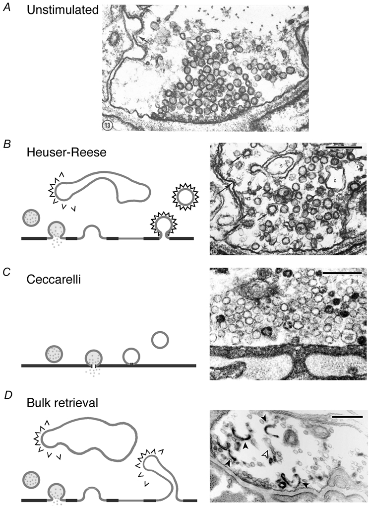
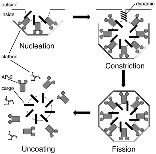
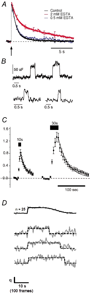
Exocytosis of neurotransmitter from a synaptic vesicle is followed by efficient retrieval of its constituent membrane and proteins. Real-time measurements indicate that fast and slow modes of retrieval operate in parallel at a number of presynaptic terminals. Two mechanisms can be distinguished by electron microscopy: clathrin-mediated retrieval of small vesicles and bulk retrieval of large cisternae. Methods that investigate the behaviour of individual vesicles have recently demonstrated a third route of retrieval: the rapid reversal of a pore-like connection between the vesicle and surface ('kiss-and-run'). Key aims for the future are to identify the molecules underlying different mechanisms of endocytosis at the synapse and the signals that select between them.


Figures

Figure 1
Figure 1. Models of synaptic vesicle retrieval at the frog NMJ
A, electron micrograph of the frog NMJ at rest. B, Heuser-Reese model for retrieval. Diagram (left) shows that vesicles fully collapse into the plasma membrane and are retrieved by CME. Coated vesicles may also form from cisternae. Electron micrograph (right) of frog NMJ following stimulation at 10 Hz for 1 min. Coated pits and vesicles (arrowed) and cisternae (c) are seen in the terminal. C, Ceccarelli model for retrieval. Diagram (left) shows a vesicle releasing neurotransmitter through a transient fusion pore by ‘kiss-and-run’; the vesicle is retrieved at the same site. Micrograph (right) of a terminal stimulated at 2 Hz for 2 h showing an absence of clathrin-coated vesicles and cisternae at low stimulation frequency. D, bulk membrane retrieval. In this model (left) large areas of membrane are internalised following complete vesicle collapse. Coated vesicles may bud from membrane invaginations or from large, internalised cisternae. Micrograph (right) shows internalised cisternae that are empty (open arrows) or filled with photoconverted FM1–43 (filled arrows). Scale bars, 250 nm in all cases. Figures reproduced from Heuser & Reese (1973). Ceccarelli et al. (1973) and Richards et al. (2000) with permission from the Rockefeller University Press and Elsevier Science/Cell Press.
Figure 2
Figure 2. Molecular mechanisms of clathrin-mediated endocytosis
Schematic diagram to show the major steps of CME in non-neuronal cells. CME comprises four major steps: nucleation, constriction, fission and uncoating. Numerous other proteins such as amphiphysin, epsin and endophilin (Brodsky et al. 2001) have been omitted for clarity, and only the core molecular players are shown here. The transmembrane cargo protein has been proposed to be synaptotagmin in the case of synaptic vesicle retrieval.
Figure 3
Figure 3. Physiological measurements of endocytosis
A, whole-cell capacitance recordings in goldfish retinal bipolar cells. A brief, 20 ms depolarisation (arrow) causes exocytosis and subsequent membrane retrieval by fast endocytosis. Introduction of EGTA to chelate intracellular calcium causes a proportion of membrane to be retrieved by slow endocytosis. B, low noise cell-attached capacitance recording in a pituitary nerve terminal reveals capacitance flickers that correspond to kiss-and-run of microvesicles. Note the similar size of the down-step following an up-step in the trace. C, average intensity of synapto-pHluorin fluorescence in hippocampal neurons. Transfected cells were imaged by confocal microscopy and stimulated at 10 Hz for 10 or 30 s. The decay of fluorescence at the end of the stimulus corresponds to reacidification of synaptic vesicles following endocytosis. D, exocytosis and endocytosis of single vesicles in hippocampal boutons. The top trace is an average of 25 events. The three individual traces below show stepwise changes in synapto-pHluorin fluorescence corresponding to exocytosis and endocytosis of individual vesicles. Figures reproduced from Neves et al. (2001). Klyachko & Jackson (2002). Sankaranarayanan & Ryan (2000) and Gandhi & Stevens (2003) with permission from the National Academy of Sciences and Nature Publishing Group.

Similar articles

Cited by

References

    1. Albillos A, Dernick G, Horstmann H, Almers W, Alvarez De Toledo G, Lindau M. The exocytotic event in chromaffin cells revealed by patch amperometry. Nature. 1997;389:509–512. - PubMed
    1. Ales E, Tabares L, Poyato JM, Valero V, Lindau M, Alvarez De Toledo G. High calcium concentrations shift the mode of exocytosis to the kiss-and-run mechanism. Nat Cell Biol. 1999;1:40–44. - PubMed
    1. Aravanis AM, Pyle JL, Tsien RW. Single synaptic vesicles fusing transiently and successively without loss of identity. Nature. 2003;423:643–647. - PubMed
    1. Ball CL, Hunt SP, Robinson MS. Expression and localization of α-adaptin isoforms. J Cell Sci. 1995;108:2865–2875. - PubMed
    1. Beutner D, Voets T, Neher E, Moser T. Calcium dependence of exocytosis and endocytosis at the cochlear inner hair cell afferent synapse. Neuron. 2001;29:681–690. - PubMed

Publication types

LinkOut - more resources