. 2003 Nov 1;552(Pt 3):961-74.
doi: 10.1113/jphysiol.2003.048009. Epub 2003 Sep 8.

Both dorsal horn and lamina VIII interneurones contribute to crossed reflexes from feline group II muscle afferents

Affiliations

Both dorsal horn and lamina VIII interneurones contribute to crossed reflexes from feline group II muscle afferents

S A Edgley et al. J Physiol. .

Abstract

Previous studies have demonstrated that group II muscle afferents exert powerful actions on contralateral motoneurones and that these actions are mediated primarily via lamina VIII commissural interneurones. We examined whether dorsal horn interneurones also contribute to these actions, as they have been shown to contribute to the actions of group II afferents on ipsilateral motoneurones. We tested the susceptibility of IPSPs and EPSPs evoked from group II afferents in contralateral motoneurones to presynaptic inhibition as an indicator of the relative contribution of dorsal horn interneurones to these PSPs, since the monosynaptic activation of dorsal horn interneurones is more weakly and more briefly depressed by presynaptic inhibition than is the monosynaptic activation of lamina VIII and other intermediate zone and ventral horn interneurones. While the earliest components of IPSPs and EPSPs evoked by group II afferents were abolished by conditioning stimulation of group II afferents, consistent with them being evoked disynaptically by commissural interneurones, trisynaptic components of these PSPs were only partly reduced and are therefore attributed to dorsal horn interneurones. The same conditioning stimuli depressed the disynaptic excitation of lamina VIII commissural interneurones by group II afferents much less effectively than they depressed monosynaptic excitation, indicating that dorsal horn interneurones contribute to this disynaptic excitation. On the basis of these observations we conclude that that dorsal horn interneurones contribute to the late actions of group II muscle afferents on contralateral motoneurones through their disynaptic actions on commissural interneurones.


Figures

Figure 1
Figure 1. Relationships between dorsal horn and commissural lamina VIII interneurones
A, the neuronal network underlying the actions of group II afferents on ipsilateral motoneurones (from Jankowska et al. 2002b). Pathways between group II afferents and ipsilateral motoneurones run either directly through intermediate zone neurones (pathway shown in black) or indirectly through dorsal horn neurones (pathway shown in grey). B, the hypothesised connections between the dorsal horn interneurones and lamina VIII commissural interneurones, following the same scheme as in A. C, diagram showing stronger and longer lasting presynaptic inhibition of transmission from group II afferents to neurones in the intermediate zone than to neurones in the dorsal horn, as estimated from changes in monosynaptic components of group II evoked field potentials at the two locations (from Jankowska et al. 2002a). Note that the dorsal horn field potentials recovered to > 50 % of control within 50 ms whereas the intermediate zone field potentials remained < 20 % of control for more than 100 ms.
Figure 2
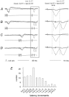
Figure 2. Effects of conditioning stimulation of contralateral group II afferents on short latency IPSPs evoked from contralateral group II afferents in motoneurones
A and B, IPSPs evoked in a GS motoneurone by single and double stimuli to contralateral group II afferents. Upper records are intracellular potentials; the lower parallel records show afferent volleys from the cord dorsum. Control (unconditioned) responses are shown in grey and conditioned responses in black. The conditioning–testing interval was 94 ms. The panels on the right show expanded records of the responses to the test stimuli (from the parts of the left hand records within the box). C and D, as A and B but with a conditioning–testing interval of 45 ms. For all traces the vertical dotted lines indicate the onsets of the conditioned and unconditioned IPSPs. The double arrow above the right panel in A shows the time window within which the areas of the IPSPs were measured. F, increments in the latency of IPSPs evoked by conditioning stimulation of Q in all cases in which IPSPs with a distinct onset were evoked (n = 101). In this and the following figures, the negativity is downwards in the microelectrode records and upwards in the records from the cord dorsum.
Figure 3
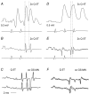
Figure 3. Effects of conditioning stimulation of contralateral group II afferents on EPSPs evoked from contralateral group II afferents in motoneurones
A and B, records from Q and PBST motoneurones respectively (upper traces) and cord dorsum potentials (lower traces). Unconditioned responses are shown in grey and conditioned (cond) responses in black. The panels on the left show records at a slow timebase, the panels to the right show the responses to the test stimuli (from the box in the lefthand panels) expanded (twice vertically four times horizontally). Dotted lines in the panels on the right indicate the onset of the unconditioned and conditioned PSPs. The double arrows in the right panels show the time windows within which the areas of the PSPs were compared.
Figure 4
Figure 4. Examples of early and late excitation of commissural neurones by group II afferents, attributable to monosynaptic and disynaptic actions
A–C and D–F, illustrate records from two different commissural neurones respectively. A and D show EPSPs evoked by two and three stimuli applied to the quadriceps nerve (Q) (upper traces) and cord dorsum potentials (lower traces). B and E show extracellular spikes, recorded just before penetration of the neurones, with cord dorsum potentials. Note the likely disynaptically induced EPSPs (the second of the dotted lines) following the monosynaptically evoked ones (first dotted lines) after the second stimulus. C and F, antidromic activation from the contralateral GS motor nucleus (co GS MN) and collision between synaptically and antidromically induced extracellular spikes confirming antidromic activation; two and three superimposed records. Voltage calibrations are 0.2 mV for A and B and 0.5 mV for D and E.
Figure 5
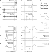
Figure 5. Depression of extracellularly recorded spike potentials and intracellularly recorded EPSPs in a commissural interneurone by conditioning stimulation
A–C, extracellularly recorded spike potentials evoked by stimulation of group II afferents in Q and the corresponding cord dorsum records. These were preceded by conditioning simulation of group II afferents (5 stimuli, 5T) to Q (B) or Sart (C), conditioning-testing interval of 80 ms. D-F, peristimulus time histograms (PSTH) of responses evoked by 20 consecutive test stimuli, including those in the areas marked by the box in A–C, unconditioned (D) and conditioned by stimulation of Q (E) or Sart (F). G, responses to stimuli applied in the contralateral GS motor nucleus (GS MN, upper trace) and collision by synaptically evoked spike potentials (lower trace) confirming antidromic activation. H, blocked antidromically evoked spike potential after penetration of the neurone. I–K, EPSPs evoked by Q group II afferents after penetration of the neurone, unconditioned (I), conditioned by Q group II afferents (3 stimuli, 5T) in J, and conditioned by sart; group II afferents (5 stimuli 5T, K). Conditioning-testing intervals were 56 ms. L–N show the responses to test stimuli of the records in I–K (in the box) on an expanded timebase. The first and second dotted lines in D–F and L–N indicate the latencies of the unconditioned spikes and EPSPs and of those induced after conditioning stimuli, respectively.
Figure 6
Figure 6. Differential effects of conditioning stimulation on activation of a commissural interneurone by group II and reticulospinal afferents
A shows three traces, from top to bottom, extracellular records from an interneurone discharged by two test stimuli to Q at 5T, the PSTH derived from a series of 20 such stimuli and the cord dorsum volleys. B, PSTH and volleys traces as in A, showing that the same test stimuli are ineffective when preceded by conditioning stimulation (Q, 4 stimuli, 4T, 56 ms interval). C, traces as in A showing that the same interneurone was discharged on stimulation of reticulospinal fibres (RF, 4 stimuli 100 μA). D shows that the reticulospinal response was not substantially altered by the same conditioning stimuli as in B. Only the PSTH and the volleys are shown. On the right the PSTH records of responses to the test stimuli from A–D are shown on an expanded timebase to facilitate comparison.
Figure 7
Figure 7. Differential effects of presynaptic inhibition on early and later components of EPSPs evoked from group II afferents in a commissural interneurone
A, control EPSP (upper trace) evoked by near maximal stimulation of Q group II afferents, which shows a decrease in amplitude and increase in latency following heterogenetic conditioning stimuli (Sart, second trace, conditioning–testing interval of 56 ms). B, horizontally expanded parts of records in boxed areas in A. C, as A but for homogenetic depression (Q) and for EPSPs evoked by double instead of single test stimuli. D, expanded parts of records in C. Dotted lines in the expanded records indicate the onset of the control and conditioned EPSPs. E, extracellular records of antidromically evoked spike potentials in the interneurone and collision by synaptically evoked spikes. F, intracellular record of the blocked antidromic spike. Bottom traces in A–D and F are from the cord dorsum. Negativity is downwards in the microelectrode records and upwards in the records from the cord dorsum. Voltage calibration in C is for all the microelectrode records.
Figure 8
Figure 8. Differential effects of presynaptic inhibition on early and later components of IPSPs evoked from group II afferents in a commissural interneurone
Records A–G were taken after the neurone was filled with an intracellular marker during which the IPSP reversed. The IPSP recorded soon after the penetration of the neurone (segmental latency 3.5–4 ms) and an antidromic spike evoked from the contralateral GS motor nucleus (latency 1 ms), are shown in H. This hyperpolarizing IPSP is superimposed (grey trace) on the reversed IPSPs in the expanded records in A and D. A, a control reversed IPSP evoked by a near threshold stimulation of group II afferents on a slow timebase in the left panel and an the expanded timebase in the right panel. B and C, reduction in the amplitude (B), and disappearance (C) of this IPSP following single and double conditioning stimuli (conditioning–testing interval 53 ms). The mean depression within a time window of 2 ms from the onset of the PSP was 4 % and 3 %. D, a control IPSP evoked at the same stimulus intensity (but larger because of the increasing depolarisation of the neurone), similarly at two timebases. E–G, increasing effects of an increasing number of conditioning stimuli (conditioning–testing interval 96 ms). The mean depression within 2 ms from the onset of the unconditioned PSP was to 6 %, 4 % and 0 %. Note that the depression of the early components was stronger than the effect on the later components, which began 0.8–2.5 ms after the onset of the control IPSPs. Bottom traces in each pair of records are from the cord dorsum. Voltage calibration in H applies to all of the microelectrode records. Note the different time scales for A–C and D–G.

Similar articles

Cited by

References

    1. Aggelopoulos NC, Burton MJ, Clarke RW, Edgley SA. Characterization of a descending system that enables crossed group II inhibitory reflex pathways in the cat spinal cord. J Neurosci. 1996;16:723–729. - PMC - PubMed
    1. Aggelopoulos NC, Edgley SA. Segmental localisation of the relays mediating crossed inhibition of hindlimb motoneurones from group II afferents in the anaesthetized cat spinal cord. Neurosci Lett. 1995;185:60–64. - PubMed
    1. Alstermark B, Kummel H. Transneuronal transport of wheat germ agglutinin conjugated horseradish peroxidase into last order spinal interneurones projecting to acromio- and spinodeltoideus motoneurones in the cat. 1. Location of labelled interneurones and influence of synaptic activity on the transneuronal transport. Exp Brain Res. 1990;80:83–95. - PubMed
    1. Arya T, Bajwa S, Edgley SA. Crossed reflex actions from group II muscle afferents in the lumbar spinal cord of the anaesthetized cat. J Physiol. 1991;444:117–131. - PMC - PubMed
    1. Bannatyne BA, Edgley SA, Hammar I, Jankowska E, Maxwell DJ. Networks of inhibitory and excitatory commissural interneurons mediating crossed reticulospinal actions identified by immunocytochemistry. Eur J Neurosci. 2003a in press. - PMC - PubMed

Publication types

LinkOut - more resources