Review
. 2003 Sep;67(3):303-42, table of contents.
doi: 10.1128/MMBR.67.3.303-342.2003.

Ancient origin of the tryptophan operon and the dynamics of evolutionary change

Affiliations
Review

Ancient origin of the tryptophan operon and the dynamics of evolutionary change

Gary Xie et al. Microbiol Mol Biol Rev. 2003 Sep.

Abstract

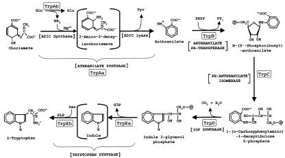
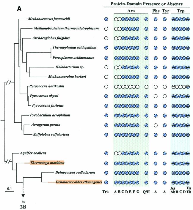
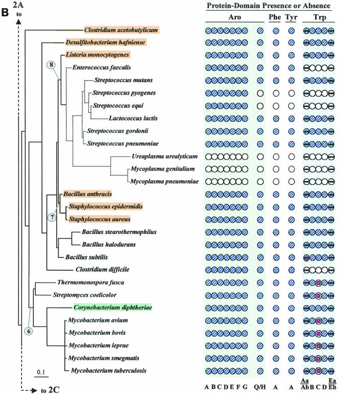
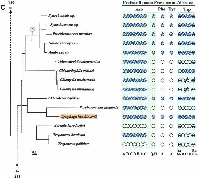
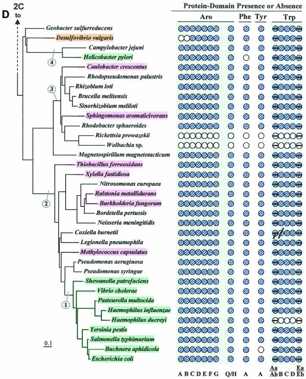
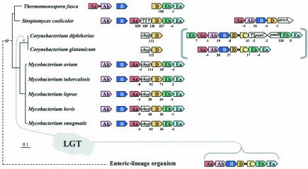
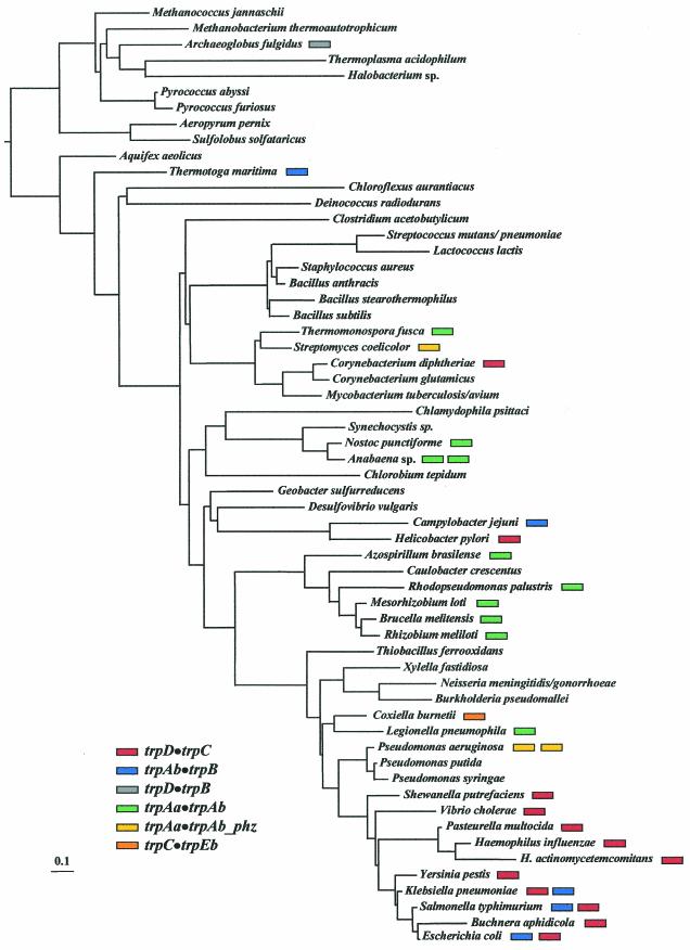
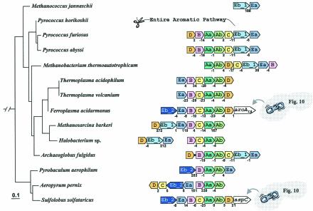
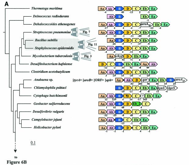
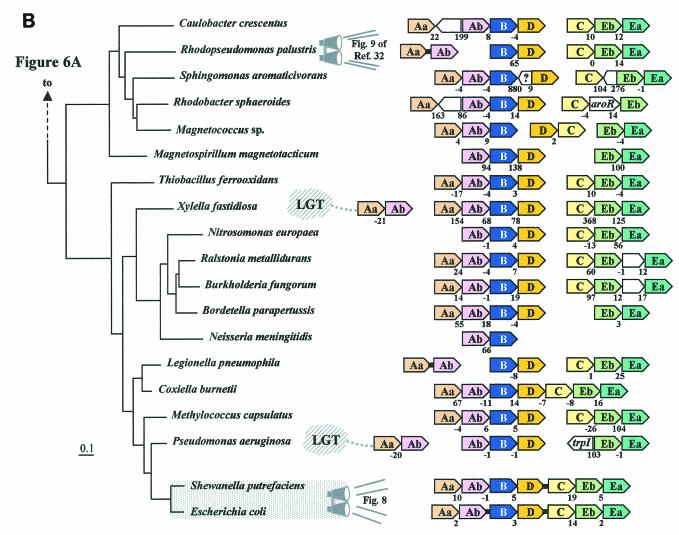
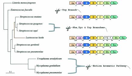
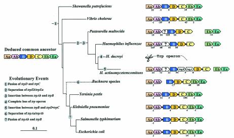
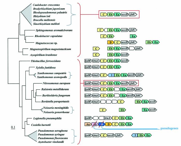
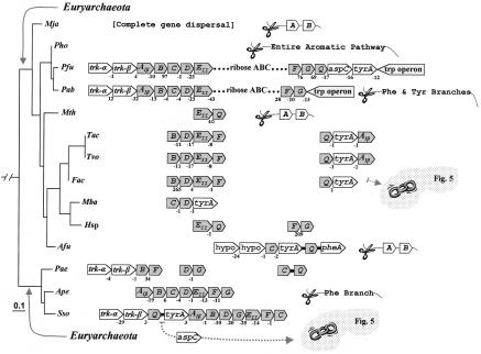
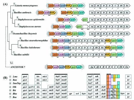
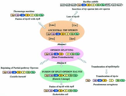
The seven conserved enzymatic domains required for tryptophan (Trp) biosynthesis are encoded in seven genetic regions that are organized differently (whole-pathway operons, multiple partial-pathway operons, and dispersed genes) in prokaryotes. A comparative bioinformatics evaluation of the conservation and organization of the genes of Trp biosynthesis in prokaryotic operons should serve as an excellent model for assessing the feasibility of predicting the evolutionary histories of genes and operons associated with other biochemical pathways. These comparisons should provide a better understanding of possible explanations for differences in operon organization in different organisms at a genomics level. These analyses may also permit identification of some of the prevailing forces that dictated specific gene rearrangements during the course of evolution. Operons concerned with Trp biosynthesis in prokaryotes have been in a dynamic state of flux. Analysis of closely related organisms among the Bacteria at various phylogenetic nodes reveals many examples of operon scission, gene dispersal, gene fusion, gene scrambling, and gene loss from which the direction of evolutionary events can be deduced. Two milestone evolutionary events have been mapped to the 16S rRNA tree of Bacteria, one splitting the operon in two, and the other rejoining it by gene fusion. The Archaea, though less resolved due to a lesser genome representation, appear to exhibit more gene scrambling than the Bacteria. The trp operon appears to have been an ancient innovation; it was already present in the common ancestor of Bacteria and Archaea. Although the operon has been subjected, even in recent times, to dynamic changes in gene rearrangement, the ancestral gene order can be deduced with confidence. The evolutionary history of the genes of the pathway is discernible in rough outline as a vertical line of descent, with events of lateral gene transfer or paralogy enriching the analysis as interesting features that can be distinguished. As additional genomes are thoroughly analyzed, an increasingly refined resolution of the sequential evolutionary steps is clearly possible. These comparisons suggest that present-day trp operons that possess finely tuned regulatory features are under strong positive selection and are able to resist the disruptive evolutionary events that may be experienced by simpler, poorly regulated operons.


Figures

FIG. 1.
FIG. 1.
Biochemical pathway of tryptophan biosynthesis. The nomenclature used in this paper for the seven catalytic domains is in boxes. See Table 1 for the alternative designations used in the literature. Anthranilate synthase catalyzes the overall reaction from chorismate to anthranilate via the half-reactions shown, whereby 2-amino-2-deoxyisochorismate (ADIC) is an enzyme-bound intermediate (62). The TrpAa/TrpAb complex functions as an amidotransferase, utilizing glutamine as the source of the o-amino group of anthranilate. TrpAa can catalyze the overall reaction alone in the presence of NH3 (thereby functioning as an aminase). TrpAb alone in some cases may be able to function as a glutaminase. As shown by McDonald et al. (59), Pseudomonas and Streptomyces species form ADIC as the product of a reaction catalyzed by PhzE. PhzE has fused domains that are homologues of TrpAa and TrpAb, which we have denoted TrpAa•TrpAb_phz (93) (see Table 1). In these organisms, ADIC can be considered a branch point that proceeds to Trp on the one hand and to phenazine pigments on the other hand. Tryptophan synthase catalyzes a second overall reaction, converting indoleglycerol phosphate to Trp in a reaction path where indole is always an intermediate. The alpha (TrpEa) and beta (TrpEb) subunits catalyze the reactions shown in which the indole intermediate is processed through a tunnel (85). PR, phosphoribosyl; IGP, indoleglycerol phosphate; G3P, glyceraldehyde 3-phosphate.
FIG.2.
FIG.2.
Distribution of aromatic-pathway catalytic domains among prokaryotes. In each panel, 16S rRNA trees are shown at the left, and the presence (shaded circles) or absence (open circles) of domains is shown at the right. Note that only the presence or absence of genes, not gene order, is indicated. Catalytic domains of the common trunk of aromatic biosynthesis (Aro), the phenylalanine branch (Phe), the tyrosine branch (Tyr), and the Trp branch are labeled across the top right; the specific letter designation for a given domain is shown at the bottom. In the Trp grouping, split circles are used to indicate the presence or absence of TrpAa (top half-circle) and TrpAb (bottom half-circle) or TrpEa (top half-circle) and TrpEa (bottom half-circle). In panel A, the presence or absence of transketolase (Trk) is indicated by the left column of circles. The connecting point of a tree segment in any given panel (A, B, C, and D) with a tree segment(s) in another panel is marked with a broken line. The scale bar corresponds to substitutions per site. Dotted lines in the Streptococcus region (B) and the Buchnera region (D) indicate our suggestion that the 16S rRNA tree shown may not reflect exactly the correct order of branching, and perhaps these organisms branch from a slightly deeper position. See Fig. 8 for the suggested branching order of Buchnera. Circled numbers indicate eight node positions from which Trp protein trees are congruent with the 16S rRNA tree. The common trunk of aromatic biosynthesis is encoded by seven genes whose corresponding gene products are named AroA through AroG. The common-pathway genes are named in exact order of pathway reactions according to the precedent implemented in references , , , , and . The chorismate mutase block is represented by homologues of either AroQ (usually) or AroH (seldom) (12). PheA refers to prephenate dehydratase, the sequence of the relatively infrequent arogenate dehydratase being currently unknown. TyrA refers to a homologue family that includes prephenate dehydrogenase, arogenate dehydrogenase, or cyclohexadienyl dehydrogenase (9, 88). See Fig. 1 for details of Trp biosynthesis. The names of organisms retaining the putative ancestral whole-pathway trp operon are shaded orange, those having the two split-pathway operons are shaded magenta, and those having operons rejoined by fusion of trpD and trpC are shaded aqua. These correspond to the major evolutionary events portrayed in Fig. 12 and indicated with the same color-coding scheme. Probable pseudogenes in chlamydiae (C) and Coxiella (21) are indicated with heavy black slash marks. Genes that function in two pathways (trpAb in Bacillus subtilis and trpC in actinomycete bacteria) are marked with magenta bull's-eyes in B. Panel A includes the Archaea and a few of the deeper-branching Bacteria at the bottom. Panel B includes the gram-positive Bacteria. Panel C includes cyanobacteria, chlamydiae, and other organisms on the 16S rRNA tree between the gram-positive organisms in panel B and the organisms in panel D, which contains the gram-negative subdivisions of the Proteobacteria. Wolbachia sp. (panel D) is an endosymbiont of Brugia malayi. A cross-index of all organisms shown in both this figure and the remaining figures is given in Table 2.
FIG.2.
FIG.2.
Distribution of aromatic-pathway catalytic domains among prokaryotes. In each panel, 16S rRNA trees are shown at the left, and the presence (shaded circles) or absence (open circles) of domains is shown at the right. Note that only the presence or absence of genes, not gene order, is indicated. Catalytic domains of the common trunk of aromatic biosynthesis (Aro), the phenylalanine branch (Phe), the tyrosine branch (Tyr), and the Trp branch are labeled across the top right; the specific letter designation for a given domain is shown at the bottom. In the Trp grouping, split circles are used to indicate the presence or absence of TrpAa (top half-circle) and TrpAb (bottom half-circle) or TrpEa (top half-circle) and TrpEa (bottom half-circle). In panel A, the presence or absence of transketolase (Trk) is indicated by the left column of circles. The connecting point of a tree segment in any given panel (A, B, C, and D) with a tree segment(s) in another panel is marked with a broken line. The scale bar corresponds to substitutions per site. Dotted lines in the Streptococcus region (B) and the Buchnera region (D) indicate our suggestion that the 16S rRNA tree shown may not reflect exactly the correct order of branching, and perhaps these organisms branch from a slightly deeper position. See Fig. 8 for the suggested branching order of Buchnera. Circled numbers indicate eight node positions from which Trp protein trees are congruent with the 16S rRNA tree. The common trunk of aromatic biosynthesis is encoded by seven genes whose corresponding gene products are named AroA through AroG. The common-pathway genes are named in exact order of pathway reactions according to the precedent implemented in references , , , , and . The chorismate mutase block is represented by homologues of either AroQ (usually) or AroH (seldom) (12). PheA refers to prephenate dehydratase, the sequence of the relatively infrequent arogenate dehydratase being currently unknown. TyrA refers to a homologue family that includes prephenate dehydrogenase, arogenate dehydrogenase, or cyclohexadienyl dehydrogenase (9, 88). See Fig. 1 for details of Trp biosynthesis. The names of organisms retaining the putative ancestral whole-pathway trp operon are shaded orange, those having the two split-pathway operons are shaded magenta, and those having operons rejoined by fusion of trpD and trpC are shaded aqua. These correspond to the major evolutionary events portrayed in Fig. 12 and indicated with the same color-coding scheme. Probable pseudogenes in chlamydiae (C) and Coxiella (21) are indicated with heavy black slash marks. Genes that function in two pathways (trpAb in Bacillus subtilis and trpC in actinomycete bacteria) are marked with magenta bull's-eyes in B. Panel A includes the Archaea and a few of the deeper-branching Bacteria at the bottom. Panel B includes the gram-positive Bacteria. Panel C includes cyanobacteria, chlamydiae, and other organisms on the 16S rRNA tree between the gram-positive organisms in panel B and the organisms in panel D, which contains the gram-negative subdivisions of the Proteobacteria. Wolbachia sp. (panel D) is an endosymbiont of Brugia malayi. A cross-index of all organisms shown in both this figure and the remaining figures is given in Table 2.
FIG. 3.
FIG. 3.
Apparent absence of trpC and an event of LGT in a lineage of actinomycete bacteria. A broader phylogenetic context can be viewed in Fig. 2B and 6A. chyp denotes a conserved hypothetical membrane protein exhibiting about 28% identity in comparison of a given Mycobacterium species with a given Corynebacterium species. Color-coded boxes pointing in the direction of transcription represent genes of Trp biosynthesis. For clarity of presentation, trpAa is shown as Aa, etc. Open boxes with question marks denote hypothetical proteins. Intergenic spacing is shown, with negative values indicating gene overlap. trpD•trpC fusions are represented by short black linker bars. On the left are 16S rRNA-based phylogenetic trees of the genomes having the gene organizations shown on the right. Orthologues that match the mycobacterial trpAa/chyp/D/Eb/Ea operon genes are aligned vertically. Contemporary trp operons in coryneform species that originated in their common ancestor by LGT of trpAa/Ab/B/D•C/Eb/Ea from a source within the enteric lineage are shown within brackets. Except for the two coryneform species, all actinomycetes have a free-standing trpB gene. The Mycobacterium spp. and Streptomyces also have a free-standing trpAb gene. The corresponding TrpB and TrpAb proteins exhibit high identity with one another but not with TrpB and TrpAb of the coryneform species. Thermomonospora has dissociated trpAa from the typical clade operon and fused it with trpAb (as also shown in Fig. 4). The trpAa/Ab/B/D/aroAII operon of S. coelicolor is known to be specifically associated with antibiotic biosynthesis (see text).
FIG. 4.
FIG. 4.
Mapping of the distribution of Trp pathway gene fusions to the 16S rRNA tree. The presence of fusion subtypes is color-coded as indicated in the legend. Although Buchnera aphidicola maps near E. coli on the 16S rRNA tree, as shown, its true point of divergence is probably prior to Yersinia, as portrayed by dotted lines in Fig. 8.
FIG. 5.
FIG. 5.
Organization of trp operon genes in the Archaea. Each trp gene is color coded differently, including the two subtypes of trpEb (Eb_1 and Eb_2) (92). trp genes that exist in the genome unlinked to any other trp genes are not shown. Archaeoglobus fulgidus has a trpD•trpB gene fusion (see Fig. 4). Intergenic spacing is shown, with negative values indicating gene overlap. Genes that are not specific trp pathway genes are in white boxes. F. acidarmanus possesses a gene encoding the aroAIβ subclass (44) of DAHP synthase. aspC in S. solfataricus is an aromatic aminotransferase of the Iγ aspartate aminotransferase type (42). This gene insertion corresponds to genes that appear to have escaped from the aro operons shown in Fig. 10. The gene order shown for Methanosarcina barkeri is the same as those in Methanosarcina acetivorans and Methanosarcina mazei. The gene order shown for S. solfataricus is the same as that for Solfolobus tokodaii.
FIG. 6.
FIG. 6.
Organization of trp operon genes in the Bacteria. Each trp gene is color coded differently, including the two subtypes of trpEb. (trpEb in this figure refers to the major trpEb_1 subtype.) The tree sections in A and B join as indicated by the dashed line. Intergenic spacing is shown, with negative values indicating gene overlap. Separations showing white space and no intergenic spacing values indicate that the gene clusters are not linked to one another. Insertions of hypothetical genes and known genes are shown as white boxes. Short black bars connecting arrows denote gene fusions. Links to zoom-in expansions of particular lineages in other figures of this paper are indicated by binoculars. In B, the gene organization shown for Rhodopseudomonas palustris is identical to those of the closely related Agrobacterium tumefaciens, Rhizobium loti, Brucella melitensis, and Sinorhizobium meliloti; that shown for Burkholderia fungorum is identical to that of Burkholderia pseudomallei and Burkholderia mallei; that shown for Bordetella parapertussis is identical to that of Bordetella pertusis and Bordetella bronchiseptica; that for Neisseria meningitidis is identical to that of Neisseria gonorrhoeae; and that for Pseudomonas aeruginosa is identical to that of Pseudomonas putida, Pseudomonas fluorescens, and Pseudomonas syringae. The apparent supraoperon of Anabaena sp. (A) has been discussed in reference . kynU and kprS on the Chlamydophila psittaci line (A) refer to genes encoding kynureninase and PRPP synthase, respectively (89). The linked trpAa/trpAb genes shown for P. aeruginosa (B) were named phnA/phnB by Essar et al. (24). because they were thought to be dedicated to phenazine biosynthesis, a conclusion shown to be incorrect by Mavrodi et al. (57). This gene pair is not within the vertical line of descent (see later section), as indicated by the LGT notation. The trpAaAb operon shown on the left for Xylella is also outside the vertical line of descent (i.e., origin by LGT) (93). Shewanella putrefaciens (B) has the newly proposed name of Shewanella oneidensis (81).
FIG. 6.
FIG. 6.
Organization of trp operon genes in the Bacteria. Each trp gene is color coded differently, including the two subtypes of trpEb. (trpEb in this figure refers to the major trpEb_1 subtype.) The tree sections in A and B join as indicated by the dashed line. Intergenic spacing is shown, with negative values indicating gene overlap. Separations showing white space and no intergenic spacing values indicate that the gene clusters are not linked to one another. Insertions of hypothetical genes and known genes are shown as white boxes. Short black bars connecting arrows denote gene fusions. Links to zoom-in expansions of particular lineages in other figures of this paper are indicated by binoculars. In B, the gene organization shown for Rhodopseudomonas palustris is identical to those of the closely related Agrobacterium tumefaciens, Rhizobium loti, Brucella melitensis, and Sinorhizobium meliloti; that shown for Burkholderia fungorum is identical to that of Burkholderia pseudomallei and Burkholderia mallei; that shown for Bordetella parapertussis is identical to that of Bordetella pertusis and Bordetella bronchiseptica; that for Neisseria meningitidis is identical to that of Neisseria gonorrhoeae; and that for Pseudomonas aeruginosa is identical to that of Pseudomonas putida, Pseudomonas fluorescens, and Pseudomonas syringae. The apparent supraoperon of Anabaena sp. (A) has been discussed in reference . kynU and kprS on the Chlamydophila psittaci line (A) refer to genes encoding kynureninase and PRPP synthase, respectively (89). The linked trpAa/trpAb genes shown for P. aeruginosa (B) were named phnA/phnB by Essar et al. (24). because they were thought to be dedicated to phenazine biosynthesis, a conclusion shown to be incorrect by Mavrodi et al. (57). This gene pair is not within the vertical line of descent (see later section), as indicated by the LGT notation. The trpAaAb operon shown on the left for Xylella is also outside the vertical line of descent (i.e., origin by LGT) (93). Shewanella putrefaciens (B) has the newly proposed name of Shewanella oneidensis (81).
FIG. 7.
FIG. 7.
Zoom-in from Fig. 6A showing instances of Trp pathway reductive evolution and expansion of intergenic space in one phylogenetic section of some gram-positive bacteria whose 16S rRNA tree relationships are shown at the left. Loss of various metabolic capabilities is indicated by scissors. Note that the order of branching of Lactococcus lactis (shown in orange) has been altered from that shown in the 16S rRNA tree of Fig. 2B. The gene order and compact spacing of Listeria innocua is the same as that shown for Listeria monocytogenes.
FIG. 8.
FIG. 8.
Zoom-in from Fig. 6B showing Trp pathway gene organization in a range of Proteobacteria defined by the presence of the trpDtrpC fusion. Deduced phylogenetic events described on the left are identified by number on the 16S rRNA tree at the evolutionary times indicated. The actual position of Buchnera on the 16S rRNA tree (as shown in Fig. 2D and Fig. 4) is closest to E. coli. However, the long branch (Fig. 4) is consistent with the more likely order of branching depicted by the dotted line for Buchnera in this figure.
FIG. 9.
FIG. 9.
Conserved genes flanking the trpC/trpEb/trpEa operon of organisms within the split-operon portion of the 16S rRNA tree. Organisms in the upper grouping are α-Proteobacteria; the cluster between Thiobacillus and Neisseria are β-Proteobacteria; and the bottom cluster is that fraction of the γ-Proteobacteria that diverged prior to the trpD•trpC fusion event. lysM and truA, conserved at the flanking gene position at the left throughout the β- and γ-Proteobacteria, are shaded grey, as are accD and folC (conserved in the flanking gene position at the right throughout the phylogenetic span portrayed in this figure). The deduced gene order of the common ancestor for each of the two major 16S rRNA clades is the same as shown for the two contemporary organisms Rhodopseudomonas palustris and Nitorosomonas europaea, as indicated by outlining in orange. Intervening genes, either hypothetical or known, are shown as open block arrows.
FIG. 10.
FIG. 10.
Linkage relationship of genes within the larger context of aromatic amino acid biosynthesis in Archaea. The tree is the same as that shown in Fig. 5, where the full organism names corresponding to the acronyms used can be viewed. Common-pathway genes are shaded and designated by the gene letter, e.g., Q = aroQ. Hypothetical genes are denoted as hypo. Genes are labeled within block arrows that point in the direction of transcription. Copies of genes encoding transketolase are designated trk-α and trk-β. Short black bars connecting arrows indicate gene fusions. Deleted genes, pathway branches, and entire pathways are indicated with scissors.
FIG. 11.
FIG. 11.
Zoom-in from Fig. 6A showing a conserved gram-positive region containing the six-gene aro operon (or remnants of it) and the trp/aro supraoperon of the B. subtilis/B. halodurans/B. stearothermophilus subgroup. (A) The aro and trp operons are mapped on a 16S rRNA tree at the far left. (The exact branching order of Oceanobacillus iheyensis has not been determined.) The Enterococcus/Streptococcus/Lactococcus grouping branches off between Listeria and the B. anthracis subgroup on a 16S rRNA tree (not shown, but see Fig. 2B and Fig. 7), but we believe from a variety of observations that it belongs just outside of the lineage shown in this figure. Shaded bracketed regions around aro operons and trp/aro supraoperons can be related to the presence of a context of conserved, flanking genes, as shown in part B. The separate aro and trp operons of a putative common ancestor are shown at the bottom of A. aro genes in B are color coded to match the genes shown in A. The conserved region to the left of aro operon genes includes eight genes (gps, hbs, hepS, menH, hepT, ndk, aroG, and aroB) that are conserved in every organism shown (heavy black overbars). Gene abbreviations: gpsA, glycerol 3-phosphate dehydrogenase; spoIVA, sporulation protein IVA; hbs, nonspecific DNA-binding protein; mtrA, GTP cyclohydrolase I; mtrB, TRAP; hepS, heptaprenyldiphosphate synthase (component I); menH, heptaprenyl naphthoquinone methyltransferase; qpt, quinone polyprenyltransferase; acd, aromatic acid decarboxylase; hypo, hypothetical gene; hepT heptaprenyldiphosphate synthase (component II); ndk, nucleoside diphosphate kinase; cheR, chemotaxis protein methyltransferase, tpr, tetratricopeptide repeat-containing protein (COG0457).
FIG.12.
FIG.12.
Schematic of the major evolutionary events (milestone I and milestone II) following the ancient establishment of a trp operon in the domain Bacteria. The ancestral trp operon has been retained by organisms such as Listeria monocytogenes (Lmo), Clostridium acetobutylicum (Cac), Streptococcus pneumoniae (Spn), and Desulfovibrio vulgaris (Dvu). The emergence of selected contemporary organisms is shown. The three stages highlighted with an orange oval, a magenta oval, and a green oval correspond to the color coding used in Fig. 2 to designate the particular contemporary organisms that have retained the exact gene organization illustrated within one of the three ovals.

References

    1. Ahmad, S., and R. A. Jensen. 1989. Utility of a bifunctional tryptophan pathway enzyme for the classification of the Herbicola-Agglomerans complex of bacteria. Int. J. Syst. Bacteriol. 39:100-104.
    1. Alifano, P., R. Fani, P. Lio, A. Lazcano, M. Bazzicalupo, M. S. Carlomagno, and C. B. Bruni. 1996. Histidine biosynthetic pathway and genes: structure, regulation, and evolution. Microbiol. Rev. 60:44-69. - PMC - PubMed
    1. Altamirano, M. M., J. M. Blackburn, C. Aguayo, and A. R. Fersht. 2000. Directed evolution of new catalytic activity with the α/β-barrel scaffold. Nature 403:617-622. - PubMed
    1. Altamirano, N., J. M. Blackburn, C. Aguayo, and A. R. Fersht. 2002. Directed evolution of new catalytic activity with the α/β-barrel scaffold: retraction. Nature 417:468. - PubMed
    1. Altschul, S. F., T. L. Madden, A. A. Schäffer, J. Zhang, Z. Zhang, W. Miller, and D. J. Lipman. 1997. Gapped Blast and PSI-Blast: a new generation of protein database search programs. Nucleic Acids Res. 25:3389-3402. - PMC - PubMed

Publication types