Review
. 2003 Sep;67(3):454-72, table of contents.
doi: 10.1128/MMBR.67.3.454-472.2003.

Transmembrane movement of exogenous long-chain fatty acids: proteins, enzymes, and vectorial esterification

Affiliations
Review

Transmembrane movement of exogenous long-chain fatty acids: proteins, enzymes, and vectorial esterification

Paul N Black et al. Microbiol Mol Biol Rev. 2003 Sep.

Abstract

The processes that govern the regulated transport of long-chain fatty acids across the plasma membrane are quite distinct compared to counterparts involved in the transport of hydrophilic solutes such as sugars and amino acids. These differences stem from the unique physical and chemical properties of long-chain fatty acids. To date, several distinct classes of proteins have been shown to participate in the transport of exogenous long-chain fatty acids across the membrane. More recent work is consistent with the hypothesis that in addition to the role played by proteins in this process, there is a diffusional component which must also be considered. Central to the development of this hypothesis are the appropriate experimental systems, which can be manipulated using the tools of molecular genetics. Escherichia coli and Saccharomyces cerevisiae are ideally suited as model systems to study this process in that both (i) exhibit saturable long-chain fatty acid transport at low ligand concentrations, (ii) have specific membrane-bound and membrane-associated proteins that are components of the transport apparatus, and (iii) can be easily manipulated using the tools of molecular genetics. In both systems, central players in the process of fatty acid transport are fatty acid transport proteins (FadL or Fat1p) and fatty acyl coenzyme A (CoA) synthetase (FACS; fatty acid CoA ligase [AMP forming] [EC 6.2.1.3]). FACS appears to function in concert with FadL (bacteria) or Fat1p (yeast) in the conversion of the free fatty acid to CoA thioesters concomitant with transport, thereby rendering this process unidirectional. This process of trapping transported fatty acids represents one fundamental mechanism operational in the transport of exogenous fatty acids.


Figures

FIG. 1.
FIG. 1.
Steps in fatty acid transport. As detailed in the text, this process is divided into five discrete steps, which can be kinetically defined (steps 1 to 5). Sites of potential protein involvement are highlighted by the thick arrows: fatty acid delivery to the membrane (steps 1 and 2), fatty acid translocation across the membrane (step 3), and fatty acid abstraction or removal from the membrane (steps 4 and 5).
FIG. 2.
FIG. 2.
Domain organization of the FATP family of proteins. (A) Amino acid sequence alignments of the ATP-AMP motif (common to all members of the adenylate-forming superfamily of enzymes) and the FATP-VLACS motif (restricted to the FATP family) from Fat1p and the six murine FATP orthologues. (B) Cartoon showing the approximate positions of the two elements of the ATP-AMP (white rectangles) and the FATP-VLACS (black rectangle) motifs in the FATP family of proteins. aa, amino acids.
FIG. 3.
FIG. 3.
Domain organization of the FACS family of enzymes. (A) Amino acid sequence alignments of the ATP-AMP motif (common to all members of the adenylate-forming superfamily of enzymes noted in Fig. 2) and the FACS motif (restricted to the FACSs) from FadD, yeast (Faa1p and Faa4p), human, mouse, and rat. (B) Cartoon showing the approximate positions of the two elements of the ATP-AMP (white rectangles) and FACS (black rectangle) motifs. aa, amino acids.
FIG. 4.
FIG. 4.
Components and energetics underlying the transport of long-chain fatty acids (LCFA) across the cellular envelope of E. coli. Long-chain fatty acids traverse the outer membrane by way of the specific transport protein FadL. Once they traverse the outer membrane, they enter the periplasmic space, where they are protonated allowing them to partition into the inner membrane. The proton electrochemical gradient across the inner membrane contributes the protons in the periplasmic space. FACS (FadD) responds to the intracellular pools of ATP and in a primed form (FadD-ATP) is hypothesized to sense the free fatty acid [LCFA(H+)] in the inner membrane. In the membrane-bound form, FadD catalyzes the formation of long-chain fatty acyl-CoA (LCFacyl CoA), rendering the process of transport unidirectional. Respiratory substrates for the generation of the electrochemical gradient across the inner membrane under conditions where cells are grown on long-chain fatty acids come from ATP synthetase and β-oxidation. Adapted from reference 11 with permission.
FIG. 5.
FIG. 5.
Predicted topology and functional predictions of the of the long-chain fatty acid transport protein FadL. (A) Cartoon showing the 20 antiparallel β-strands of FadL, which are presumed to form a fatty acid-specific β-barrel within the outer membrane; noted are R93 and R384, which are externally exposed, and the location of the insertion mutation fadLH3, a 2-amino-acid insertion in a periplasmic exposed loop that causes the FadL channel to be in an open conformation. (B) Cartoon illustrating ligand-induced conformational change in FadL, thereby facilitating long-chain fatty acid movement across the outer membrane. 1, FadL in a closed conformation; 2, FadL-specific binding of long-chain fatty acids requires both the acyl chain and the carboxylate of the fatty acid; 3, long-chain fatty acid induced conformational change allowing the fatty acid to traverse the membrane and enter the periplasmic space. Reprinted from reference with permission.
FIG. 6.
FIG. 6.
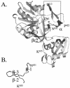
(A) Model of the E. coli FACS developed using the Swiss-Model Protein Modeling Server and visualized using MolScript. The predicted structure begins with residue T213 of the native enzyme. Residues that comprise the peptide modified with APNA and continuing into the FACS signature motif are indicated (P422 to K455). α denotes the predicted α-helix upstream from the FACS signature motif; β-1, β-2, and β-3 denote the β, β-turn-β structure of the FACS signature motif (Fig. 3); and L denotes the linker between the large N-terminal and small C-terminal domains of the enzyme. The boxed region denotes the β-loop β structure that comprises the first sequence element of the ATP-AMP signature motif, while the residue identified by an asterisk is E361 (within the second sequence element of the ATP-AMP signature motif), which is conserved in all adenylate-forming enzymes. (B) α-Carbon tracing of the FACS signature motif (N431 to K455) highlighting the β,β-turn-βstructure, which is proposed to contribute to the fatty acid binding pocket within the enzyme. Reproduced from reference with permission.
FIG. 7.
FIG. 7.
Patterns of C1-BODIPY-C12 uptake in fat1Δ and wild-type (FAT1) cells of S. cerevisiae. Reproduced from reference with permission.
FIG. 8.
FIG. 8.
Directed mutagenesis of FAT1. Asterisk indicate the residues within the ATP-AMP and FATP-VLACS signature motifs (Fig. 2) within Fat1p subjected to mutagenesis. Highlights of these mutants (140) are presented in the text.
FIG. 9.
FIG. 9.
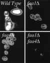
Patterns of C1-BODIPY-C12 uptake in wild-type, faa1Δ, faa4Δ and faa1Δ faa4Δ cells of S. cerevisiae. Reprinted from reference with permission.
FIG. 10.
FIG. 10.
Coimmunoprecipitation of Fat1p and Faa1p or Faa4p indicate Fat1p and a cognate FACS form a functional complex at the plasma membrane. (A) Anti-T7 antibody (α-T7) was used to pull down full-length T7Fatlp in extracts prepared from cells coexpressing T7Fatlp and V5Faa1p or V5Faa4p as indicated. The proteins were separated by sodium dodecyl sulfate-polyacrylamide gel electrophoresis, and subsequent Western blots were probed with anti-V5 (α-V5) antibody to detect V5Faa1p or V5Faa4p as shown. (B) Similarly, anti-V5 was used as the precipitating antibody to pull down V5Faa1p or V5Faa4p following coexpression of T7Fat1p and V5Faa1p or V5Faa4p, and the resultant blot was probed with anti-T7. IP, immunoprecipitating antibody; IB, antibody used in the immunoblot. Lanes: T, total-cell extract; IP, samples immunoprecipitated with the indicated antibody; Beads, protein A-Sepharose alone without an immunoprecipitating antibody. Anti-Pma1p was used as a control protein specific to a yeast plasma membrane protein but unrelated to Fat1p, Faa1p or Faa4p. Reprinted from reference with permission.
FIG. 11.
FIG. 11.
Vectorial acylation in yeast. Cartoon showing the interaction between Fat1p and FACS (either Faa1p or Faa4p), which function to promote the coupled transport and activation of exogenous fatty acids. Fatty acids (FA) partition into the plasma membrane and flip between the membrane surfaces. It is unclear whether Fat1p enhances this process. Fat1p (illustrated as a dimer within the membrane) and a cognate FACS (illustrated as a dimer) form a complex, which functions to abstract the fatty acid from the membrane concomitant with esterification to CoA thioesters (FA-CoA) for further metabolism.

References

    1. Abumrad, N. A., R. C. Perkins, J. H. Park, and C. R. Park. 1981. Mechanism of long chain fatty acid permeation in the isolated adipocyte. J. Biol. Chem. 256:9183-9191. - PubMed
    1. Abumrad, N. A., J. H. Park, and C. R. Park. 1984. Permeation of long-chain fatty acid into adipocytes. Kinetics, specificity, and evidence for involvement of a membrane protein. J. Biol. Chem. 259:8945-8953. - PubMed
    1. Abumrad, N. A., C. C. Forest, D. M. Regen, and S. Sanders. 1991. Increase in membrane uptake of long-chain fatty acids early during preadipocyte differentiation. Proc. Natl. Acad. Sci. USA 88:6008-6012. - PMC - PubMed
    1. Abumrad, N., C. Harmon, and A. Ibrahimi. 1998. Membrane transport of long-chain fatty acids: evidence for a facilitated process. J. Lipid Res. 39:2309-2318. - PubMed
    1. Abumrad, N. A., M. R. el-Maghrabi, E. Z. Amri, E. Lopez, and P. A. Grimaldi. 1993. Cloning of a rat adipocyte membrane protein implicated in binding or transport of long-chain fatty acids that is induced during preadipocyte differentiation. Homology with human CD36. J. Biol. Chem. 268:17665-17668. - PubMed

Publication types

LinkOut - more resources