. 2003 Nov;307(2):450-9.
doi: 10.1124/jpet.103.054965. Epub 2003 Sep 9.

Discrete cell gene profiling of ventral tegmental dopamine neurons after acute and chronic cocaine self-administration

Affiliations

Discrete cell gene profiling of ventral tegmental dopamine neurons after acute and chronic cocaine self-administration

Eric Backes et al. J Pharmacol Exp Ther. 2003 Nov.

Abstract

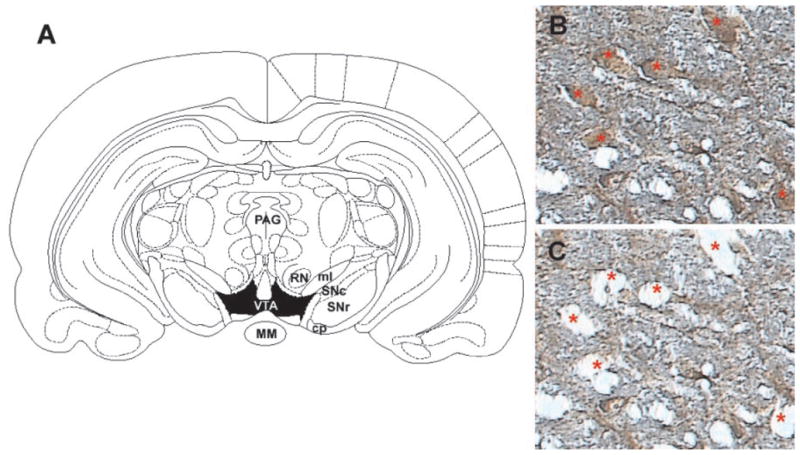
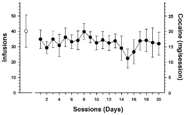
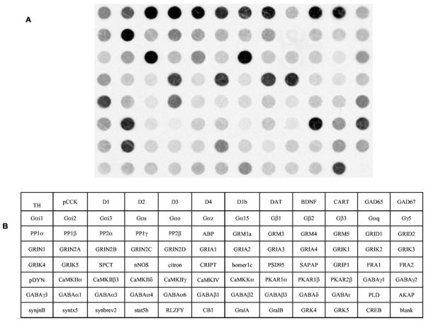
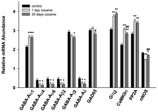
Chronic cocaine administration induces a number of biochemical alterations within the mesolimbic dopamine system that may mediate various aspects of the addictive process such as sensitization, craving, withdrawal, and relapse. In the present study, rats were allowed to self-administer cocaine (0.5 mg/infusion) for 1 or 20 days. Tyrosine hydroxylase immunopositive cells were microdissected from the ventral tegmental area (VTA) using laser capture microdissection, and changes in the abundances of 95 mRNAs were assessed using cDNA macroarrays. Five GABA-A receptor subunit mRNAs (alpha4, alpha6, beta2, gamma2, and delta) were down-regulated at both 1 and 20 days of cocaine self-administration. In contrast, the catalytic subunit of protein phosphatase 2A (PP2alpha), GABA-A alpha1, and Galphai2 were significantly increased at both time points. Additionally, calcium/calmodulin-dependent protein kinase IIalpha mRNA levels were increased initially followed by a slight decrease after 20 days, whereas neuronal nitric-oxide synthase mRNA levels were initially decreased but returned to near control levels by day 20. These results indicate that alterations of specific GABA-A receptor subtypes and other signal transduction transcripts seem to be specific neuroadaptations associated with cocaine self-administration. Moreover, as subunit composition determines the functional properties of GABA-A receptors, the observed changes may indicate alterations in the excitability of dopamine transmission underlying long-term biochemical and behavioral effects of cocaine.


Figures

Fig. 1
Fig. 1
A, schematic of region for laser capture microdissection. Rat coronal midbrain section at −5.30 mm (relative to bregma) showing boundaries for VTA (shaded in black) from which tyrosine hydroxylase immunopositive cells were dissected. Midbrain section immunolabeled with anti-TH antibody (B) and the same section after microdissection of the indicated neurons (C) under 20× magnification. Note the specificity of the dissections and the minimal disruption of surrounding neuropil. MM, mammillary nucleus; PAG, periaqueductal gray area; SNc, substantia nigra-pars compacta; SNr, substantia nigra-pars reticulata; RN, red nucleus; ml, medial lemniscus; cp, cerebral peduncle.
Fig. 2
Fig. 2
The mean (± S.E.M.) number of infusions for cocaine self-administration for 1 (open circle) or 20 days (filled circles). There was no significant difference in the number of infusions on day one between the two groups.
Fig. 3
Fig. 3
Representative cDNA macroarray (95 clones and one blank spot) demonstrating the expression profile of VTA dopamine neurons. The signal intensity of each spot is proportional to the abundance of the particular mRNA in the sample. CCK, cholecystokinin; D1, dopamine receptor 1; D2, dopamine receptor 2; D3, dopamine receptor 3; D4, dopamine receptor 4; D1b, dopamine receptor 1b; DAT, dopamine transporter; BDNF, brain-derived neurotrophic factor; CART, cocaine and amphetamine regulated transcript; GAD65, glutamate decarboxylase 65 kDa; GAD67, glutamate decarboxylase 65 kDa; Gαi1, G protein á inhibiting activity polypeptide 1; Gαi2, G protein á inhibiting activity polypeptide 2; Gαi3, G protein á inhibiting activity polypeptide 3; Gαs, G protein á stimulating activity polypeptide; Gαo, G protein á activating activity polypeptide O; Gαz, G protein á z polypeptide; Gα15, G protein á 15 (Gq class); Gβ1, G protein β polypeptide 1; Gβ2, G protein β polypeptide 2; Gβ3, G protein β polypeptide 3; Gαq, G protein Q polypeptide; Gγ5, = G protein γ polypeptide 5; PP1α, protein phosphatase 1, catalytic subunit, á isoform; PP1β, protein phosphatase 1, catalytic subunit, β isoform; PP2α, protein phosphatase 2, catalytic subunit, á isoform; PP1γ, protein phosphatase 1, catalytic subunit, γ isoform; PP2β, protein phosphatase 2, catalytic subunit, β isoform; ABP, α-amino-3-hydroxy-5-methyl-4-isoxazolepropionic acid receptor binding protein; GRM1a, metabotropic glutamate receptor 1a; GRM3, metabotropic glutamate receptor 3; GRM4, metabotropic glutamate receptor 4; GRM5, metabotropic glutamate receptor 5; GluRδ1, glutamate receptor subunit delta1; GluRδ2, glutamate receptor subunit; GRIN1, glutamate receptor subunit, ionotropic, N-methyl-D-aspartate 1; GRIN2A, glutamate subunit receptor, ionotropic, N-methyl-D-aspartate 2A; GRIN2B, glutamate subunit receptor, ionotropic, N-methyl-D-aspartate 2B; GRIN2C, glutamate subunit receptor, ionotropic, N-methyl-D-aspartate 2C; GRIN2D, glutamate subunit receptor, ionotropic, N-methyl-D-aspartate 2D; GRIA1, glutamate receptor subunit, ionotropic, α-amino-3-hydroxy-5-methyl-4-isoxazolepro-pionic acid 1; GRIA2, glutamate receptor subunit, ionotropic, α-amino-3-hydroxy-5-methyl-4-isoxazolepropionic acid 2; GRIA3, glutamate receptor subunit, ionotropic, α-amino-3-hydroxy-5-methyl-4-isoxazolepropionic acid 3; GRIA4, glutamate receptor subunit, ionotropic, α-amino-3-hydroxy-5-methyl-4-isoxazolepropionic acid 4; GRIK1, glutamate receptor subunit, ionotropic, α-amino-3-hydroxy-5-methyl-4-isoxazolepropionic acid 5; GRIK2, glutamate receptor subunit, ionotropic, α-amino-3-hydroxy-5-methyl-4-isoxazolepropionic acid 6; GRIK3, glutamate receptor subunit, ionotropic, α-amino-3-hydroxy-5-methyl-4-isoxazolepropionic acid 7; GRIK4, glutamate receptor subunit, ionotropic, kainate 4; GRIK5, glutamate receptor subunit, ionotropic, kainate 5; SPCT, spectrin; citron; cript, cysteine-rich interactor of PDZ3; hom1c, homer 1c; PSD95, postsynaptic density protein SAPAP =/PSD-95-associated protein; GRIP1, glutamate receptor interacting protein 1; FRA1, fos-related antigen 1; FRA2, fos-related antigen 2; pDYN, prodynorphin; CaMKIIα, calcium/calmodulin-dependent protein kinase 2á; CaMKIIβ3, calcium/calmodulin-dependent protein kinase 2β 3 subunit; CaMKIIδ, calcium/calmodulin-dependent protein kinase 2δ subunit; CaMKIIγ, calcium/calmodulin-dependent protein kinase 2γ subunit; CaMKIV, calcium/calmodulin-dependent protein kinase; CaMKKα, calcium/calmodulin-dependent protein kinase kinase α subunit; PKR1α, cAMP-dependent protein kinase type I-α regulatory chain; PKR1β, cAMP-dependent protein kinase type I-β regulatory chain; PKR2β, cAMP-dependent protein kinase type II-β regulatory chain; GABAγ1, GABA-A receptor subunit γ1; GABAγ2, GABA-A receptor subunit γ2; GABAγ3, GABA-A receptor subunit γ3; GABAα1, GABA-A receptor subunit α1; GABAα3, GABA-A receptor subunit α3; GABAα4, GABA-A receptor subunit α4; GABAα6, GABA-A receptor subunit α6; GABAβ1, GABA-A receptor subunit β1; GABAβ2, GABA-A receptor subunit β2; GABAβ3, GABA-A receptor subunit β3; GABAδ, GABA-A receptor subunit δ; GABAε, GABA-A receptor subunit ε; PLD, phospholipase D; AKAP, protein kinase A anchor protein; synj2, synaptojanin 2; syntx5, syntaxin 5; synbv2, synaptobrevin 2; stat5b, signal transducer and activator of transcription 5B; RLZF-Y, rat lung zinc finger-Y; CB1, cannabinoid receptor 1; GralA, GTP-binding protein ral A; GralB, GTP-binding protein ral B; GRK4, G protein-dependent receptor kinase 4; grk5, G protein-dependent receptor kinase 5; CREB, blank.
Fig. 4
Fig. 4
Comparisons of gene expression changes in VTA tyrosine-hydroxylase immunopositive neurons after 1 or 20 days of cocaine self-administration. mRNA expression values correspond to hybridization intensity for individual transcripts and represent the relative abundance of mR-NAs normalized to the summated signal intensities for all spots on the blot (minus background; see Materials and Methods).*, p < 0.05; **, p < 0.01, compared with controls; #, p < 0.05; ##, p < 0.01 compared with 1-day cocaine.

References

    1. Ang E, Chen J, Zagouras P, Magna H, Holland J, Schaeffer E, Nestler EJ. Induction of nuclear factor-kappaB in nucleus accumbens by chronic cocaine administration. J Neurochem. 2001;79:221–224. - PubMed
    1. Carlezon WA, Jr, Thome J, Olson VG, Lane-Ladd SB, Brodkin ES, Hiroi N, Duman RS, Neve RL, Nestler EJ. Regulation of cocaine reward by CREB. Science (Wash DC) 1998;282:2272–2275. - PubMed
    1. Churchill L, Swanson CJ, Urbina M, Kalivas PW. Repeated cocaine alters glutamate receptor subunit levels in the nucleus accumbens and ventral tegmental area of rats that develop behavioral sensitization. J Neurochem. 1999;72:2397–2403. - PubMed
    1. Ciani E, Guidi S, Bartesaghi R, Contestabile A. Nitric oxide regulates cGMP-dependent cAMP-responsive element binding protein phosphorylation and Bcl-2 expression in cerebellar neurons: implication for a survival role of nitric oxide. J Neurochem. 2002;82:1282–1289. - PubMed
    1. Eberwine J, Yeh H, Miyashiro K, Cao Y, Nair S, Finnell R, Zettel M, Coleman P. Analysis of gene expression in single live neurons. Proc Natl Acad Sci USA. 1992;89:3010–3014. - PMC - PubMed

Publication types