

Early changes in apoptosis and proliferation following primary chemotherapy for breast cancer
- PMID: 12966422
- PMCID: PMC2376965
- DOI: 10.1038/sj.bjc.6601173
Early changes in apoptosis and proliferation following primary chemotherapy for breast cancer
Abstract
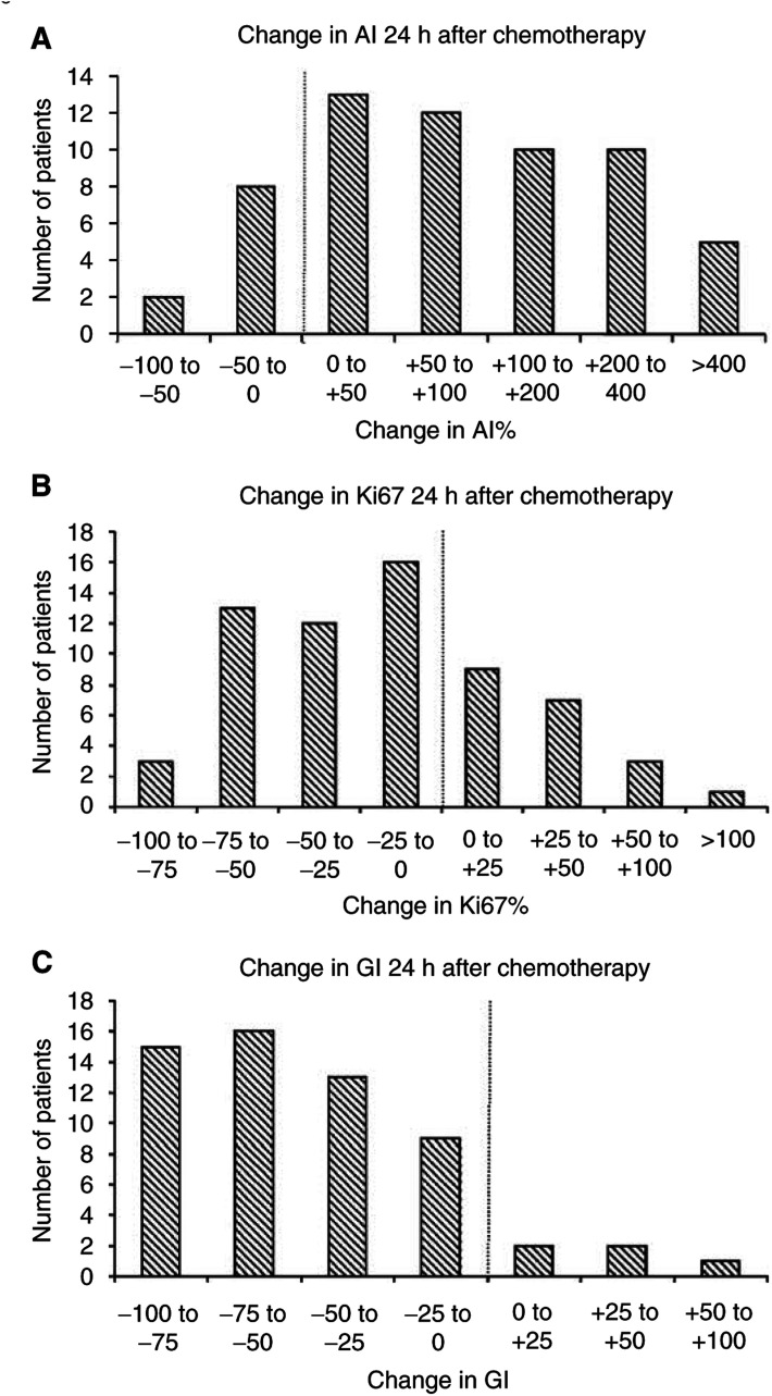
Patients undergoing primary chemotherapy for invasive breast cancer consented to a core biopsy of the invasive breast primary pre- and 24 h postchemotherapy. The resulting tissue was analysed for apoptosis, Ki67, ER and HER-2 using immunohistochemical techniques. These data were then used to evaluate the relationship between these biological markers and response to chemotherapy and overall survival. Response rate to chemotherapy in this group was 86%, 16 patients (25%) achieved a clinical complete response and 41 (63%) a partial response. Prechemotherapy there was a significant correlation between Ki67 and apoptotic index (AI), r=0.6, (P<0.001). A significant rise in AI (P<0.001), and fall in Ki67 (P=0.002) was seen 24 h following chemotherapy. No relationship was seen between pretreatment AI and clinical response, but higher Ki67 and growth index (Ki67/AI ratio, GI) did correlate with clinical response (both r=0.31, P<0.025). No correlation was seen between the change in AI or Ki67 at 24 h and clinical response or survival. Significant changes in apoptosis and proliferation can be demonstrated 24 h following chemotherapy, but these changes do not relate to clinical response or outcome in this study. Pretreatment proliferation and GI are however predictive of response to chemotherapy in breast cancer.
Figures


References
-
- Assersohn L, Gangi L, Zha Y, Dowsett M, Simon R, Powles TJ, Liu ET (2002) The feasibility of using fine needle aspirations from primary breast cancers for cDNA microarray analysis. Clin Cancer Res 3: 794–801 - PubMed
-
- Bonadonna G, Valagussa P, Brambilla C, Ferrari L, Moliterni A, Terenziani M, Zambetti M (1998) Primary chemotherapy in operable breast cancer: eight-year experience at the Milan Cancer Institute. J Clin Oncol 16: 93–100 - PubMed
-
- Bonetti A, Zaninelli M, Rodella S, Molino A, Sperotto L, Piubello Q, Bonetti F, Nortilli R, Turazza M, Cetto GL (1996) Tumor proliferative activity and response to first-line chemotherapy in advanced breast carcinoma. Breast Cancer Res Treat 38: 289–297 - PubMed
-
- Boyer MJ, Tannock IF (1998) Cellular and molecular basis of chemotherapy. In The Basic Science of Oncology, Tannock IF, Hill RP (eds) Vol. 1, pp 353–355. New York: McGraw-Hill
Publication types
MeSH terms
Substances
LinkOut - more resources
Full Text Sources
Other Literature Sources
Medical
Research Materials
Miscellaneous
