Aberrant expression of minichromosome maintenance protein-2 and Ki67 in laryngeal squamous epithelial lesions
- PMID: 12966424
- PMCID: PMC2376943
- DOI: 10.1038/sj.bjc.6601234
Aberrant expression of minichromosome maintenance protein-2 and Ki67 in laryngeal squamous epithelial lesions
Abstract
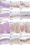
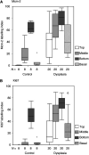
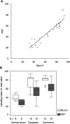
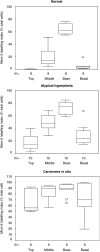
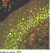
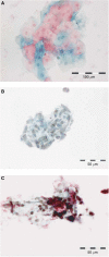
Histological classification of laryngeal epithelial lesions is highly subjective, and methods of cytological detection are not well developed. Improved determination of aberrant cell cycle entry may allow increased objectivity in histological assessment and enable the development of less invasive diagnostic cytology tests. Sections of normal larynx (n=10), laryngeal dysplasia (n=20) and laryngeal squamous cell carcinoma (SCC) (n=10) were classified according to the Ljubljana classification and stained for markers of cell cycle entry, minichromosome maintenance protein-2 (Mcm-2) and Ki67. Expression patterns were compared using double labelling confocal microscopy. There was a correlation between Mcm-2 and Ki67 labelling indices (rho=0.93; 95% CI [0.84, 0.97]) and both markers showed increased expression from normal epithelium to SCC (Mcm-2, P=0.001; Ki67, P=0.0002). Importantly, there was minimal expression of Mcm-2 or Ki67 in the most superficial layers of normal larynx and abnormal or atypical hyperplasia, in contrast to carcinoma in situ and SCC. Clusters of Mcm-2/5-positive cells were present in cytological preparations from SCC, but not from those showing atypical hyperplasia or inflammation in non-neoplastic tissue. Minichromosome maintenance protein-2 staining may increase the objectivity and reliability of histological grading of laryngeal epithelial lesions. Laryngeal brushings, combined with immuno-enhanced liquid-based cytology, could be useful, as a less invasive approach, to the detection of laryngeal malignant and premalignant lesions.
Figures
Similar articles
-
Potential role of minichromosome maintenance protein 2 as a screening biomarker in esophageal cancer high-risk population in China.Hum Pathol. 2011 Jun;42(6):808-16. doi: 10.1016/j.humpath.2010.04.022. Epub 2011 Jan 15. Hum Pathol. 2011. PMID: 21237484
-
Immunohistochemical estimation of cell cycle phase in laryngeal neoplasia.Br J Cancer. 2006 Aug 7;95(3):314-21. doi: 10.1038/sj.bjc.6603262. Epub 2006 Jul 11. Br J Cancer. 2006. PMID: 16832409 Free PMC article.
-
Expression of cyclin-dependent kinases inhibitors p21(WAF1) and p27(KIP1) in benign, premalignant and malignant laryngeal lesions. correlation with cell cycle regulatory proteins.In Vivo. 2004 Nov-Dec;18(6):719-24. In Vivo. 2004. PMID: 15646812
-
Laryngeal Dysplasia, Squamous Cell Carcinoma, and Variants.Surg Pathol Clin. 2017 Mar;10(1):15-33. doi: 10.1016/j.path.2016.10.003. Surg Pathol Clin. 2017. PMID: 28153131 Review.
-
The Ljubljana classification: a practical strategy for the diagnosis of laryngeal precancerous lesions.Adv Anat Pathol. 2000 Jul;7(4):240-51. doi: 10.1097/00125480-200007040-00006. Adv Anat Pathol. 2000. PMID: 10907809 Review.
Cited by
-
Gene expression profiling via bioinformatics analysis reveals biomarkers in laryngeal squamous cell carcinoma.Mol Med Rep. 2015 Aug;12(2):2457-64. doi: 10.3892/mmr.2015.3701. Epub 2015 Apr 29. Mol Med Rep. 2015. PMID: 25936657 Free PMC article.
-
MCM2 in human cancer: functions, mechanisms, and clinical significance.Mol Med. 2022 Oct 27;28(1):128. doi: 10.1186/s10020-022-00555-9. Mol Med. 2022. PMID: 36303105 Free PMC article. Review.
-
Immunocytochemical detection of minichromosome maintenance protein 2 as a potential urinary-based marker of bladder cancer: A prospective observational study.Indian J Urol. 2020 Jan-Mar;36(1):32-36. doi: 10.4103/iju.IJU_128_19. Indian J Urol. 2020. PMID: 31983824 Free PMC article.
-
Molecular basis for advances in cervical screening.Mol Diagn. 2005;9(3):129-42. doi: 10.1007/BF03260081. Mol Diagn. 2005. PMID: 16271014 Review.
-
MCM immunocytochemistry as a first line cervical screening test in developing countries: a prospective cohort study in a regional cancer centre in India.Br J Cancer. 2007 Apr 10;96(7):1107-11. doi: 10.1038/sj.bjc.6603679. Epub 2007 Mar 6. Br J Cancer. 2007. PMID: 17342084 Free PMC article.
References
-
- Biscotti CV, O'Brien DL, Gero MA, Gramlich TL, Kennedy AW, Easley KA (2002) Thin-layer Pap test vs conventional Pap smear. Analysis of 400 split samples. J Reprod Med 47: 9–13 - PubMed
-
- Brandwein MS, Nuovo GJ, Biller H (1993) Analysis of prevalence of human papillomavirus in laryngeal carcinomas. Study of 40 cases using polymerase chain reaction and consensus primers. Ann Otol Rhinol Laryngol 102: 309–313 - PubMed
-
- Davies RJ, Freeman A, Morris LS, Bingham S, Dilworth S, Scott I, Laskey RA, Miller R, Coleman N (2002) Analysis of minichromosome maintenance proteins as a novel method for detection of colorectal cancer in stool. Lancet 359: 1917–1919 - PubMed
-
- Freeman A, Morris LS, Mills AD, Stoeber K, Laskey RA, Williams GH, Coleman N (1999) Minichromosome maintenance proteins as biological markers of dysplasia and malignancy. Clin Cancer Res 5: 2121–2132 - PubMed
-
- Gale N, Kambic V, Michaels L, Cardesa A, Hellquist HB, Zidar N, Poljak M (2000) The Ljubljana classification: a practical strategy for the diagnosis of laryngeal precancerous lesions. Adv Anat Pathol 7: 240–251 - PubMed
Publication types
MeSH terms
Substances
LinkOut - more resources
Full Text Sources
Other Literature Sources
Research Materials
Miscellaneous

