. 2003 Sep 15;89(6):1108-15.
doi: 10.1038/sj.bjc.6601223.

Potentiation of tumour apoptosis by human growth hormone via glutathione production and decreased NF-kappaB activity

Affiliations

Potentiation of tumour apoptosis by human growth hormone via glutathione production and decreased NF-kappaB activity

C Cherbonnier et al. Br J Cancer. .

Abstract

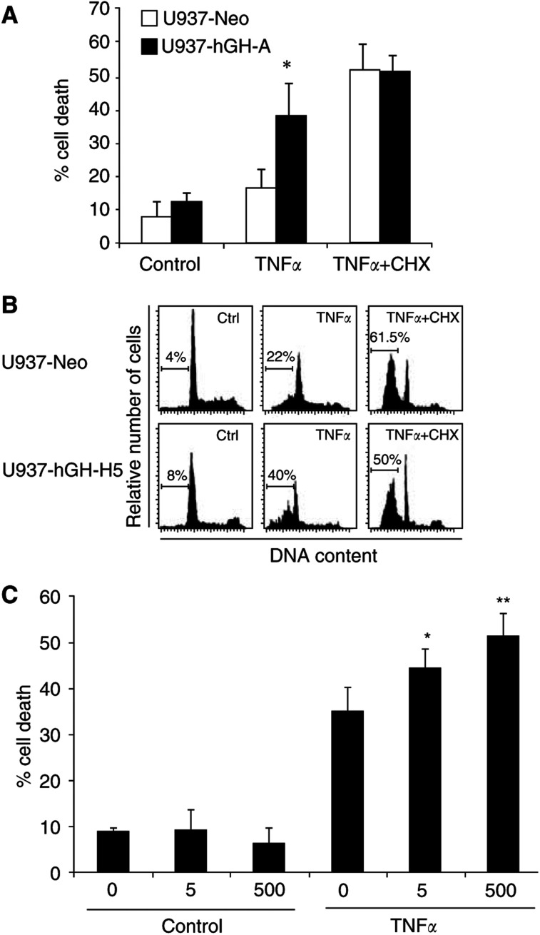
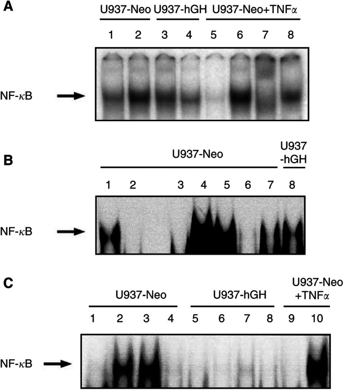
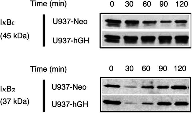
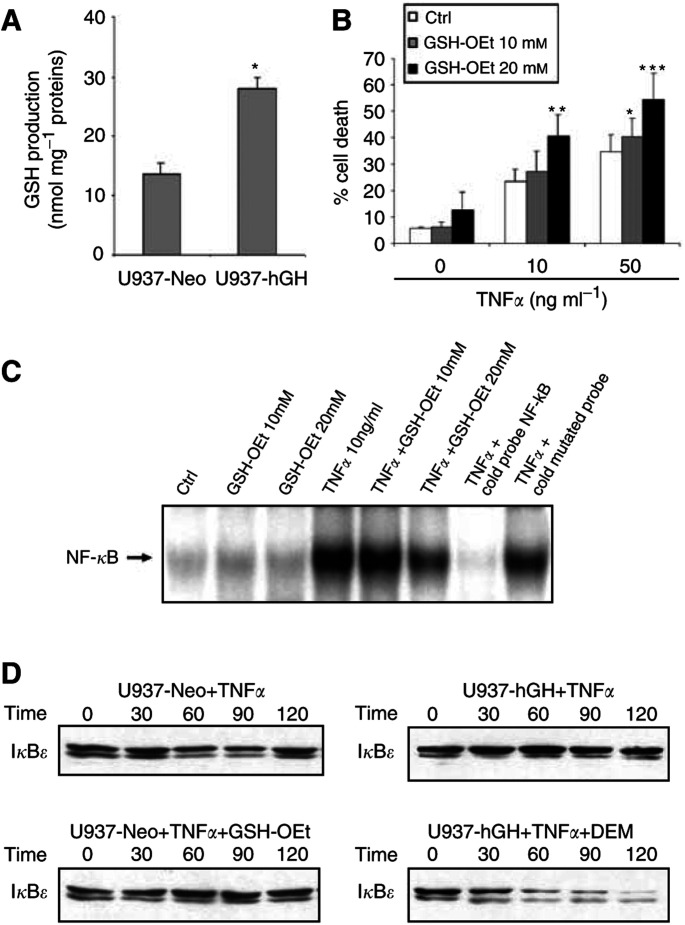
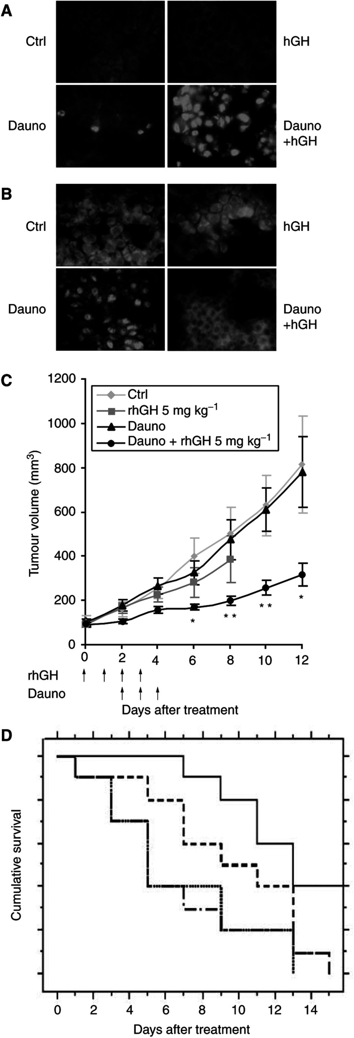
In addition to its primary role as growth factor, human growth hormone (hGH) can also participate in cell survival, as already documented by its protective effect on human monocytes or human promyelocytic leukaemia U937 cells exposed to a Fas-mediated cell death signal. However, despite similarities in the molecular events following Fas and TNF-alpha receptor engagement, we report that U937 cells, genetically engineered to constitutively produce hGH, were made more sensitive to TNF-alpha-induced apoptosis than parental cells. This was due to overproduction of the antioxidant glutathione, which decreased the nuclear factor (NF)-kappaB activity known to control the expression of survival genes. These findings were confirmed in vivo, in nude mice bearing U937 tumours coinjected with recombinant hGH and the NF-kappaB -inducing anticancer drug daunorubicin, to avoid the in vivo toxicity of TNF-alpha. This study therefore highlights one of the various properties of hGH that may have potential clinical implications.


Figures

Figure 1
Figure 1
Effect of hGH on TNF-α-induced apoptosis. U937-Neo and U937-hGH cell lines were cultured in the presence or absence of TNF-α (10 ng ml−1) with or without CHX (400 ng ml−1) (A) The percentage of dead cells was measured by PI staining. The values represent the mean±s.e.m. of six independent experiments (*P=0.001). (B) The percentage of cells with hypodiploid DNA content was detected by PI staining of ethanol-permeabilised cells. Linear scales are represented. (C) U937 cells were cultured for 48 h in the presence or absence of 50 ng ml−1 TNF-α with or without exogenous recombinant hGH at 5 or 500 ng ml−1. The percentage of dead cells was measured by trypan blue exclusion. The values represent the mean±s.e.m. of four experiments (*P<0.05, **P<0.01).
Figure 2
Figure 2
Effect of hGH on NF-κB activation. (A) An EMSA was performed on nuclear extracts as described in Materials and Methods. Control U937 or hGH-transfected cells were either nonstimulated (lanes 1 and 3, respectively) or stimulated with TNF-α (10 ng ml−1) (lanes 2 and 4, respectively). Nuclear factor-κB migration was assessed by migration of nuclear extracts from U937-Neo cells stimulated with TNF-α, coincubated with nonlabelled NF-κB-specific (lane 5) or NF-κB-mutated probes (lane 6), or with anti-P65 (lane 7) and anti-P50 (lane 8) NF-κB subunit antibodies. (B) In a separate experiment, an EMSA was performed on nuclear extracts from U937-Neo cells cultured in the absence or in the presence of TNF-α (10 ng ml−1) (lanes 3 and 4, respectively), coincubated with 5, 50, or 500 ng ml−1 of rhGH (lanes 5, 6 and 7, respectively). U937-hGH cells were also stimulated with TNF-α (10 ng ml−1) (lane 8). Nuclear factor-κB migration was assessed by migration of nuclear extracts from U937-Neo cells stimulated with TNF-α, and coincubated with nonlabelled NF-κB-specific (lane 2) or NF-κB-mutated (lane 1) probes. (C) In another experiment, U937-Neo and U937-hGH cells were either nonstimulated (lanes 1 and 5, respectively), stimulated with TNF-α (10 ng ml−1) (lanes 2 and 6, respectively), stimulated with TNF-α and CHX (400 ng ml−1) (lanes 3 and 7, respectively) or stimulated with anti-Fas mAbs (lanes 4 and 8, respectively). Specificity was assessed by migration of nuclear extracts from U937-Neo cells stimulated with TNF-α, and coincubated with the probes described above, nonlabelled NF-κB-specific (lane 9) or NF-κB-mutated (lane 10) probes.
Figure 3
Figure 3
Effect of hGH on IκB degradation. Whole-cell extracts from the indicated cell lines stimulated with TNF-α (10 ng ml−1) for 0–120 min were subjected to Western blotting using anti-IκB Abs under the conditions described in Materials and Methods.
Figure 4
Figure 4
(A) Effect of hGH on GSH production. The glutathione level was measured in U937-Neo and U937-hGH cells under the conditions described in Materials and Methods. The values represent the mean±s.e.m. of four independent experiments (*P<0.05). (B) Effect of GSH-OEt on TNF-α-induced apoptosis. U937-Neo cells were stimulated with TNF-α in the presence or absence of GSH-OEt and under the conditions described in Materials and Methods: the percentage of dead cells was measured by PI staining. The values represent the mean±s.e.m. of three independent experiments (*P<0.04, **P<0.03, ***P<0.02), when comparing GSH-OEt treated cells to GSH-OEt non-treated cells in each group. (C) Effect of GSH-OEt on NF-κB activation. Nuclear extracts were analysed by EMSA for measurement of NF-κB activation, as described in Figure 2. (D) Effect of GSH on IκB degradation. Whole-cell extracts were subjected to Western blotting using anti-IκB Abs, as described in Figure 3.
Figure 5
Figure 5
In vivo effect of hGH on daunorubicin treatment of engrafted tumours. Small tumour fragments obtained from U937-Neo tumours were transplanted subcutaneously in previously irradiated nude Swiss mice. Tumour-bearing mice either received i.p. injections of saline solution or subcutaneous injections of rhGH alone, or the highest subtoxic dose of daunorubicin (1.5 mg kg−1) with or without 5 mg kg−1 of rhGH. (A) Tissue sections from mice treated as above were submitted to TUNEL assay. Note the significant number of stained nuclei in tumours from rhGH- and daunorubicin-treated animals, indicating a high apoptosis rate in these animals. (B) The same tumours were assessed for NF-κB activation, as evidenced by the presence of the P65 subunit in the nuclei. Note the lower number of stained nuclei in tumours from mice treated with rhGH and daunorubicin, compared with daunorubicin-treated animals. (C) Tumour volumes were measured in each group of mice. The measurement was stopped when a mouse died or was killed because of a tumour volume exceeding 2000 mm3. Each line represents the mean±s.e.m. of the tumour volumes (*P<0.05 and **P<0.01, when comparing daunorubicin-treated mice to mice receiving daunorubicin and rhGH). (D) Kaplan-Meier curve comparing the different groups of mice with a tumour volume below 300 mm3. PBS-injected mice (….), daunomycine-injected mice (.-.-.-.-.) hGH-injected mice (- - -) and daunomycine+hGH-injected mice (---).

Similar articles

Cited by

References

    1. Argetsinger LS, Campbell GS, Yang X, Witthuhn BA, Silvennoinen O, Ihle JN, Carter-Su C (1996) Mechanism of signaling by growth hormone receptor. Cell 76: 1089–1107 - PubMed
    1. Arlt A, Vorndamm J, Breitenbroich M, Folsch UR, Kalthoff H, Schmidt WE, Schafer H (2001) Inhibition of NF-kappaB sensitizes human pancreatic carcinoma cells to apoptosis induced by etoposide (VP16) or doxorubicin. Oncogene 20: 859–868 - PubMed
    1. Baeuerle PA, Baltimore D (1988) IκB: a specific inhibitor of the NF-κB transcription factor. Science 242: 540–546 - PubMed
    1. Budd RC (2002) Death receptors couple to both cell proliferation and apoptosis. J Clin Invest 109: 437–441 - PMC - PubMed
    1. Cherbonnier C, Déas O, Vassal G, Merlin J-L, Haeffner A, Senik A, Charpentier B, Dürrbach A, Bénard J, Hirsch F (2002) Human growth hormone gene transfer into tumor cells may improve cancer chemotherapy. Cancer Gene Ther 9: 497–504 - PubMed

Publication types

MeSH terms