. 2004 Jan;286(1):C8-21.
doi: 10.1152/ajpcell.00428.2002. Epub 2003 Sep 10.

Rho-kinase-mediated Ca2+-independent contraction in rat embryo fibroblasts

Affiliations

Rho-kinase-mediated Ca2+-independent contraction in rat embryo fibroblasts

Daniel A Emmert et al. Am J Physiol Cell Physiol. 2004 Jan.

Abstract

Thus far, determining the relative contribution of Ca2+/calmodulin-dependent myosin light chain kinase (MLCK) and Ca2+-independent Rho-kinase pathways to myosin II activation and contraction has been difficult. In this study, we characterize the role of Rho-kinase in a rat embryo fibroblast cell line (REF-52), which contains no detectable MLCK. No endogenous MLCK could be detected in REF-52 cells by either Western or Northern blot analysis. In the presence or absence of Ca2+, thrombin or lysophosphatidic acid (LPA) increased RhoA activity and Rhokinase activity, correlating with isometric tension development and myosin II regulatory light chain (RLC) phosphorylation. Resting tension is associated with a basal phosphorylation of 0.31 +/- 0.02 mol PO4/mol RLC, whereas upon LPA or thrombin treatment myosin II RLC phosphorylation increases to 1.08 +/- 0.05 and 0.82 +/- 0.05 mol PO4/mol RLC, respectively, within 2.5 min. Ca2+ chelation has minimal effect on the kinetics and magnitude of isometric tension development and RLC phosphorylation. Treatment of REF-52 cells with the Rho-kinase-specific inhibitor Y-27632 abolished thrombin- and LPA-stimulated contraction and RLC phosphorylation. These results suggest that Rho-kinase is sufficient to activate myosin II motor activity and contraction in REF-52 cells.


Figures

Fig. 1
Fig. 1
Expression of Rho-kinase and myosin light chain kinase (MLCK) in rat embryo fibroblast (REF)-52 cells. REF-52 cell pellets (2 × 10⁶ cells) were lysed and extracted as outlined in expertmental procedures, electrophoresed on 10% SDS-PAGE, transferred to 0.45-μm polyvinylidene difluoride (PVDF) membrane, and probed with rabbit polyclonal antibody to Rho-kinase. A: REF-52 cell extracts probed with a rabbit polyclonal antibody to Rho-kinase. Rho-kinase was found to have a molecular mass of 160 kDa. B: REF-52 cell extracts probed with an affinity-purified rabbit polyclonal antibody to MLCK155, which recognizes both isoforms of MLCK. No endogenous MLCK was detected in REF-52 cells (lane 1). Lanes 2 and 3 are recombinant MLCK220 and MLCK155 standards, respectively. C: Northern analysis of MLCK mRNAs in rat cells and tissues. RNA (15 μg total RNA/lane) was isolated from rat uterus (Rt Ut; lane 1), rat aorta (lane 2), rat embryo fibroblasts (REF; lane 3), and rat embryonic thoracic aorta smooth muscle cells (A10; lane 4) and probed as outlined in experimental procedures. The positions of the RNA molecular weight standards (kB) are shown at left. Bottom: photograph documenting the amounts of 18S rRNA present in each lane.
Fig. 2
Fig. 2
RhoA and Rho-kinase activity. A and B: representative Western blots demonstrate RhoA activation. REF-52 cells 4 days postconfluence were stimulated with 1 U/ml thrombin (A) or 1 μM lysophosphatidic acid (LPA; B) for 0, 1, 2.5, 5, or 10 min. Cell lysates were incubated with glutathione S-transferase-Rho-binding domain (GST-RBD) fusion protein for 1 h at 4°C, electrophoresed, transferred to PVDF, and probed with RhoA antibody. RhoA activity was determined as the amount of RBD-bound active RhoA (GTP-bound RhoA) normalized to the total amount of RhoA in the cell lysate. Below the blot is shown a whole cell lysate for total RhoA. C and D: thrombin and LPA induced Rho-kinase activation. REF-52 monolayers were stimulated with 1 U/ml thrombin (C) or 1 μM LPA (D) for the indicated time intervals, cells were lysed, and Rho-kinase was immunoprecipitated. Immunopurified Rho-kinase was used for in vitro phosphorylation reactions using purified myosin II regulatory light chain (RLC) as substrate as described in experimental procedures. Phosphorylated myosin II RLCs were analyzed on 12.5% SDS gels and exposed to a PhosphorImager plate. Thrombin and LPA treatment resulted in an 11- and 8-fold increase in Rho-kinase catalytic activity, respectively.
Fig. 3
Fig. 3
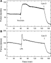
Agonist-induced isometric contraction in REF-52 collagen gels. Representative tension tracings were produced by REF-52 cells treated with thrombin or LPA. Collagen gels were mounted on force transducers in HEPES-buffered saline (HBS) and allowed 60 min to establish a stable baseline force before being stimulated with either 1 U/ml thrombin (A) or 1 μM LPA (B). Both agonists rapidly increased isometric tension, which remained elevated for over 2 h. Addition of cytochalasin D (cyto D) ablated cellular tension.
Fig. 4
Fig. 4
Agonist-induced MLC phosphorylation. REF-52 monolayers were preincubated for 1 h at room temperature in HBS. Cultures were stimulated with 1 U/ml thrombin (A)or1 μM LPA (B) for 0, 2.5, 5, 15, 30, 60, 90, or 120 min. Samples were separated by glycerol-urea gel electrophoresis, transferred to nitrocellulose, and probed with anti-myosin RLC antibody. The unphosphorylated (Un), monophosphorylated (P1), and diphosphorylated (P2) states of myosin II RLC are represented.
Fig. 5
Fig. 5
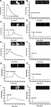
Ca2+ fluorescence signal in REF-52 cells. REF-52 cells were incubated in 37°C HBS containing 1 μM Cell-Tracker orange for 45 min, followed by an additional 45-min incubation at room temperature in HBS/2 μM fluo 4-AM/0.04% (wt/vol) Pluronic F-127 supplemented with (A, C, and E) or without 1.8 mM Ca2+ (B, D, and F). For chelation of Ca2+, BAPTA-AM (10 μM) and thapsigargin (1 μM) were added to monolayers in a Ca2+-free medium (B, D, and F). Cells were visualized, and images were captured using a Wallac UltraView spinning disk confocal laser microscope fitted onto an Olympus IX70 inverted fluorescence microscope. Cells were maintained at 37°C with a heated stage, and images were collected every 500 ms with an AstroCam cooled charge-coupled device camera. Pixel density was calculated from whole cell averages, less background intensity. Cells were localized by Cell-Tracker orange cytoplasmic dye (at 568 nm), and intracellular Ca2+ signal was measured with fluo 4 (at 488 nm). A and B demonstrate the effects of 10 μM ionomycin to stimulate Ca2+ influx in 1.8 mM Ca2+ and Ca2+-free conditions, respectively. C and D demonstrate the effects of 1 U/ml thrombin to stimulate Ca2+ influx in 1.8 mM Ca2+ and Ca2+-free conditions, respectively. E and F demonstrate the results for 1 μM LPA. The 3 insets in each panel, from left to right, display Ca2+ fluorescence images before stimulation, at peak Ca2+ influx, and after cell recovery. G–J show representative measurements of REF-52 cell-populated collagen gels loaded with fluo 4 in 1.8 mM Ca2+ (G and I) or under Ca2+ chelation conditions (H and J) as outlined in experimental procedures. Collagen gels stimulated with 1 U/ml thrombin (G) or 1 μM LPA (I) evoked an influx of extracellular Ca2+ similar to the response shown in monolayer cultures (C and E). As with monolayer cultures, BAPTA-AM/thapsigargin blocked the rise in cytosolic Ca2+ shown with thrombin (H) and LPA (J).
Fig. 6
Fig. 6
Ca2+-independent RhoA and Rho-kinase activity. A and B: representative Western blots of Ca2+-independent RhoA activity. REF-52 cultures were preincubated for 1 h at room temperature in Ca2+-free HBS containing 10 μM BAPTA-AM/1 μM thapsigargin. Cells were stimulated with 1 U/ml thrombin (A) or 1 μM LPA (B) for the indicated time intervals, monolayers were lysed, and cell lysates were incubated with GST-RBD for 1 h. Samples were electrophoresed, transferred to PVDF, and probed with RhoA antibody. Below the blot is shown a whole cell lysate for total RhoA to indicate equal loading. C and D: thrombin and LPA induced Ca2+-independent Rho-kinase activity. REF-52 monolayers were preincubated for 1 h at room temperature in Ca2+-free HBS containing 10 μM BAPTA-AM/1 μM thapsigargin. Cells were stimulated with 1 U/ml thrombin (C) or 1 μM LPA (D) for the indicated time intervals, cells were lysed, and Rho-kinase was immunoprecipitated. Immunopurified Rho-kinase was used for in vitro phosphorylation reactions with purified myosin II RLC as substrate as described in experimental procedures. Phosphorylated myosin II RLCs were analyzed on 12.5% SDS gels and exposed to a PhosphorImager plate. Both thrombin and LPA upregulated Ca2+-independent Rho-kinase catalytic activity.
Fig. 7
Fig. 7
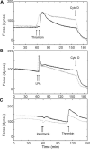
Ca2+-independent agonist-induced isometric contraction in REF-52 collagen gels. REF-52 cell-populated collagen gels were mounted on isometric force transducers and incubated for 1 h in either HBS (dashed lines) or Ca2+-free HBS supplemented with 10 μM BAPTA-AM and 1 μM thapsigargin (solid lines). After 1 h, the medium was replaced with fresh, inhibitor-free 37°C HBS or Ca2+-free HBS. Basal tension was monitored for 1 h before the addition of 1 U/ml thrombin (A), 1 μM LPA (B), or 1 μM ionomycin (C).
Fig. 8
Fig. 8
Ca2+-independent agonist-induced MLC phosphorylation. Monolayers were preincubated for1hat room temperature in Ca2+-free HBS containing 10 μM BAPTA-AM/1 μM thapsigargin. Cells were stimulated with 1 U/ml thrombin (A) or 1 μM LPA (B) for the indicated time intervals. Samples were separated by glycerol-urea gel electrophoresis, transferred to nitrocellulose, and probed with anti-myosin RLC antibody. Unphosphorylated (Un), monophosphorylated (P1), and diphosphorylated (P2) states of myosin II RLC are represented.
Fig. 9
Fig. 9
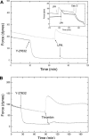
Effects of Rho-kinase inhibitor Y-27632 on basal and agonist-induced isometric tension. REF-52 cell-populated collagen gels were incubated with 10 μM Y-27632 for 1 h before the addition of agonists in the presence of Y-27632 (solid lines). A: effects of Y-27632 on LPA-induced isometric tension development. Y-27632 caused a rapid drop in basal tension, establishing a new baseline tension within 10 min. Pretreatment with Y-27632 inhibited the LPA-induced isometric tension development. Inset: reversibility of Y-27632. Organ baths were drained and replaced with media without Y-27632. Tension steadily increased, establishing a steady baseline tension within 3 h. Addition of a second dose of LPA resulted in a rapid rise in tension (inset). B: Y-27632 also inhibited thrombin-induced isometric tension. Results are representative of 3 experiments.
Fig. 10
Fig. 10
Effects of Rho-kinase inhibitor Y-27632 on basal and agonist-stimulated MLC phosphorylation. REF-52 cultures were pretreated with 10 μM Y-27632 for 15 min (A and B, lanes 3 and 4) or without Y-27632 (A and B, lanes 1 and 2). Monolayers were stimulated with 1 U/ml thrombin (A, lanes 2 and 4) or 1 μM LPA (B, lanes 2 and 4) for 5 min. Samples were separated by glycerol-urea gel electrophoresis, transferred to nitrocellulose, and probed with anti-myosin RLC antibody. Unphosphorylated (Un), monophosphorylated (P1), and diphosphorylated (P2) states of myosin II RLC are represented. A and B, lane 1, represent unstimulated control cells.

References

    1. Amano M, Chihara K, Nakamura N, Fukata Y, Yano T, Shibata M, Ikebe M, Kaibuchi K. Myosin II activation promotes neurite retraction during the action of Rho and Rho-kinase. Genes Cells. 1998;3:177–188. - PubMed
    1. Amano M, Ito M, Kimura K, Fukata Y, Chihara K, Nakano T, Matsuura Y, Kaibuchi K. Phosphorylation and activation of myosin by Rho-associated kinase (Rho-kinase). J Biol Chem. 1996;271:20246–20249. - PubMed
    1. Birukov KG, Schavocky JP, Shirinsky VP, Chibalina MV, Van Eldik LJ, Watterson DM. Organization of the genetic locus for chicken myosin light chain kinase is complex: multiple proteins are encoded and exhibit differential expression and localization. J Cell Biochem. 1998;70:402–413. - PubMed
    1. Bishop AL, Hall A. Rho GTPases and their effector proteins. Biochem J. 2000;348:241–255. - PMC - PubMed
    1. Blue EK, Goeckeler ZM, Jin Y, Hou L, Dixon SA, Herring BP, Wysolmerski RB, Gallagher PJ. 220- and 130-kDa MLCKs have distinct tissue distributions and intracellular localization patterns. Am J Physiol Cell Physiol. 2002;282:C451–C460. - PMC - PubMed

Publication types

MeSH terms

LinkOut - more resources