. 2003 Sep 10;23(23):8221-30.
doi: 10.1523/JNEUROSCI.23-23-08221.2003.

Reciprocal interactions between neurons and glia are required for Drosophila peripheral nervous system development

Affiliations

Reciprocal interactions between neurons and glia are required for Drosophila peripheral nervous system development

Katharine J Sepp et al. J Neurosci. .

Abstract

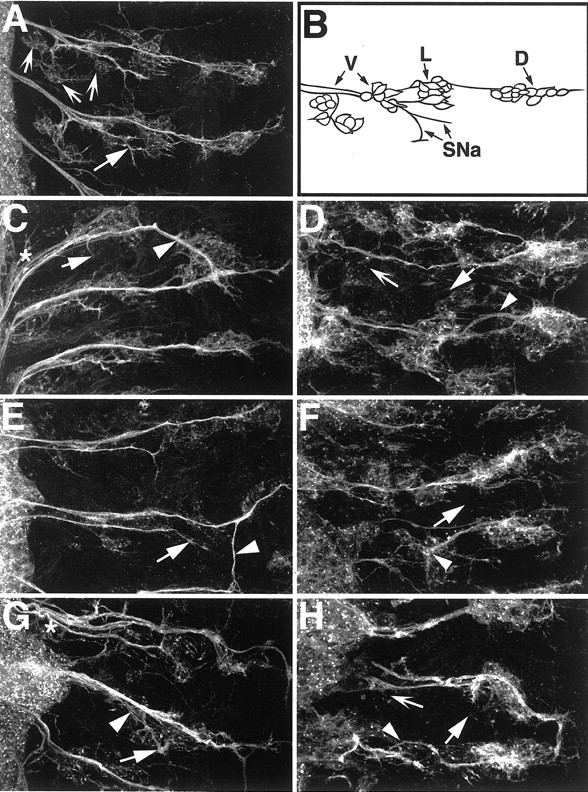
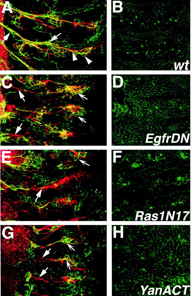
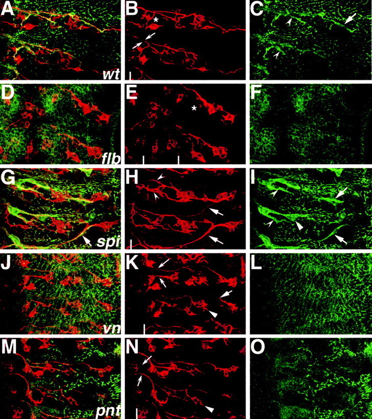
A major developmental role of peripheral glia is to mediate sensory axon guidance; however, it is not known whether sensory neurons influence peripheral glial development. To determine whether glia and neurons reciprocally interact during embryonic development, we ablated each cell type by overexpressing the apoptosis gene, grim, and observed the effects on peripheral nervous system (PNS) development. When neurons are ablated, glial defects occur as a secondary effect, and vice versa. Therefore glia and neurons are codependent during embryogenesis. To further explore glial-neuronal interactions, we genetically disrupted glial migration or differentiation and observed the secondary effects on sensory neuron development. Glial migration and ensheathment of PNS axons was blocked by overexpression of activated Rho GTPase, a regulator of actin dynamics. Here, sensory axons extended to the CNS without exhibiting gross pathfinding errors. In contrast, disrupting differentiation by expression of dominant-negative Ras GTPase in glia resulted in major sensory axon pathfinding errors, similar to those seen in glial ablations. Glial overexpression of transgenic components of the epidermal growth factor receptor (EGFR) signaling pathway yielded similar sensory neuron defects and also downregulated the expression of the glial marker Neuroglian. Mutant analysis also suggested that the EGFR ligands Spitz and Vein play roles in peripheral glial development. The observations support a model in which glia express genes necessary for sensory neuron development, and these genes are potentially under the control of the EGFR/Ras signaling pathway.


Figures

Figure 4.
Figure 4.
Activated MAP kinase in peripheral glia during embryonic development. Embryos were labeled with anti-diphospho ERK (black) in all panels and costained with anti-fasciclin II (brown) B-F, Anterior is to the top, and CNS is to the left in all panels except E, where it is centered. A, Activated MAP kinase is detected in the nuclei of peripheral glia (arrows) along with the strong staining in the muscle attachment sites (arrowheads). B-D, The nuclei of peripheral glia are associated with labeled neurons (brown) and are positive for activated MAP kinases (black). E, At the lateral edges of the CNS, the intersegmental glia and exit glia are also positive for activated MAP kinase (arrows) as are the longitudinal glia (not shown in this focal plane). F, High magnification of a peripheral glia nucleus. A, 200× magnification; B-E, 630× magnification; F, 1000× magnification.
Figure 1.
Figure 1.
Early ablation of neurons causes loss of glia and vice versa in the peripheral nervous system. For glial nuclear labeling, embryos were stained with anti-Repo (blue). Neurons were counterstained with anti-Futsch (brown). Anterior is to the top, midline (ventral) is off to the left, and dorsal is on the right. The border between the CNS and periphery is marked with a dashed line. A, A wild-type, stage 16 embryo shows glial nuclei (arrows) associating with two major sensory tracts, the anterior fascicles (af) and the posterior fascicles (pf). B, An elav::grim embryo where neurons are targeted for ablation shows severe neuronal loss at lower segments. Glial nuclear staining (arrows) is reduced most in segments with strongest neuronal ablation. Faded glial nuclei are also detected (arrowheads). C, A repo::grim embryo where glia are targeted for ablation. Glial nuclei remain at the lateral edge of the CNS and are faded (arrowheads), similar to those in B. A loss of sensory neurons (asterisk) is seen in those segments with severe glial loss. D-F, repo::grim embryos at higher magnification to show specific defects in Futsh-positive sensory neurons. The lack of Futsch-positive sensory neurons is not caused by displacement (compare D, E, which are two focal planes of the same region). Most segments had less than the usual cluster of five Futsch-positive chordontonals (D, F, asterisks). G, H, elav::grim embryos at higher magnification to show specific defects in Repo-positive peripheral glia. Loss of all the sensory neurons resulted in little or no Repo-positive glial cells being present in the affected segments of the peripheral nervous system. The presence of more sensory axons (H) correlated with a greater incidence of Repo-positive glia being present.
Figure 2.
Figure 2.
Late death of glia and neurons: effects on pathfinding. For glial nuclear labeling, embryos were stained with anti-Repo (blue). Neurons were counterstained with anti-Futsch (brown). Anterior is to the top, and midline (ventral) is off to the left. A, A wild-type stage 16 embryo. Neuron-ablated elav::grim embryos (B, D) and a glial-ablated repo::grim embryo (C) show similar sensory axon extension and pathfinding defects. They include axon stalls (solid arrows), total failure of axogenesis (concave arrowheads), and gross misguidance errors that include axons pathfinding across segment boundaries (concave arrows). B, In cases in which no axons traverse the CNS-PNS transition zone in the ventral region, peripheral glial nuclei appear stalled in their birthplace (long arrows).
Figure 3.
Figure 3.
Continuous contact of glial sheath by growth cones is not required for sensory axon migration. For glial staining, embryos expressing glial actin-GFP were labeled with anti-GFP (green). Sensory neurons were counterstained with anti-Futsch (red). Anterior is at the top, and midline is to the left. A, A wild-type stage 16 embryo shows complete glial ensheathment of sensory neuronal tracts. The stereotypic position of the glia nuclei, which can be detected by their characteristic shape and lack of actin-GFP, are shown (asterisks). B, repo::Ras1N17 embryo shows incomplete peripheral glial ensheathment of nerves (arrows) as well as sensory axons crossed incorrectly to the neighboring anterior segment (arrowhead). Glia nuclei are clustered at the CNS-PNS boundary (asterisks) C, D, Two examples of repo::RhoV14 embryos are shown, in which glial extension of cytoplasmic processes is blocked. Sensory axon pathways to the CNS are normal and organized into two tracts (arrows) as in wild-type hemisegments. The tracts are defasciculated (arrowheads), which is likely caused by the loss of the glial sheath.
Figure 5.
Figure 5.
Downregulation of EGFR signals in glia causes neuronal defects. For sensory and motor neuronal staining, embryos were labeled with anti-HRP. Anterior is to the top, and CNS is to the left. A, A mature wild-type embryo. The segmental nerve a (SNa) motor branch (solid arrow) and the ventral cluster sensory neurons (concave arrows) are indicated. B, A schematic diagram of sensory neurons and motor neurons that are labeled by the anti-HRP antibody. Sensory neurons are organized into ventral (V), lateral (L), and dorsal (D) clusters, and their cell bodies project axons to the CNS. Motor neurons whose cell bodies are not shown project axons from the CNS to the somatic musculature in the periphery. The SNa motor branch is indicated. For simplicity, the other motor branches of the PNS are not shown. In the ventral region, motor and sensory axons are bundled together into combined fascicles by peripheral glia. C, D, repo::EgfrDN embryos. E, F, repo::Ras1N17 embryos. G, H, repo::YanACT embryos. All mutant embryos show similar classes of phenotypes, and the genotypes cannot be distinguished from each other if they are observed “blind.” The PNS patterns in all mutants (C-H) do not match the wild type (A). Sensory neuron cell bodies are generally misplaced, and their axons show frequent misrouting and stalls (C-H, arrowheads). The SNa motor axon branches often have abnormal projections or are missing (C-H, solid arrows). Fasciculation of sensory-motor bundles is abnormal (C, G, asterisks). There is a loss of sensory neurons in the ventral regions of severely affected embryos (D, H, concave arrows).
Figure 6.
Figure 6.
Glial Neuroglian expression is downregulated in EGFR pathway mutants. Embryos were labeled with mAb 1B7 (anti-Nrg, green) and anti-HRP (red). Confocal stacks of wild-type and mutant embryos were split and projected such that the nervous system and the epidermis are shown on the left- and right-hand columns, respectively. A, Wild-type mature embryo shows peripheral glial expression of Nrg (solid arrows) in a profile similar to that in Figure 3A. Nrg is also expressed by sensory neurons (arrowheads). B, The epidermis of wild-type embryos has strong Nrg staining around the circumference of the cells. C, D, repo::EgfrDN embryo. E, F, repo::Ras1N17 embryo. G, H, repo::YanACT embryo. C, E, G, Peripheral glial Nrg expression in mutant embryos is weak (solid arrows); however, sensory neuronal Nrg staining can be detected (concave arrows). D, F, H, The epidermal expression level of Nrg is not visibly downregulated.
Figure 7.
Figure 7.
EGFR pathway loss-of-function mutants have neuronal and glial PNS defects. Embryos were labeled with anti-Futsch (red) and anti-Gliotactin (green), and PNS hemisegments were imaged with confocal microscopy. Anterior is to the top, and CNS is to the left of the white lines in H, K, and N and between the white lines in E. A-C, Wild-type mature embryo shows stereotyped arrangement of sensory neurons projecting axons along two main fascicles to the CNS (B, arrows). Asterisk in B shows region of ventral cluster sensory neurons. C, Peripheral glial anti-Gliotactin staining (arrows) overlies punctate epidermal expression. D-F, EgfrF2 mutant shows a significant loss of Futsch-positive sensory neurons (E, compare asterisks with B). Peripheral glial expression of Gliotactin cannot be detected (F). G-H, The spi1 homozygote shows aberrant sensory axon pathfinding across segment boundaries (H, solid arrows). Peripheral glial wrapping profiles correspond to misguided axon patterns (G, I, compare arrows). Wrapping morphology does not match narrow wild-type profile (I, solid arrowhead); however, ventral peripheral glia separate correctly from main nerve trunk (concave arrowheads; compare I, C). J-L, The vnL6 homozygotes show axonal stalling (K, solid arrow), and peripheral glial Gliotactin expression is not detected (L). M-O, The pntΔ88 homozygotes have disrupted Gliotactin expression (O). All of the spitz group mutants analyzed have a characteristic loss of lateral chordotonal sensory neurons (E, H, K, N, solid arrowheads). Abnormal separation of anterior and posterior peripheral nerve fascicles was noted in spi, vn, and pnt mutants as compared with the wild type (B, H, K, N, concave arrows).

Similar articles

Cited by

References

    1. Auld VJ ( 1999) Glia as mediators of growth cone guidance: studies from insect nervous systems. Cell Mol Life Sci 55 : 1377-1385. - PMC - PubMed
    1. Auld VJ, Fetter RD, Broadie K, Goodman CS ( 1995) Gliotactin, a novel transmembrane protein on peripheral glia, is required to form the blood-nerve barrier in Drosophila Cell 81 : 757-767. - PubMed
    1. Bergmann A, Tugentman M, Shilo B-Z, Steller H ( 2002) Regulation of cell number by MAPK-dependent control of apoptosis: a mechanism for trophic survival signaling. Dev Cell 2 : 159-170. - PubMed
    1. Bieber AJ, Snow PM, Hortsch M, Patel NH, Jacobs JR, Traquina ZR, Schilling J, Goodman CS ( 1989) Drosophila Neuroglian: a member of the immunoglobulin superfamily with extensive homology to the vertebrate neural adhesion molecule L1. Cell 59 : 447-460. - PubMed
    1. Booth GE, Kinrade EFV, Hidalgo A ( 2000) Glia maintain follower neuron survival during Drosophila CNS development. Development 127 : 237-244. - PubMed

Publication types

MeSH terms