. 2003 Sep 10;23(23):8302-9.
doi: 10.1523/JNEUROSCI.23-23-08302.2003.

Metabotropic glutamate 5 receptor blockade alleviates akinesia by normalizing activity of selective basal-ganglia structures in parkinsonian rats

Affiliations

Metabotropic glutamate 5 receptor blockade alleviates akinesia by normalizing activity of selective basal-ganglia structures in parkinsonian rats

Nathalie Breysse et al. J Neurosci. .

Abstract

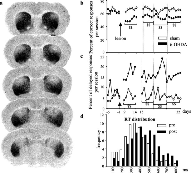
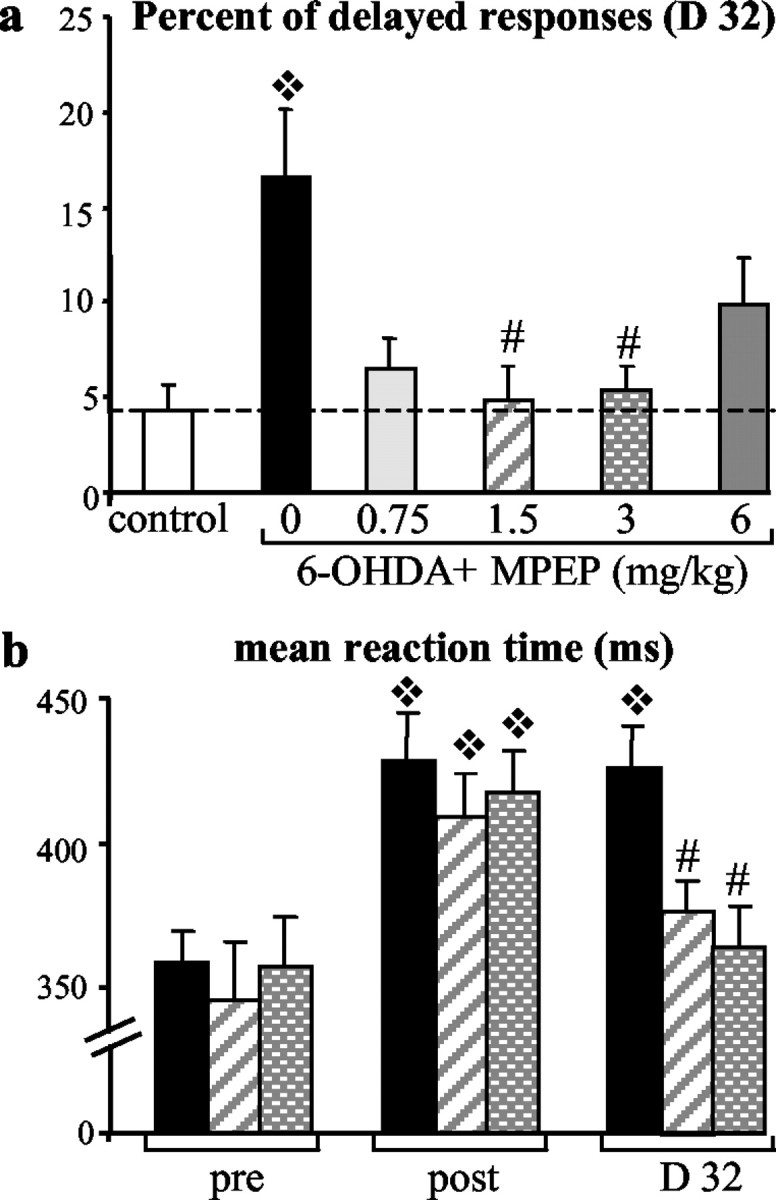
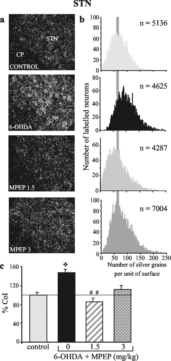
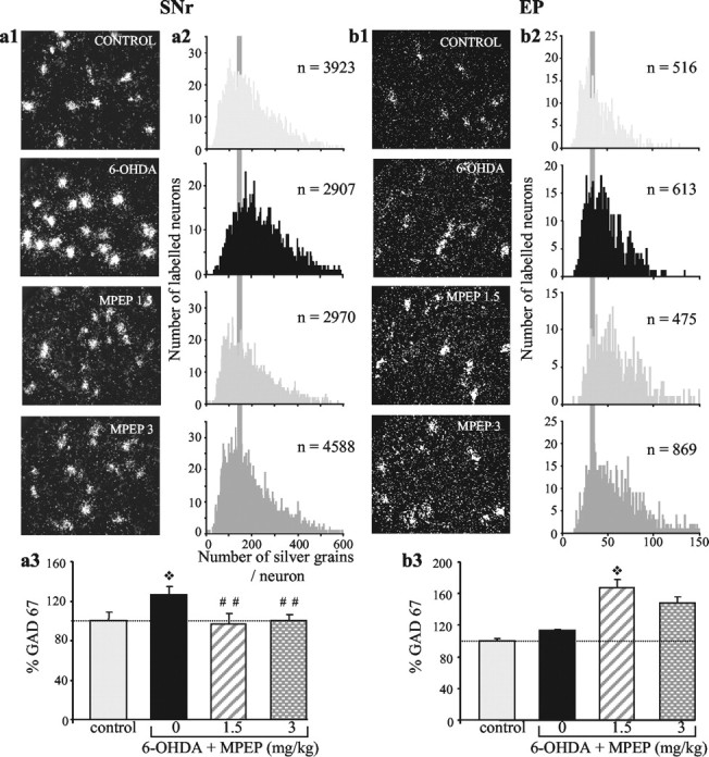
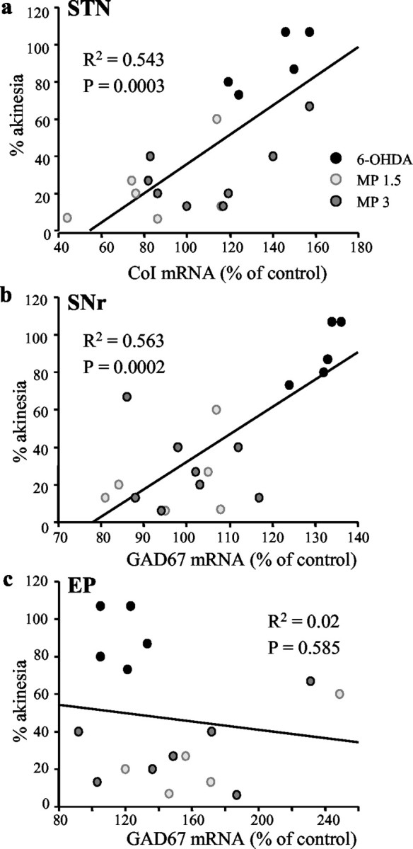
Glutamate overactivity within the basal ganglia has been shown to be central to the expression of motor symptoms in advanced stages of Parkinson's disease, and metabotropic glutamate receptors (mGluRs) represent promising targets for new therapeutic strategies in this pathology. Little is known, however, about the cellular and behavioral changes occurring in the early stages of the disease when dopamine depletion is moderate. Here, we report that rats with partial bilateral dopamine lesions exhibit akinetic deficits associated with dramatically increased neuronal metabolic activity in selective structures of the basal ganglia such as the subthalamic nucleus and the substantia nigra pars reticulata, but not in the entopeduncular nucleus. Furthermore, chronic treatment with the mGluR5 antagonist 2-methyl-6-(phenylethylnyl)-pyridine alleviated the akinesia and was associated with a normalization of the activity of these two overactive structures. These data stress the therapeutic potential of mGluR5 antagonists in the treatment of parkinsonian patients in the early stages of the disease.


Figures

Figure 1.
Figure 1.
DA lesion extent and behavioral effects in the RT task. a, Digitized autoradiographic images of striatal 3H-mazindol binding to DA uptake sites showing the antero-posterior extent of the 6-OHDA-induced striatal denervation in a typical animal case. Note the partial and symmetrical loss of DA nerve terminals (lack of labeling) restricted to the dorsal striatum. b, c, Preoperative and postoperative performance of sham-operated (n = 6) and 6-OHDA-lesioned animals (n = 7) in the RT task for correct and delayed responses. The mean number of each type of responses per session is illustrated for six preoperative sessions (days -6 to -1) and 24 postoperative sessions (days 9-32). Animals were tested every day in a 100-trial session. As shown, the partial bilateral lesion induced a long-lasting effect in the RT task by reducing the level of correct responses (b) and increasing the number of delayed responses (c). d, Distribution of RTs (longer than 100 msec and shorter than 800 msec) for one preoperative session (day -3) and one postoperative session (day 14). Each bar represents the mean number of RTs for all 6-OHDA-treated animals recorded by 50 msec intervals during the prelesion (white bars) and postlesion (black bars) session. DA depletion produced a shift toward long values of RTs. $$, Significant difference from sham group (treated no-lesioned animals; p < 0.001; Scheffé's test after significant ANOVA).
Figure 2.
Figure 2.
Effects of chronic MPEP treatment on behavioral parameters measured in the reaction time task. a, Percentage of delayed responses for each experimental group ± SEM (sham, n = 6; 6-OHDA, n = 7; 0.75 mg/kg MPEP, n = 7; 1.5 mg/kg MPEP, n = 6; 3 mg/kg MPEP, n = 9; 6 mg/kg MPEP, n = 10) measured for the last RT session (day 32). Whereas MPEP chronic treatment at the lowest and highest doses (0.75 and 6 mg/kg) just partially improved recovery of performance, MPEP at doses of 1.5 and 3 mg/kg allowed a total recovery. ❖ , Significant difference from control group (p < 0.05; Scheffé's test after significant ANOVA); #, significant difference from 6-OHDA-nontreated group (p < 0.05; Scheffé's test after significant ANOVA). b, Mean RT ± SEM for one preoperative session (day -1) and two postoperative sessions corresponding to the lesion effect without treatment (day 14) in comparison with the last day of chronic treatment (day 32) and measured for the 6-OHDA, 1.5 mg/kg MPEP, and 3 mg/kg MPEP groups. At day 32, mean RTs were shortened by MPEP chronic treatment and significantly differed from the control-lesioned group injected with vehicle. ❖, Significant difference from preoperative performance for each group (p < 0.05; Scheffé's test after significant ANOVA); #, significant difference from 6-OHDA group at D32 (p < 0.05; Scheffé's test after significant ANOVA).
Figure 3.
Figure 3.
Chronic treatment with MPEP reverses the DA lesion-induced increase in CoI gene expression in the STN. Photomicrographs (a) taken under dark-field epi-illumination and histograms (b) of frequency distribution of labeling, showing the effects of DA lesion without or with subsequent treatment with MPEP. The lesion (n = 6) induces an increase in intraneuronal labeling, confirmed by a shift to the right of the distribution curve (toward the higher levels), that is no longer observed after MPEP treatment at the doses of 1.5 mg/kg (n = 6) and 3 mg/kg (n = 8). c, Histograms representing the mean number of silver grains per labeled neuron in each experimental condition expressed as percentage ± SEM of control (n = 6). Statistical comparisons were performed using a one-way ANOVA, followed by Scheffé's test. ❖p < 0.01, compared with the control group; ##p < 0.01, compared with values obtained in animals with the DA lesion alone.
Figure 4.
Figure 4.
Chronic treatment with MPEP differentially affects gene expression of GAD67 in the SNr and EP of animals with the DA lesion. Photomicrographs (a1, b1) taken under dark-field epi-illumination and histograms (a2, b2) of frequency distribution of labeling, showing the effects of DA lesion without or with subsequent treatment with MPEP. The lesion induces an increase in intraneuronal labeling in the SNr (n = 7), confirmed by a shift to the right of the distribution curve (toward the higher levels), without affecting labeling in EP neurons (n = 7). MPEP treatment reverses the lesion-induced increase in the SNr (n = 6 at 1.5 mg/kg MPEP and n = 9 at 3 mg/kg MPEP; distribution curve similar to control) while producing an overexpression in EP neurons (n = 5 at 1.5 mg/kg MPEP and n = 8 at 3 mg/kg MPEP; distribution shifted to the right). a3, b3, Histograms representing the mean number of silver grains per labeled neuron in each experimental condition expressed as percentage ± SEM of corresponding controls (n = 7 for SNr and n = 6 for EP). Statistical comparisons were performed using a one-way ANOVA, followed by Scheffé's test. ❖p < 0.05, compared with control values; ##p < 0.01, compared with values obtained in animals with the DA lesion alone.
Figure 5.
Figure 5.
Correlations between akinetic deficit, expressed as the number of delayed responses, and changes in gene expression of CoI mRNA in the STN (a) and of GAD67 in the SNr (b) and EP (c). Each point represents the difference in the number of delayed responses between experimental and control scores, expressed as percentage of control, with respect to percentage of change in mRNA levels in individual animals from the different experimental groups. Cellular changes in the STN and SNr, but not in EP, are significantly correlated with the behavioral deficit.

Similar articles

Cited by

References

    1. Amalric M, Berhow M, Polis I, Koob GF ( 1993) Selective effects of low-dose D2 dopamine receptor antagonism in a reaction-time task in rats. Neuropsychopharmacology 8 : 195-200. - PubMed
    1. Amalric M, Baunez C, Nieoullon A ( 1995a) Does the blockade of excitatory amino acid transmission in the basal ganglia simply reverse reaction time deficits induced by dopamine inactivation? Behav Pharmacol 6 : 508-519. - PubMed
    1. Amalric M, Moukhles H, Nieoullon A, Daszuta A ( 1995b) Complex deficits on reaction time performance following bilateral intrastriatal 6-OHDA infusion in the rat. Eur J Neurosci 7 : 972-980. - PubMed
    1. Anderson JJ, Rao SP, Rowe B, Giracello DR, Holtz G, Chapman DF, Tehrani L, Bradbury MJ, Cosford NDP, Varney MA ( 2002) [3H] Methoxymethyl-3-[(2-methyl-1,3-thiazol-4-yl)ethynyl]pyridine binding to metabotropic glutamate receptor subtype 5 in rodent brain: in vitro and in vivo characterization. J Pharmacol Exp Ther 303 : 1044-1051. - PubMed
    1. Awad H, Hubert GW, Smith Y, Levey AI, Conn PJ ( 2000) Activation of metabotropic glutamate receptor 5 has direct excitatory effects and potentiates NMDA receptor currents in neurons of the subthalamic nucleus. J Neurosci 20 : 7871-7879. - PMC - PubMed

Publication types

MeSH terms

LinkOut - more resources