. 2003 Sep 10;23(23):8423-31.
doi: 10.1523/JNEUROSCI.23-23-08423.2003.

Modulation of synaptic transmission by the BCL-2 family protein BCL-xL

Affiliations

Modulation of synaptic transmission by the BCL-2 family protein BCL-xL

Elizabeth A Jonas et al. J Neurosci. .

Abstract

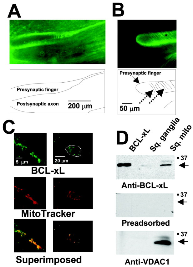
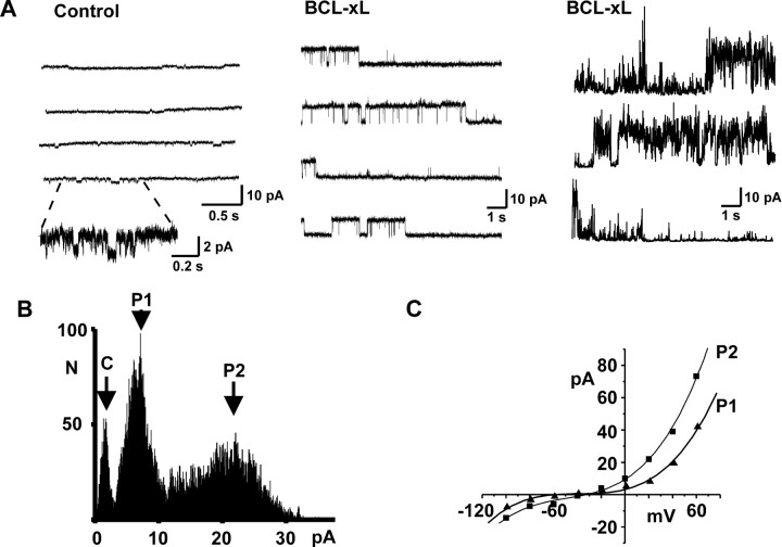
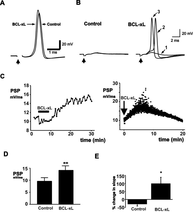
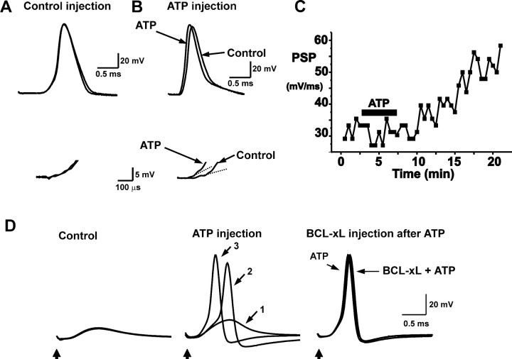
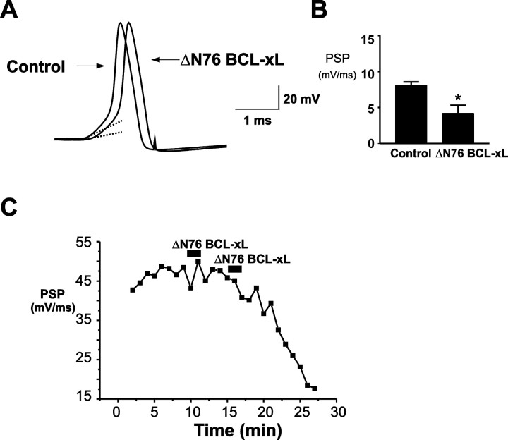
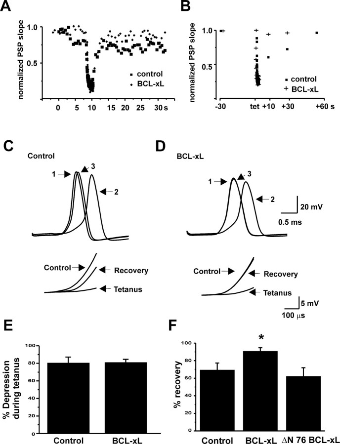
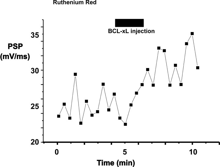
BCL-2 family proteins are known to regulate cell death during development by influencing the permeability of mitochondrial membranes. The anti-apoptotic BCL-2 family protein BCL-xL is highly expressed in the adult brain and localizes to mitochondria in the presynaptic terminal of the adult squid stellate ganglion. Application of recombinant BCL-xL through a patch pipette to mitochondria inside the giant presynaptic terminal triggered multiconductance channel activity in mitochondrial membranes. Furthermore, injection of full-length BCL-xL protein into the presynaptic terminal enhanced postsynaptic responses and enhanced the rate of recovery from synaptic depression, whereas a recombinant pro-apoptotic cleavage product of BCL-xL attenuated postsynaptic responses. The effect of BCL-xL on synaptic responses persisted in the presence of a blocker of mitochondrial calcium uptake and was mimicked by injection of ATP into the terminal. These studies indicate that the permeability of outer mitochondrial membranes influences synaptic transmission, and they raise the possibility that modulation of mitochondrial conductance by BCL-2 family proteins affects synaptic stability.


Figures

Figure 1.
Figure 1.
Presence of BCL-xL immunoreactivity in neuronal mitochondria. A, BCL-xL immunoreactivity in a section through the giant presynaptic terminal. B, At higher magnification, staining appears more punctate, and striations in staining (dashed arrows) are apparent. C, Colocalization of BCL-xL immunoreactivity with mitochondria at even higher magnification in the presynaptic terminal (left panels) and in neuronal somata (right panels) of the stellate ganglion. Mitochondria were stained using MitoTracker Red (middle), and the bottom panels show superposition of BCL-xL and MitoTracker staining. The outline of a single soma is marked with the white dotted line in the top panel. D, Immunoblot analysis of recombinant human BCL-xL protein, squid stellate ganglion (Sq. ganglia), and mitochondria (Sq. mito; purified from squid stellate ganglia) was performed using anti-BCL-xL antibody (top). The middle panel shows an immunoblot of the same samples using preadsorbed antibody. The bottom panel shows immunoblot analysis using an anti-VDAC1 antibody.
Figure 2.
Figure 2.
Induction of multiconductance channel activity on mitochondrial membranes by BCL-xL. A, Typical recordings of intracellular membrane activity in a squid presynaptic terminal using patch pipettes containing control intracellular solution (left) or intracellular solution containing 8.0 μg/ml BCL-xL (middle and right). Part of the control trace (left) is also shown on an expanded time and current scale to illustrate typical small conductance openings detected in control recordings. The center panel shows BCL-xL-induced activity that is maintained at a single conductance level, whereas the right panel shows typical transitions between conductance levels. Recordings were made at patch potentials of 100 (left), 40 (middle), and 100 mV (right). B, Amplitude histogram of multiconductance activity induced by BCL-xL (+20 mV) in a different patch from those shown in A. The closed state is marked C, and two prominent peaks in the histogram are labeled P1 and P2. C, Current-voltage relationship of unitary currents for the two conductance states shown in B. Amplitudes were determined from peaks in the amplitude histograms at each potential. In this patch, the mean slope conductances over the voltage range 0-60 mV were 240 and 462 pS for P1 and P2, respectively.
Figure 3.
Figure 3.
BCL-xL injection enhances synaptic transmission. A, Evoked postsynaptic responses before and 20 min after presynaptic injection of BCL-xL (20 μg/ml in pipette solution). B, Effects of BCL-xL injection into the presynaptic terminal in a synapse in which the postsynaptic response had failed to reach threshold for action potential firing. The left panel shows three superimposed postsynaptic responses before BCL-xL injection. The right panel shows responses at increasing times (1, 2, and 3) after injection up to 17 min. C, Typical time courses of effect of BCL-xL injection on the slope of the postsynaptic response evoked by stimulation at 0.033 Hz (left) or 2.0 Hz (right). D, Bar graphs of all experiments showing the peak postsynaptic response before and after BCL-xL injection (n = 10; **p < 0.005). E, Percentage change in the slope of the postsynaptic response over 10 min in untreated synapses (n = 6) compared with the percentage change in response of BCL-xL-injected terminals (20 min after injection; *p < 0.015).
Figure 7.
Figure 7.
ATP injection mimics BCL-xL injection. A, Postsynaptic responses shown before and 20 min after injection of squid intracellular solution without ATP. Bottom traces display the rate of rise of the postsynaptic responses on an expanded scale and show that there is no change in the postsynaptic response. B, Postsynaptic responses shown before and 20 min after injection of 20 mm ATP in squid intracellular solution. Bottom traces display postsynaptic responses on an expanded scale, and the superimposed dashed lines are the fits to the initial slope of the postsynaptic responses. C, Time course of response to ATP injection. D, BCL-xL fails to enhance transmission in the presence of injected ATP. The left panel shows three superimposed postsynaptic responses before ATP injection in a synapse with a postsynaptic response that had failed to reach threshold for action potential firing. The middle panel shows responses at increasing times (5, 8, and 20 min corresponding to traces 1, 2, and 3, respectively) after injection. The third panel shows the response immediately before BCL-xL injection [20 min after ATP injection (200 mm)], together with the traces recorded 5 and 10 min after introduction of BCL-xL. No enhancement of synaptic potentials in response to BCL-xL injection was detected.
Figure 4.
Figure 4.
Injection of ΔN 76 BCL-xL into the presynaptic terminal depresses synaptic transmission. A, Evoked postsynaptic responses before and 10 min after presynaptic injection of ΔN 76 BCL-xL (20 μg/ml in pipette solution) in a neuron with a strong synaptic response. Dashed lines are fitted to the initial slope of the postsynaptic responses. B, Bar graphs of all experiments showing the rates of rise of postsynaptic responses before and after ΔN 76 BCL-xL injection (n = 6; *p < 0.03). C, Typical time course of effect of ΔN 76 BCL-xL injection on the slope of the postsynaptic response.
Figure 5.
Figure 5.
Effects of BCL-xL injection on recovery from a tetanus. A, Time course of synaptic depression and recovery from depression in response to a tetanus (50 Hz, 2 sec) given during basal stimulation at 2 Hz, before and 9 min after injection of BCL-xL. B, Time course of recovery from depression in response to a tetanus given during basal stimulation at 0.033 Hz, before and after injection of BCL-xL. Postsynaptic responses were evoked at 10, 30, or 60 sec after the tetanus. C, Postsynaptic responses before (1), during (2), and 30 sec after (3) a tetanus during basal stimulation at 0.033 Hz. Data were obtained before the effect of BCL-xL. The inset shows rates of rise of postsynaptic responses on an expanded scale. D, Postsynaptic responses for the same cell recorded 50 min after injection of BCL-xL. E, Bar graphs comparing the pretetanus rate of rise of postsynaptic responses with that of the last response in the train for controls and synapses injected with BCL-xL. F, Bar graphs showing percentage recovery of postsynaptic responses 30 sec after a tetanus. BCL-xL responses were significantly increased over control (*p < 0.03). For controls, n = 10; for BCL-xL, n = 6; for ΔN 76 BCL-xL, n = 4.
Figure 6.
Figure 6.
Ruthenium red does not block the enhancement of the postsynaptic response by injection of BCL-xL. The time course of the rate of rise of postsynaptic responses is shown in a synapse with strong basal transmission. The preparation was treated with ruthenium red (3 μm). Period of injection with BCL-xL (20 μm in pipette solution) is shown by a bar.

References

    1. Antonsson B, Conti F, Ciavatta A, Montessuit S, Lewis S, Martinou I, Bernasconi L, Bernard A, Mermod JJ, Mazzei G, Maundrell K, Gambale F, Sadoul R, Martinou JC ( 1997) Inhibition of Bax channel-forming activity by Bcl-2. Science 277 : 370-372. - PubMed
    1. Augustine GJ ( 2001) How does calcium trigger neurotransmitter release? Curr Opin Neurobiol 11 : 320-326. - PubMed
    1. Babcock DF, Hille B ( 1998) Mitochondrial oversight of cellular Ca2+ signaling. Curr Opin Neurobiol 8 : 398-404. - PubMed
    1. Basanez G, Nechushtan A, Drozhinin O, Chanturiya A, Choe E, Tutt S, Wood K, Hsu Y, Zimmerberg J, Youle RJ ( 1999) Bax, but not Bcl-xL, decreases the lifetime of planar phospholipid bilayer membranes at subnanomolar concentrations. Proc Natl Acad Sci USA 96 : 5492-5497. - PMC - PubMed
    1. Basanez G, Zhang J, Chau BN, Maksaev GI, Frolov VA, Brandt TA, Burch J, Hardwick JM, Zimmerberg J ( 2001) Pro-apoptotic cleavage products of Bcl-xL form cytochrome c-conducting pores in pure lipid membranes. J Biol Chem 276 : 31083-31091. - PubMed

Publication types

MeSH terms

LinkOut - more resources