

Diversification of furanocoumarin-metabolizing cytochrome P450 monooxygenases in two papilionids: Specificity and substrate encounter rate
- PMID: 12968082
- PMCID: PMC304124
- DOI: 10.1073/pnas.1934643100
Diversification of furanocoumarin-metabolizing cytochrome P450 monooxygenases in two papilionids: Specificity and substrate encounter rate
Abstract
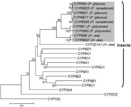
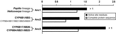
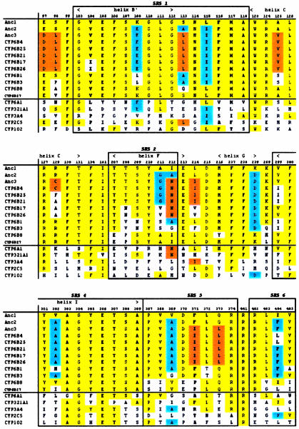
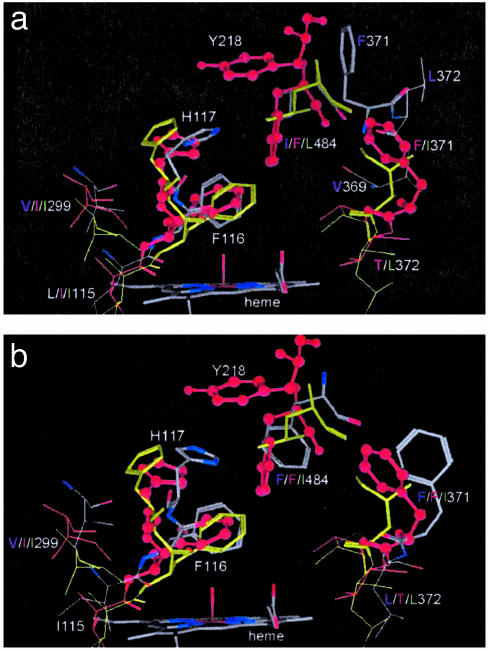
Diversification of cytochrome P450 monooxygenases (P450s) is thought to result from antagonistic interactions between plants and their herbivorous enemies. However, little direct evidence demonstrates the relationship between selection by plant toxins and adaptive changes in herbivore P450s. Here we show that the furanocoumarin-metabolic activity of CYP6B proteins in two species of swallowtail caterpillars is associated with the probability of encountering host plant furanocoumarins. Catalytic activity was compared in two closely related CYP6B4 and CYP6B17 groups in the polyphagous congeners Papilio glaucus and Papilio canadensis. Generally, P450s from P. glaucus, which feeds occasionally on furanocoumarin-containing host plants, display higher activities against furanocoumarins than those from P. canadensis, which normally does not encounter furanocoumarins. These P450s in turn catalyze a larger range of furanocoumarins at lower efficiency than CYP6B1, a P450 from Papilio polyxenes, which feeds exclusively on furanocoumarin-containing host plants. Reconstruction of the ancestral CYP6B sequences using maximum likelihood predictions and comparisons of the sequence and geometry of their active sites to those of contemporary CYP6B proteins indicate that host plant diversity is directly related to P450 activity and inversely related to substrate specificity. These predictions suggest that, along the lineage leading to Papilio P450s, the ancestral, highly versatile CYP6B protein presumed to exist in a polyphagous species evolved through time into a more efficient and specialized CYP6B1-like protein in Papilio species with continual exposure to furanocoumarins. Further diversification of Papilio CYP6Bs has likely involved interspersed events of positive selection in oligophagous species and relaxation of functional constraints in polyphagous species.
Figures
Similar articles
-
Molecular analysis of multiple CYP6B genes from polyphagous Papilio species.Insect Biochem Mol Biol. 2001 Sep;31(10):999-1011. doi: 10.1016/s0965-1748(01)00048-0. Insect Biochem Mol Biol. 2001. PMID: 11483436
-
CYP6B cytochrome p450 monooxygenases from Papilio canadensis and Papilio glaucus: potential contributions of sequence divergence to host plant associations.Insect Mol Biol. 2002 Dec;11(6):543-51. doi: 10.1046/j.1365-2583.2002.00363.x. Insect Mol Biol. 2002. PMID: 12421412
-
Metabolism of linear and angular furanocoumarins by Papilio polyxenes CYP6B1 co-expressed with NADPH cytochrome P450 reductase.Insect Biochem Mol Biol. 2003 Sep;33(9):937-47. doi: 10.1016/s0965-1748(03)00100-0. Insect Biochem Mol Biol. 2003. PMID: 12915185
-
Insect cytochromes P450: diversity, insecticide resistance and tolerance to plant toxins.Comp Biochem Physiol C Pharmacol Toxicol Endocrinol. 1998 Nov;121(1-3):147-55. doi: 10.1016/s0742-8413(98)10035-x. Comp Biochem Physiol C Pharmacol Toxicol Endocrinol. 1998. PMID: 9972456 Review.
-
The marmoset cytochrome P450 superfamily: Sequence/phylogenetic analyses, genomic structure, and catalytic function.Biochem Pharmacol. 2020 Jan;171:113721. doi: 10.1016/j.bcp.2019.113721. Epub 2019 Nov 18. Biochem Pharmacol. 2020. PMID: 31751534 Review.
Cited by
-
Aliphatic esters as targets of esterase activity in the parsnip webworm (Depressaria pastinacella).J Chem Ecol. 2012 Feb;38(2):188-94. doi: 10.1007/s10886-012-0073-2. Epub 2012 Feb 16. J Chem Ecol. 2012. PMID: 22350520
-
Genes involved in the evolution of herbivory by a leaf-mining, Drosophilid fly.Genome Biol Evol. 2012;4(9):900-16. doi: 10.1093/gbe/evs063. Epub 2012 Jul 19. Genome Biol Evol. 2012. PMID: 22813779 Free PMC article.
-
CYP6AE gene cluster knockout in Helicoverpa armigera reveals role in detoxification of phytochemicals and insecticides.Nat Commun. 2018 Nov 16;9(1):4820. doi: 10.1038/s41467-018-07226-6. Nat Commun. 2018. PMID: 30446639 Free PMC article.
-
The genetic basis of a plant-insect coevolutionary key innovation.Proc Natl Acad Sci U S A. 2007 Dec 18;104(51):20427-31. doi: 10.1073/pnas.0706229104. Epub 2007 Dec 11. Proc Natl Acad Sci U S A. 2007. PMID: 18077380 Free PMC article.
-
Defence mechanisms of Ficus: pyramiding strategies to cope with pests and pathogens.Planta. 2019 Mar;249(3):617-633. doi: 10.1007/s00425-019-03098-2. Epub 2019 Jan 28. Planta. 2019. PMID: 30689053 Review.
References
-
- Lewis, D. F. V., Watson, E. & Lake, B. G. (1998) Mutat. Res. 410, 245-270. - PubMed
-
- Schuler, M. A. (1996) Crit. Rev. Plant Sci. 15, 235-284.
-
- Chapple, C. (1998) Annu. Rev. Plant Physiol. Plant Mol. Biol. 49, 311-343. - PubMed
-
- Feyereisen, R. (1999) Annu. Rev. Entomol. 44, 507-533. - PubMed
-
- Scott, J. G. (1999) Insect Biochem. Mol. Biol. 29, 757-777. - PubMed
Publication types
MeSH terms
Substances
Grants and funding
LinkOut - more resources
Full Text Sources
