. 2004 Jan 1;103(1):85-92.
doi: 10.1182/blood-2003-05-1446. Epub 2003 Sep 11.

Safety and efficacy of factor IX gene transfer to skeletal muscle in murine and canine hemophilia B models by adeno-associated viral vector serotype 1

Affiliations

Safety and efficacy of factor IX gene transfer to skeletal muscle in murine and canine hemophilia B models by adeno-associated viral vector serotype 1

Valder R Arruda et al. Blood. .

Abstract

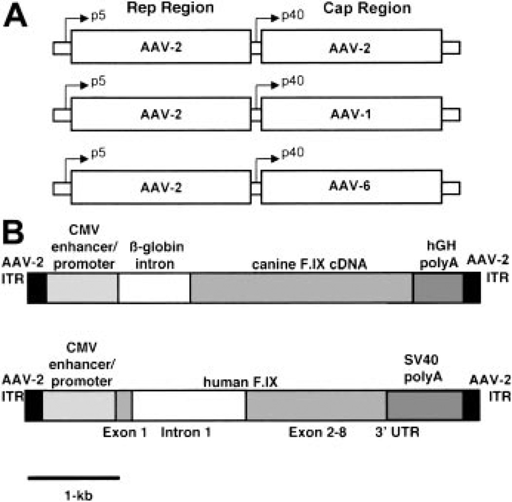
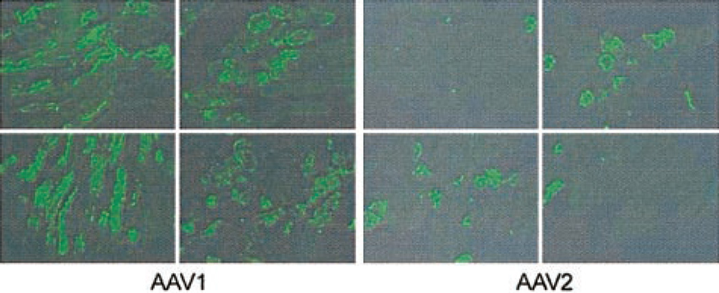
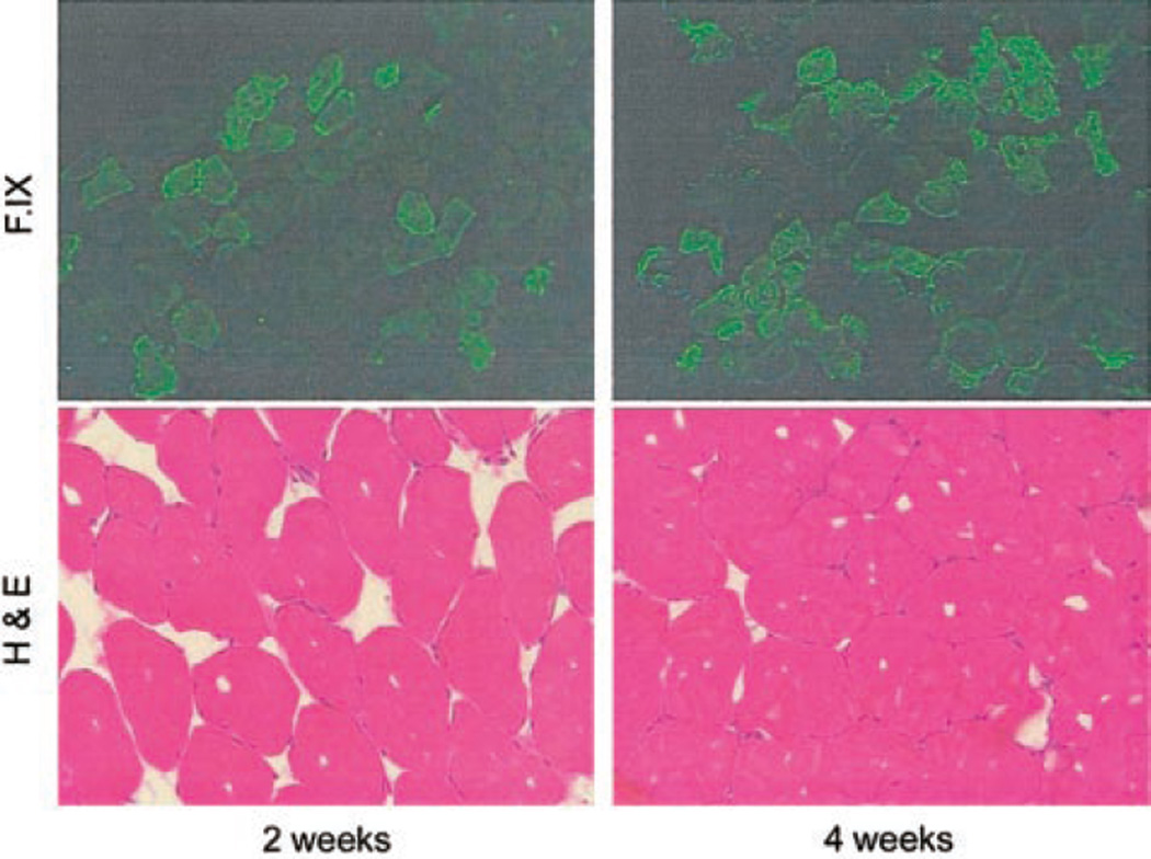
Adeno-associated viral (AAV) vectors (serotype 2) efficiently transduce skeletal muscle, and have been used as gene delivery vehicles for hemophilia B and for muscular dystrophies in experimental animals and humans. Recent reports suggest that AAV vectors based on serotypes 1, 5, and 7 transduce murine skeletal muscle much more efficiently than AAV-2, with reported increases in expression ranging from 2-fold to 1000-fold. We sought to determine whether this increased efficacy could be observed in species other than mice. In immunodeficient mice we saw 10- to 20-fold higher levels of human factor IX (hF.IX) expression at a range of doses, and in hemophilic dogs we observed approximately 50-fold higher levels of expression. The increase in transgene expression was due partly to higher gene copy number and a larger number of cells transduced at each injection site. In all immunocompetent animals injected with AAV-1, inhibitory antibodies to F.IX developed, but in immunocompetent mice treated with high doses of vector, inhibitory antibodies eventually disappeared. These studies emphasize that the increased efficacy of AAV-1 vectors carries a risk of inhibitor formation, and that further studies will be required to define doses and treatment regimens that result in tolerance rather than immunity to F.IX.


Figures

Figure 1
Figure 1. Constructs used in vector preparation
(A) Helper plasmids contain rep gene sequences fromAAV-2 and cap gene sequences fromAAV-2,AAV-1, andAAV-6 respectively. (B) The canine transgene cassette contains the cF.IX cDNA, the human β-globin intron 1, the CMV enhancer/promoter, and the human growth hormone polyadenylation signal, flanked by the AAV-2 inverted terminal repeats. The human transgene cassette contains the human F.IX cDNA interrupted by a 1.4-kb fragment of hF.IX intron 1, the CMV enhancer/promoter, and the SV40 polyadenylation signal, also flanked by the AAV-2 inverted terminal repeats.
Figure 2
Figure 2. Time course of hF.IX expression in C57Bl/6 CD4 knockout mice
Mice were injected at 2 × 10¹¹ vg/kg at 4 intramuscular sites in the hindlimbs. Each line represents average values for the cohort (n = 4 mice). Circles represent mice injected with AAV-6–F.IX vector; triangles, mice injected with AAV-1–F.IX; squares, mice injected with AAV-2–F.IX.
Figure 3
Figure 3. Gene copy number in AAV-injected muscle tissue
(A) Southern blot analysis of genomic DNA extracted from murine skeletal muscle injected with AAV vectors. The first 3 lanes are from mice injected with AAV-1–F.IX at the doses listed beneath each lane; the next 3 lanes are from mice injected with AAV-2–F.IX. Copy number standards were prepared by adding 10, 100, or 1000 pg plasmid to 20 µg murine genomic DNA.
Figure 4
Figure 4. Immunofluorescent staining for hF.IX in AAV-injected mouse muscle
Animals were injected in the hindlimbs with 4 × 10¹² vg/kg AAV-1 or AAV-2, divided equally among 4 injection sites. Twelve weeks later mice were killed and injected muscle was resected for immunofluorescent staining. Positive cells were scored by observers blinded to vector type. Representative sections from the quadriceps are shown for AAV-1– and AAV-2–injected muscle; average percent of positive cells was 54% for AAV-1, 22% for AAV-2.
Figure 5
Figure 5. Paired immunofluorescent and hemotoxylin and eosin staining of AAV-1–injected murine muscle at early time points after injection
Figure 6
Figure 6. Human F.IX expression and anti–F.IX antibody measurements as a function of dose and time in AAV-1–injected immunocompetent hemophilic mice
C57Bl/6 hemophilia B mice were injected at a high (1.6 × 10¹³ vg/kg, n = 5), medium (4 × 10¹² vg/kg, n = 2), or low (6.5 × 10¹¹ vg/kg, n = 3) dose at intramuscular sites. Panels A–D refer to medium- and high-dose cohorts, panel E to low-dose cohort. Each line represents one mouse. (A) Human F.IX levels were determined by ELISA. Levels in high-dose animals range from 300 ng/mL to 2200 ng/mL at 8 weeks, with levels of approximately 100 ng/mL at the same time point in medium-dose animals. After the initial lag, F.IX levels remain stable for the duration of the experiment. (B) Anti–F.IX-specific IgG1 antibodies. These were detected in all animals, peaked at 2 to 4 weeks after injection, markedly decreased by 8 weeks, and disappeared by 12 weeks after injection. (C) Bethesda titers. Inhibitory antibody is first detected at 2 weeks after injection, reaches a maximum at 4 weeks, then gradually diminishes. (D) Western blot to detect anti–F.IX antibody. Lanes 1 to 5 contain serum from the mice treated at a high dose, lanes 6 to 7 conatin serum from mice treated at a lower dose. At the 4-week time point, antibody was detected in 5 of 7 mice. At the 8-week time point it was detected in only 2 of 7 mice. (E) Left panel represents hF.IX levels determined by ELISA. Center and right panels represent antibodies to F.IX detected in all 3 mice by specific IgG to F.IX or by Bethesda assay, respectively. *Denotes the death of one animal after week 8.
Figure 7
Figure 7. Coagulation testing and antibody studies in dog E35
(A) Canine F.IX antigen levels as a function of time after injection. F.IX level peaked 2 weeks after injection and rapidly declined to undetectable. Arrows denote time points at which cyclophosphamide was administered. (B) WBCT as a function of time after injection. The WBCT fell into the normal range by day 6 and began to prolong again by day 15. By day 23 it had returned to the baseline of more than 60 minutes. (C) Bethesda titer as a function of time. An inhibitory antibody of 8.2 BU was first detected at day 27, and persisted for the remainder of the animal’s lifespan. (D) Anti–F.IX antibody measured by subclass as a function of time. IgG2 was first detected at high levels (2225 ng/mL) on day 15 after vector injection, prior to appearance of inhibitory antibody or to infusion of canine plasma. IgG1 rose gradually beginning about 35 days after vector injection and continued to rise throughout the course of the experiment.
Figure 8
Figure 8. Coagulation testing and antibody studies in dog E57
(A) Canine F.IX antigen levels as a function of time after injection. Arrows denote time points at which cyclophosphamide was administered; asterisks denote infusions of canine plasma. F.IX level peaked 55 days after injection and fell rapidly after cyclophosphamide was discontinued. (B) WBCT as a function of time after injection. The WBCT fell to the normal range about 1 week after vector injection. Levels remained reduced into or near the normal range until approximately day 70 when they were again prolonged. (C) Bethesda titer as a function of time after injection. Bethesda titer was undetectable until day 77. The titer was initially low but subsequently rose to approximately 8 BU. (D) Anti–F.IX antibody subclasses as a function of time. Both subclasses remain at baseline through the duration of cyclophosphamide therapy, but IgG1 rises rapidly, and IgG2 more slowly, after cyclophosphamide discontinuation.

Similar articles

Cited by

References

    1. Wagner JA, Moran ML, Messner AH, et al. A phase I/II study of tgAAV-CF for the treatment of chronic sinusitis in patients with cystic fibrosis. Hum Gene Ther. 1998;9:889–909. - PubMed
    1. Manno CS, Chew AJ, Hutchison S, et al. AAV-mediated factor IX gene transfer to skeletal muscle in patients with severe hemophilia B. Blood. 2003;101:2963–2972. - PubMed
    1. Stedman H, Wilson JM, Finke R, Kleckner AL, Mendell J. Phase I clinical trial utilizin gene therapy for limb girdle muscular dystrophy: alpha-, beta-, gamma-, or delta-sarcoglycan gene delivered with intramuscular instillations of adeno- associated vectors. Hum Gene Ther. 2000;11:777–790. - PubMed
    1. Chao H, Liu Y, Rabinowitz J, Li C, Samulski RJ, Walsh CE. Several log increase in therapeutic transgene delivery by distinct adeno-associated viral serotype vectors. Mol Ther. 2000;2:619–623. - PubMed
    1. Duan D, Yan Z, Yue Y, Ding W, Engelhardt JF. Enhancement of muscle gene delivery with pseudotyped adeno-associated virus type 5 correlates with myoblast differentiation. J Virol. 2001;75:7662–7671. - PMC - PubMed

Publication types

MeSH terms