Comparative Study
. 2003 Sep;140(2):413-21.
doi: 10.1038/sj.bjp.0705443. Epub 2003 Aug 11.

Protein kinase C-mediated Ca2+ entry in HEK 293 cells transiently expressing human TRPV4

Affiliations
Comparative Study

Protein kinase C-mediated Ca2+ entry in HEK 293 cells transiently expressing human TRPV4

Feng Xu et al. Br J Pharmacol. 2003 Sep.

Abstract

1. We investigated whether protein kinase C (PKC) activation stimulates Ca2+ entry in HEK 293 cells transfected with human TRPV4 cDNA and loaded with fura-2. 2. Phorbol 12-myristate 13-acetate (PMA), a PKC-activating phorbol ester, increased the intracellular Ca2+ concentration ([Ca2+]i) in a dose-dependent manner, with an EC50 value of 11.7 nm. Exposure to a hypotonic solution (HTS) after PMA further increased [Ca2+]i. Two other PKC-activating phorbol esters, phorbol 12,13-didecanoate (PDD) and phorbol 12,13-dibutyrate, also caused [Ca2+]i to increase. 3. The inactive isomer 4alpha-PMA was less effective and the peak [Ca2+]i increase was significantly smaller than that induced by PMA. In contrast, 4alpha-PDD produced a monophasic or biphasic [Ca2+]i increase with a different latency, while 4alpha-phorbol had no effect. 4. The PMA-induced [Ca2+]i increase was abolished by prior exposure to bisindolylmaleimide (BIM), a PKC-specific inhibitor, and suppressed by the nonspecific PKC inhibitor 1-(5-isoquinolinesulphonyl)-2-methylpiperazine. The [Ca2+]i increase induced by 4alpha-PMA, 4alpha-PDD or HTS was not significantly affected by BIM. 5. These results suggest that both PKC-dependent and -independent mechanisms are involved in the phorbol ester-induced activation of TRPV4, and the PKC-independent pathway is predominant in HTS-induced Ca2+ entry.


Figures

Figure 1
Figure 1
Effects of the PKC-activating phorbol ester PMA on intracellular calcium concentration ([Ca2+]i; expressed as F340/F380 values) in hTRPV4-transfected HEK 293 cells. (a) Representative trace showing the effect of 300 nM PMA in hTRPV4-transfected or pIRES2-EGFP vector-transfected (mock) HEK 293 cells. (b) Summary of [Ca2+]i increases caused by PMA. Each column represents mean±s.e.m.; numbers of cells examined are indicated above each column. **P<0.01 vs mock. (c) Role of extracellular calcium in the PMA-induced calcium increase in hTRPV4-transfected HEK 293 cells. Cells were exposed to 300 nM PMA in the presence or absence of 1 mM extracellular calcium. (d) Concentration–response relationship of the PMA-induced Ca2+ influx in HEK 293 cells expressing hTRPV4. Responses were measured as peak increases in fluorescence ratio minus basal level, and are expressed relative to the maximal response evoked by HTS after PMA application. The continuous line was fitted with the Hill equation. Number of observations per point was 6–11.
Figure 2
Figure 2
Effects of the PKC-activating phorbol esters PDD and PDBu on [Ca2+]i in hTRPV4-transfected HEK 293 cells. (a) Representative trace showing the effect of PDD at 300 nM (i) and 0.1 nM (ii). (b) Representative trace showing effect of 300 nM PDBu. (c) Summary of [Ca2+]i increases caused by the phorbol esters. Each column represents mean±s.e.m.; numbers of cells examined are indicated above each column. **P<0.01 vs PMA.
Figure 3
Figure 3
Effects of non-PKC-activating phorbol esters on intracellular calcium concentration ([Ca2+]i; expressed as F340/F380 values) in hTRPV4- or mock-transfected HEK 293 cells. Representative traces showing the effect of 300 nM 4α-PMA (a), 300 nM 4α-PDD (b) and 1000 nM 4α-phorbol (c) on [Ca2+]i. (ai) Typical trace showing that in 16 out of 27 cells expressing TRPV4, 300 nM 4α-PMA induced no significant [Ca2+]i increase. (aii) Trace showing that in six cells expressing TRPV4, 300 nM 4α-PMA induced a biphasic [Ca2+]i increase. HTS used in the present experiment was 200 mOsm/kg H2O. Ionomycin (1 μM) plus 10 mM CaCl2 was applied to obtain maximal [Ca2+]i. (b) 4α-PDD (300 nM) induced monophasic (bi) and biphasic (bii) [Ca2+]i increases in cells expressing TRPV4. (d) Summary of [Ca2+]i increases caused by non-PKC-activating phorbol esters. Each column represents mean±s.e.m.; numbers of cells examined are indicated above each column. *P<0.05 and **P<0.01 vs 300 nM PMA. The comparison between the effects of 4α-PMA and PMA was made using the Mann–Whitney test.
Figure 4
Figure 4
Effect of inhibition of PKC on the phorbol ester-induced calcium increase. Representative traces showing [Ca2+]i responses to PMA (a), 4α-PMA (b) and 4α-PDD (c) without (i) or with (ii) 100 nM BIM treatment. BIM was applied 60 min prior to the start of [Ca2+]i measurement, and was present throughout the experiment. (d) Summary of [Ca2+]i increases caused by phorbol esters with or without BIM. Each column represents mean±s.e.m.; numbers of cells examined are indicated above each column. **P<0.01.
Figure 5
Figure 5
Effect of H-7 on the PMA- and 4α-PDD-induced calcium increases. Traces showing the PMA-induced (a) and 4α-PDD-induced (b) [Ca2+]i increase without (i) or with (ii) 30 μM H-7. H-7 was applied 60 min before stimulation with phorbol esters, and was present throughout the experiments. Data are representative of three experiments.
Figure 6
Figure 6
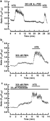
Effect of the specific PKC inhibitor BIM on the hypotonicity-dependent calcium increase. (a) Representative traces showing [Ca2+]i responses to HTS in hTRPV4- and mock-transfected cells. (b) Summary of [Ca2+]i increases caused by HTS in TRPV4- and mock-transfected cells. Each column represents mean±s.e.m.; numbers of cells examined are indicated above each column. (c) Representative trace showing the effect of extracellular Ca2+ on the HTS-induced calcium increase in hTRPV4-transfected HEK 293 cells; extracellular Ca2+ concentration 1 mM; 1 μM ionomycin plus 10 mM CaCl2 was applied to obtain maximal [Ca2+]i. (d) Whne cells were preincubated with 300 nM PMA, HTS still caused a robust [Ca2+]i increase. (e) Representative traces showing the PMA- and HTS-induced [Ca2+]i increases in the presence of BIM applied 60 min before and during stimulation with phorbol esters or HTS. (f) Summary of [Ca2+]i increases caused by HTS without or with BIM. Each column represents mean±s.e.m.; numbers of cells examined are indicated above each column.
Figure 7
Figure 7
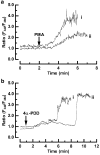
(a) Traces showing the effects of preincubation with 3 μM PMA on HTS- and 4α-PDD-induced calcium entry in hTRPV4-transfected cells. Only the 4α-PDD-induced calcium increase was abolished. (b) Traces showing the [Ca2+]i increase induced in the absence (i) or presence of PD98059 (ii) applied 60 min before and during stimulation with PMA and HTS. Results are representative of three experiments.

Similar articles

Cited by

References

    1. ALTAMIRANO J., BRODWICK M.S., ALVAREZ-LEEFMANS F.J. Regulatory volume decrease and intracellular Ca2+ in murine neuroblastoma cells studied with fluorescent probes. J. Gen. Physiol. 1998;112:145–160. - PMC - PubMed
    1. BRAKEMEIER S., EICHLER I., HOPP H., KOHLER R., HOYER J. Up-regulation of endothelial stretch-activated cation channels by fluid shear stress. Cardiovasc. Res. 2002;53:209–218. - PubMed
    1. CHEN Y., SIMASKO S.M., NIGGEL J., SIGURDSON W.J., SACHS F. Ca2+ uptake in GH3 cells during hypotonic swelling: the sensory role of stretch-activated ion channels. Am. J. Physiol. 1996;270:C1790–C1798. - PubMed
    1. CHENG J.-J., WUNG B.-S., CHAO Y.-J., WANG D.L. Sequential activation of protein kinase C (PKC)-α and PKC-ɛ contributes to sustained Raf/ERK1/2 activation in endothelial cells under mechanical strain. J. Biol. Chem. 2001;276:31368–31375. - PubMed
    1. DELANY N.S., HURLE M., FACER P., ALNADAF T., PLUMPTON C., KINGHORN I., SEE C.G., COSTIGAN M., ANAND P., WOOLF C.J., CROWTHER D., SANSEAU P., TATE S.N. Identification and characterization of a novel human vanilloid receptor-like protein, VRL-2. Physiol. Genomics. 2001;4:165–174. - PubMed

Publication types

MeSH terms

LinkOut - more resources