. 2003 Sep;140(2):339-46.
doi: 10.1038/sj.bjp.0705453. Epub 2003 Aug 26.

A model for receptor-peptide binding at the glucagon-like peptide-1 (GLP-1) receptor through the analysis of truncated ligands and receptors

Affiliations

A model for receptor-peptide binding at the glucagon-like peptide-1 (GLP-1) receptor through the analysis of truncated ligands and receptors

Suleiman Al-Sabah et al. Br J Pharmacol. 2003 Sep.

Abstract

1. The receptor for glucagon-like peptide-1 (GLP-1) can be activated by both its physiological hormone and a peptide discovered in the venom of the Gila Monster, exendin-4, which shows promise as an antidiabetic agent. 2. Exendin-4 displays receptor-binding properties not observed for GLP-1. Firstly, exendin-4 can be truncated by up to eight residues at its N-terminus without a significant loss of affinity. Secondly, exendin-4 maintains high affinity for the isolated N-terminal domain of the receptor, suggesting that exendin-4 makes additional contacts with this domain of the receptor, which nullify the requirement for ligand-receptor interactions involving the extracellular loops and/or transmembrane helices of the receptor's core domain. 3. In order to further understand the nature of the receptor-peptide interaction, a variety of full length and truncated peptide analogues were used to quantify the contribution of each distinct region of exendin-4 and GLP-1 to receptor affinity. 4. Our data show that, for both exendin-4 and GLP-1, the primary interaction is between the putative helical region of the peptide and the extracellular N-terminal domain of the receptor. 5. However, we demonstrate that the contribution to receptor affinity provided by the N-terminal segment of GLP-1 is greater than that of exendin-4, while the C-terminal nine residue extension of exendin-4, absent in GLP-1, forms a compensatory interaction with the N-terminal domain of the receptor. 6. We describe a peptide-receptor binding model to account for these data.


Figures
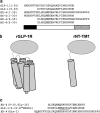
Figure 1
Figure 1
Peptides and receptor constructs used in this work. (a) An alignment of the full-length and truncated peptides used in this study. In order to facilitate direct comparison with EX-4, the numbering of GLP-1 has been modified from the conventional system, so that its first residue is His-1. The three distinct regions of the peptides are highlighted by the bar below the sequences, such that the central helix is shown as white, the N-terminal region as black and the C-terminal region of EX-4 as hatched. This pictorial shorthand system will be used in Figures 2, 3, 4 and 5. All the peptides were C-terminally amidated. (b) Pictorial representation of the full-length (rGLP-1) and truncated (rNT-TM1) receptors used in this study. The extracellular N-terminal domain is shown by a shaded oval, while the TM helices are depicted as cylinders. (c) An alignment of three modified peptides based upon GLP-1 and EX-4. The modifications to the sequence are underlined. All the three peptides were C-terminally amidated.
Figure 2
Figure 2
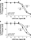
Binding of the helical regions of EX-4 and GLP-1 to rGLP-1R and rNT-TM1. 125I-exendin-4(9–39) competition-binding assays for (a) rGLP-1R and (b) rNT-TM1 with EX-4(9–30) and GLP-1(9–30). The figures are representative of one of at least three independent experiments to assess the affinity of the central helical region of EX-4 and GLP-1. pIC50 values from Table 1 are given next to the symbol of each peptide.
Figure 3
Figure 3
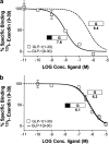
Binding of N-terminal region of GLP-1 to rGLP-1R, and rNT-TM1. 125I-exendin-4(9–39) competition-binding assays for (a) rGLP-1R and (b) rNT-TM1 with GLP-1(1–30). The figures are representative of one of at least three independent experiments to assess the affinity of the N-terminal region of GLP-1 by comparing GLP-1(1–30) with GLP-1(9–30). The dotted lines represent the GLP-1(9–30) curves from Figure 2. pIC50 values from Table 1 are given next to the symbol of each peptide. The figure highlights the large improvement in affinity at rGLP-1R, resulting from the addition of the N-terminal region of GLP-1 in (a) (significantly different, P<0.0001). The difference in pIC50 at rNT-TM1 (b) is not significant (P<0.05).
Figure 4
Figure 4
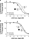
Binding of the N-terminal region of EX-4 to rGLP-1R and rNT-TM1. 125I-exendin-4(9–39) competition-binding assays for (a) rGLP-1R and (b) rNT-TM1 with EX-4(1–30). The figures are representative of one of at least three independent experiments to assess the affinity of the N-terminal region of EX-4 by comparing EX-4(1–30) with EX-4(9–30). The dotted lines represent the EX-4(9–30) curves from Figure 2. pIC50 values from Table 1 are given next to the symbol of each peptide. Panel (a) highlights the small improvement in affinity resulting from the addition of the N-terminal region of EX-4 (significantly different, P<0.01). This small enhancement of affinity is of the same magnitude and significance to that observed when comparing EX-4(1–39) with EX-4(9–39) (see Table 1).
Figure 5
Figure 5
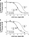
Binding of the C-terminal region of EX-4 to rGLP-1R and rNT-TM1. 125I-exendin-4(9–39) competition-binding assays for (a) rGLP-1R and (b) rNT-TM1 with EX-4(9–39). The figures are representative of one of at least three independent experiments to assess the affinity of the C-terminal region of EX-4 by comparing EX-4(9–39) with EX-4(9–30). The dotted lines represent the EX-4(9–30) curves from Figure 2. pIC50 values from Table 1 are given next to the symbol of each peptide. The figure highlights the large improvement in affinity at both rGLP-1R and rNT-TM1 (significantly different, P<0.0001 and P<0.0005, respectively) resulting from the addition of the C-terminal region of EX-4. The enhancement of affinity is of a similar magnitude to that observed when comparing EX-4(1–39) with EX-4(1–30) (significantly different for both receptors, P<0.0001; see Table 1).
Figure 6
Figure 6
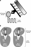
A model for peptide–receptor binding. (a) Schematic model for the H interaction between a putative groove on the N-terminal domain of the receptor and the conserved face of the helical region of the peptides. (b) Two schematic diagrams depicting the binding of GLP-1 (left) and EX-4 (right) to GLP-1R. The receptor is shown as consisting of two domains, the ‘N-terminal domain' and the ‘core domain'. The peptides are displayed between the circled symbols N and C, with their putative helical regions shown as cylinders. GLP-1 binding (left) involves an interaction H between its helical region and the receptor's N-terminal domain, which accounts for approximately 82% of the total binding energy of GLP-1 (see Discussion). The remaining binding energy comes from the interaction Ng between the N-terminal sequence of GLP-1 and the core domain. The interaction between EX-4 and GLP-1R (right) is also predominantly via the peptide's helical region and the receptor's N-terminal domain, with this H interaction accounting for approximately 79% of the total binding energy of EX-4. However, the Nex only contributes 5% of the binding energy of the full-length receptor. In addition, EX-4 forms an additional Ex interaction via its C-terminal region and the N-terminal domain of the receptor. This Ex interaction accounts for approximately 16% of the total binding energy of EX-4. The magnitude of the Ng interaction is equivalent to that of the Ex interaction.

Similar articles

Cited by

References

    1. BERGWITZ C., GARDELLA T.J., FLANNERY M.R., POTTS J.T., KRONENBERG H.M., GOLDRING S.R., JÜPPNER H. Full activation of chimeric receptors by hybrids between parathyroid hormone and calcitonin. Evidence for a common pattern of ligand–receptor interaction. J. Biol. Chem. 1996;271:26469–26472. - PubMed
    1. DAKIN C.L., GUNN I., SMALL C.J., EDWARDS C.M.B., HAY D.L., SMITH D.M., GHATEI M.A., BLOOM S.R. Oxyntomodulin inhibits food intake in the rat. Endocrinology. 2001;142:4244–4250. - PubMed
    1. GÖKE R., FEHMANN H.C., LINN T., SCHMIDT H., KRAUSE M., ENG J., GÖKE B. Exendin-4 is a high potency agonist and truncated exendin-(9–39)-amide an antagonist at the glucagon-like peptide-1(7–36)-amide receptor on insulin secreting cells. J. Biol. Chem. 1993;268:19650–19655. - PubMed
    1. GUTNIAK M., ØRSKOV C., HOLST J.J., AHRÉN B., EFFENDIC S. Antidiabetogenic effect of glucagon-like peptide-1(7–36)amide in normal subjects and patients with diabetes mellitus. N. Engl. J. Med. 1992;326:1316–1322. - PubMed
    1. HJORTH S.A., ADELHORST K., PEDERSEN B.B., KIRK O., SCHWARTZ T.W. Glucagon and glucagon-like peptide 1: selective receptor recognition via distinct peptide epitopes. J. Biol. Chem. 1994;269:30121–30124. - PubMed

Publication types

MeSH terms

LinkOut - more resources