Rosiglitazone and 15-deoxy-Delta12,14-prostaglandin J2, ligands of the peroxisome proliferator-activated receptor-gamma (PPAR-gamma), reduce ischaemia/reperfusion injury of the gut
- PMID: 12970094
- PMCID: PMC1574022
- DOI: 10.1038/sj.bjp.0705419
Rosiglitazone and 15-deoxy-Delta12,14-prostaglandin J2, ligands of the peroxisome proliferator-activated receptor-gamma (PPAR-gamma), reduce ischaemia/reperfusion injury of the gut
Expression of concern in
-
EXPRESSION OF CONCERN: Rosiglitazone and 15-deoxy-Δ12,14-prostaglandin J2, ligands of the peroxisome proliferator-activated receptor-γ (PPAR-γ), reduce ischaemia/reperfusion injury of the gut.Br J Pharmacol. 2025 Feb;182(3):925. doi: 10.1111/bph.17421. Epub 2024 Dec 2. Br J Pharmacol. 2025. PMID: 39623604 No abstract available.
Abstract
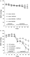
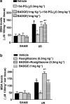
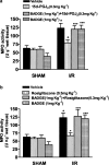
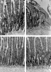
1. The peroxisome proliferator-activated receptor-gamma (PPAR-gamma) is a member of the nuclear receptor superfamily of ligand-dependent transcription factors related to retinoid, steroid and thyroid hormone receptors. The thiazolidinedione rosiglitazone and the endogenous cyclopentenone prostaglandin (PG)D2 metabolite, 15-deoxy-Delta12,14-PGJ2 (15d-PGJ2), are two PPAR-gamma ligands, which modulate the transcription of target genes. 2. The aim of this study was to investigate the effect of rosiglitazone and 15d-PGJ2 on the tissue injury caused by ischaemia/reperfusion (I/R) of the gut. 3. I/R injury of the intestine was caused by clamping both the superior mesenteric artery and the coeliac trunk for 45 min, followed by release of the clamp allowing reperfusion for 2 or 4 h. This procedure results in splanchnic artery occlusion (SAO) shock. 4. Rats subjected to SAO developed a significant fall in mean arterial blood pressure, and only 10% of the animals survived for the entire 4 h reperfusion period. Surviving animals were killed for histological examination and biochemical studies. Rats subjected to SAO displayed a significant increase in tissue myeloperoxidase (MPO) activity and malondialdehyde (MDA) levels, significant increases in plasma tumour necrosis factor (TNF)-alpha and interleukin (IL)-1beta levels and marked injury to the distal ileum. 5. Increased immunoreactivity to nitrotyrosine was observed in the ileum of rats subjected to SAO. Staining of sections of the ileum obtained from SAO rats with anti-intercellular adhesion molecule (ICAM-1) antibody resulted in diffuse staining. 6. Administration at 30 min prior to the onset of gut ischaemia of the two PPAR-gamma agonists (rosiglitazone (0.3 mg kg-1 i.v.) and 15d-PGJ2 (0.3 mg kg-1 i.v.)) significantly reduced the (i) fall in mean arterial blood pressure, (ii) mortality rate, (iii) infiltration of the reperfused intestine with polymorphonuclear neutrophils (MPO activity), (iv) lipid peroxidation (MDA levels), (v) production of proinflammatory cytokines (TNF-alpha and IL-1beta) and (vi) histological evidence of gut injury. Administration of rosiglitazone and 15d-PGJ2 also markedly reduced the nitrotyrosine formation and the upregulation of ICAM-1 during reperfusion. 7. In order to elucidate whether the protective effects of rosiglitazone and 15d-PGJ2 are related to the activation of the PPAR-gamma receptor, we also investigated the effect of a PPAR-gamma antagonist, bisphenol A diglycidyl ether (BADGE), on the protective effects of rosiglitazone and 15d-PGJ2. BADGE (1 mg kg-1 administered i.v. 30 min prior to the treatment of rosiglitazone or 15d-PGJ2) significantly antagonised the effect of the two PPAR-gamma agonists and thus abolished the protective effect against gut I/R. 8. These results demonstrate that the two PPAR-gamma agonists, rosiglitazone and 15d-PGJ2, significantly reduce I/R injury of the intestine.
Figures


Similar articles
-
WY 14643, a potent exogenous PPAR-alpha ligand, reduces intestinal injury associated with splanchnic artery occlusion shock.Shock. 2004 Oct;22(4):340-6. doi: 10.1097/01.shk.0000136704.26372.2d. Shock. 2004. PMID: 15377889
-
Effect of rosiglitazone and 15-deoxy-Delta12,14-prostaglandin J2 on bleomycin-induced lung injury.Eur Respir J. 2005 Feb;25(2):225-34. doi: 10.1183/09031936.05.00049704. Eur Respir J. 2005. PMID: 15684285
-
Ligands of the peroxisome proliferator-activated receptors (PPAR-gamma and PPAR-alpha) reduce myocardial infarct size.FASEB J. 2002 Jul;16(9):1027-40. doi: 10.1096/fj.01-0793com. FASEB J. 2002. PMID: 12087064
-
Hormone regulation of microglial cell activation: relevance to multiple sclerosis.Brain Res Brain Res Rev. 2005 Apr;48(2):322-7. doi: 10.1016/j.brainresrev.2004.12.020. Epub 2005 Jan 19. Brain Res Brain Res Rev. 2005. PMID: 15850670 Free PMC article. Review.
-
PPAR and immune system--what do we know?Int Immunopharmacol. 2002 Jul;2(8):1029-44. doi: 10.1016/s1567-5769(02)00057-7. Int Immunopharmacol. 2002. PMID: 12349941 Free PMC article. Review.
Cited by
-
Peroxisome Proliferator-Activated Receptors: Experimental Targeting for the Treatment of Inflammatory Bowel Diseases.Front Pharmacol. 2020 May 27;11:730. doi: 10.3389/fphar.2020.00730. eCollection 2020. Front Pharmacol. 2020. PMID: 32536865 Free PMC article. Review.
-
The cyclopentenone prostaglandin 15-deoxydelta(12,14)-prostaglandin J2 attenuates the development of zymosan-induced shock.Intensive Care Med. 2005 May;31(5):693-700. doi: 10.1007/s00134-005-2596-2. Epub 2005 Mar 15. Intensive Care Med. 2005. PMID: 15868139
-
Novel pharmacological approaches to the treatment of renal ischemia-reperfusion injury: a comprehensive review.Naunyn Schmiedebergs Arch Pharmacol. 2007 Oct;376(1-2):1-43. doi: 10.1007/s00210-007-0183-5. Epub 2007 Sep 22. Naunyn Schmiedebergs Arch Pharmacol. 2007. PMID: 18038125 Review.
-
PPARγ agonists promote the resolution of myelofibrosis in preclinical models.J Clin Invest. 2021 Jun 1;131(11):e136713. doi: 10.1172/JCI136713. J Clin Invest. 2021. PMID: 33914703 Free PMC article.
-
New insights into the role of peroxisome proliferator-activated receptors in regulating the inflammatory response after tissue injury.PPAR Res. 2012;2012:728461. doi: 10.1155/2012/728461. Epub 2012 Feb 29. PPAR Res. 2012. PMID: 22481914 Free PMC article.
References
-
- BECKMAN J.S. Oxidative damage and tyrosine nitration from peroxynitrite. Chem. Res. Toxicol. 1996;9:836–844. - PubMed
-
- BITTERMAN H., KINARTY A., LAZAROVICH H., LAHAT N. Acute release of cytokines is proportional to tissue injury induced by surgical trauma and shock in rats. J. Clin. Immunol. 1991;11:184–192. - PubMed
-
- BITTERMANN H., LEFER A.M. Use of a novel peptide leukotriene receptor antagonist, Ly 163441, in splanchnic artery occlusion shock. Prostagland. Leukotr. Essent. Fatty Acids. 1988;32:63–68. - PubMed
-
- BULKLEY G.B.Mediators of splanchnic organ injury: overview and perspective Splanchnic Ischaemia and Multiple Organ Failure 1989London: Edward Arnold Publ; 191–193.ed. Marston, A., Bulkley, G., Fiddian-Green, R.G. & Haglund, U.H. pp
-
- BUTCHER E.C. Leukocyte–endothelial cell adhesion as an active, multi-step process: a combinatorial mechanism for specificity and diversity in leukocyte targeting. Adv. Exp. Med. Biol. 1992;323:181–194. - PubMed
MeSH terms
Substances
LinkOut - more resources
Full Text Sources
Research Materials
Miscellaneous

