Comparative Study
. 2003 Sep;140(2):285-94.
doi: 10.1038/sj.bjp.0705438. Epub 2003 Aug 26.

Possible involvement of IGF-1 receptor and IGF-binding protein in insulin-induced enhancement of noradrenaline response in diabetic rat aorta

Affiliations
Comparative Study

Possible involvement of IGF-1 receptor and IGF-binding protein in insulin-induced enhancement of noradrenaline response in diabetic rat aorta

Tsuneo Kobayashi et al. Br J Pharmacol. 2003 Sep.

Abstract

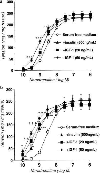
1. We investigated the mechanisms underlying the changes in vascular contractile responsiveness induced by chronic treatment with insulin in controls and established streptozotocin (STZ)-induced diabetic rats. 2. The aortic contractile response to noradrenaline (NA) showed no significant difference between controls and diabetics, but it was significantly greater in insulin-treated diabetic rats than in the other groups. To investigate the mechanism, we examined the changes in NA-induced contractility following treatment with insulin and insulin-like growth factor-1 (IGF-1) in organ-cultured control and diabetic aortas. 3. The contractile response to NA in organ-cultured diabetic rat aortas treated with insulin (500 ng ml-1, 16 h) or IGF-1 (20 ng ml-1, 16 h) was significantly greater than the corresponding values for (a) diabetic rat aortas cultured in serum-free medium, and (b) control aortas incubated with insulin or IGF-1. Incubating control aortas with insulin or IGF-1 had no significant effect on the contraction induced by NA. 4. The expressions of the IGF-1 receptor mRNA and protein were increased in STZ-induced diabetic aortas and further increased in insulin-treated diabetics. The mRNA expressions of IGF-binding protein (IGFBP)-2 and IGFBP-3 were normal in diabetic aortas. In contrast, those of IGFBP-4 and IGFBP-5 were significantly decreased in diabetic aortas, and not restored by insulin treatment. 5. These results suggest that the insulin deficiency and chronic hyperinsulinemia in diabetes upregulate the IGF-1 receptor and downregulate IGFBP-4 and IGFBP-5 in the aorta. This may be a major cause of the increased vascular contractility induced by insulin administration and by hyperinsulinemia in established diabetes, resulting in hypertension.


Figures

Figure 1
Figure 1
Concentration – response curves for NA-induced contractions in aortic strips with endothelial cells (a) or without endothelial cells (b). Aortic strips were taken from age-matched controls, untreated diabetic rats, insulin-treated controls, and insulin-treated diabetic rats. Ordinate shows increase in tension (expressed in mg tension mg tissue−1) measured at the peak of the response. Each data point represents the mean±s.e.m. of six to eight experiments; the s.e.m. is included only when it exceeds the dimension of the symbol used. *P<0.05, ***P<0.001 insulin-treated control vs insulin-treated diabetic. †††P<0.001, diabetic vs insulin-treated diabetic.
Figure 2
Figure 2
Concentration – response curves for NA-induced contractions in endothelium-denuded strips of cultured aortas. Aortas from age-matched controls (a) and untreated diabetic (b) rats were cultured in serum-free medium or in the presence of insulin (500 ng ml−1) or IGF-1 (20 ng ml−1, or 50 ng ml−1) for 16 h. Ordinate shows the increase in tension (expressed in mg tension mg tissue−1) measured at the peak of the response. Each data point represents the mean±s.e.m. of six to eight experiments; the s.e.m. is included only when it exceeds the dimension of the symbol used. *P<0.05, **P<0.01, ***P<0.001 diabetic aortas cultured with insulin vs diabetic aortas in serum-free medium. P<0.05, ††P<0.01, †††P<0.001 diabetic aortas cultured with IGF-1 vs diabetic aortas in serum-free medium. ##P<0.01, ###P<0.001 control aortas cultured with 50 ng ml−1 IGF-1 vs control aortas in serum-free medium.
Figure 3
Figure 3
Concentration – response curves for NA-induced contractions in endothelium-denuded aortic strips in modified KHS. Aortas from age-matched controls and untreated diabetic rats were incubated in KHS or in the presence of IGF-1 (20 ng/ml) for 16 h. Ordinate shows the increase in tension (expressed in mg tension mg tissue−1) measured at the peak of the response. Each data point represents the mean±s.e.m. of six to eight experiments; the s.e.m. is included only when it exceeds the dimension of the symbol used.
Figure 4
Figure 4
RT – PCR assay of expression of mRNA for IGF-1 receptor in control, diabetic, insulin-treated control, and insulin-treated diabetic rat aortas. (a) Expression of mRNA for IGF-1 receptor assayed by RT – PCR. (b) Quantitative analysis of expression of mRNA for IGF-1 receptor by scanning densitometry. Control rats (n=8, open column); STZ-induced diabetic rats (n=8, closed column); insulin-treated control rats (n=8, hatched column); insulin-treated diabetic rats (n=8, stippled column). Values are each the mean±s.e.m. of eight determinations (IGF-1 receptor GAPDH−1). **P<0.01, vs control. P<0.05, insulin-treated diabetic vs diabetic, ##P<0.01; insulin-treated diabetic vs insulin-treated control.
Figure 5
Figure 5
RT – PCR assay of expressions of mRNAs for IGFBP-2 and IGFBP-3 in control, diabetic, insulin-treated control, and insulin-treated diabetic rat aortas. (a) Expressions assayed by RT – PCR. (b) Quantitative analysis of expressions by scanning densitometry. Control rats (n=8, open column); STZ-induced diabetic rats (n=8, closed column); insulin-treated control rats (n=8, hatched column); insulin-treated diabetic rats (n=8, stippled column). Values are each the mean±s.e.m. of eight determinations (IGFBP GAPDH−1).
Figure 6
Figure 6
RT – PCR assay of expressions of mRNAs for IGFBP-4 and IGFBP-5 in control, diabetic, insulin-treated control, and insulin-treated diabetic rat aortas. (a) Expressions assayed by RT – PCR. (b) Quantitative analysis of expressions by scanning densitometry. Control rats (n=8, open column); STZ-induced diabetic rats (n=8, closed column); insulin-treated control rats (n=8, hatched column); insulin-treated diabetic rats (n=8, stippled column). Values are each the mean±s.e.m. of eight determinations (IGFBP GAPDH−1). *P<0.05, **P<0.01, ***P<0.001, vs control. #P<0.05, ###P<0.001, insulin-treated control vs insulin-treated diabetic.
Figure 7
Figure 7
Immunoblots showing protein expressions for IGF-1 receptor (95 kDa), IGFBP-4 (34 kDa) and IGFBP-5 (36 kDa) in control, diabetic, insulin-treated control, and insulin-treated diabetic rat aortas. (a) Expressions assayed by Western blotting. (b) Quantitative analysis of expressions by scanning densitometry. Control rats (n=4, open column); STZ-induced diabetic rats (n=4, closed column); insulin-treated control rats (n=4, hatched column); insulin-treated diabetic rats (n=4, stippled column). Values are each the mean±s.e.m. of four determinations. *P<0.05, **P<0.01, vs control. ††P<0.01, insulin-treated diabetic vs diabetic. ##P<0.01, ###P<0.001, insulin-treated diabetic vs insulin-treated control.

Similar articles

Cited by

References

    1. ABE H., YAMADA N., KAMATA K., KUWAKI T., SHIMADA M., OSUGA J., SHIONOIRI F., YAHAGI N., KADOWAKI T., TAMEMOTO H., ISHIBASHI S., YAZAKI Y., MAKUUCHI M. Hypertension, hypertriglyceridemia, and impaired endothelium-dependent vascular relaxation in mice lacking insulin receptor substrate-1. J. Clin. Invest. 1998;101:1784–1788. - PMC - PubMed
    1. ANWAR A., ZAHID A.A., PHILLIPS L., DELAFONTAINE P. Insulin-like growth factor binding protein-4 expression is decreased by angiotensin II and thrombin in rat aortic vascular smooth muscle cells. Arterioscler. Thromb. Vasc. Biol. 2000;20:370–376. - PubMed
    1. BAYES-GENIS A., CONOVER C.A., SCHWARTZ R.S. The insulin-like growth factor axis: a review of atherosclerosis and restenosis. Circ. Res. 2000;86:125–130. - PubMed
    1. BLAKESLEY V.A., SCRIMGEOUR A., ESPOSITO D., LE ROITH D. Signaling via the insulin-like growth factor-I receptor: does it differ from insulin receptor signaling. Cytokine Growth Factor Rev. 1996;7:153–159. - PubMed
    1. BORNFELDT K.E., ARNQVIST H.J., CAPRON L. In vivo proliferation of vascular smooth muscle in relation to diabetes mellitus, insulin-like growth factor I and insulin. Diabetologia. 1992;35:104–108. - PubMed

Publication types

MeSH terms