. 2003 Oct;52(10):1465-71.
doi: 10.1136/gut.52.10.1465.

The Cdx2 homeobox gene has a tumour suppressor function in the distal colon in addition to a homeotic role during gut development

Affiliations

The Cdx2 homeobox gene has a tumour suppressor function in the distal colon in addition to a homeotic role during gut development

C Bonhomme et al. Gut. 2003 Oct.

Abstract

Background: During development, the homeobox gene Cdx2 exerts a homeotic function, providing the positional information necessary for correct specification of the midgut endoderm. This is illustrated by the non-neoplastic gastric-type heteroplasias present at birth in the pericaecal region of Cdx2(+/-) mice. Furthermore, intestinal expression of Cdx2 continues throughout life but diminishes in colorectal cancers compared with adjacent normal tissue, suggesting a role in tumorigenesis.

Aim: To investigate the consequence of altered Cdx2 expression on colon tumour initiation and/or progression.

Methods: Heterozygous Cdx2(+/-) mice were analysed for spontaneous malignant tumours and for tumour development after treatment with a DNA mutagen, azoxymethane.

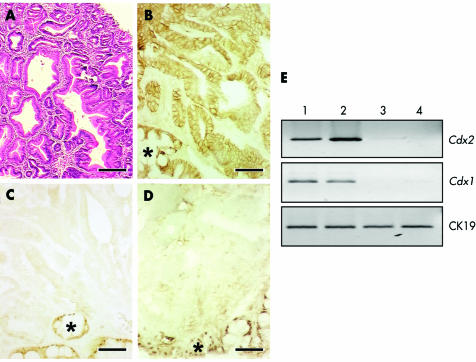
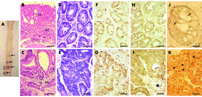
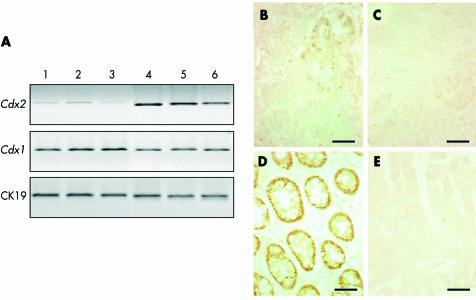
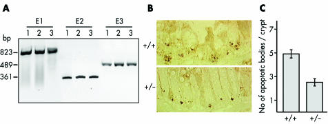
Results: Cdx2(+/-) mice did not spontaneously develop malignant tumours. After azoxymethane treatment, the gastric-like heteroplasias in the pericaecal region did not evolve into cancer indicating that they are not precancerous lesions. However, azoxymethane treated Cdx2(+/-) mice developed tumours specifically in the distal colon 12 weeks after azoxymethane treatment whereas no tumours were found in wild-type littermates at this stage. Histopathological and molecular analyses indicated that these tumours were invasive adenocarcinomas that recapitulated the malignant sequence observed in the majority of sporadic colorectal cancers in human. In addition, we found that the colonic epithelium was less sensitive to radiation induced apoptosis in Cdx2(+/-) than in wild-type mice.

Conclusion: This study provides the first experimental evidence that Cdx2 is a tumour suppressor gene involved in cancer progression in the distal colon. This action in adults is functionally and geographically distinct from its homeotic role during gut development.


Figures

Figure 1
Figure 1
Gastric-like heteroplasias in the caecum of azoxymethane (AOM) treated Cdx2+/− mice. (A) Haematoxylin and eosin staining of antral-type heteroplasias in the caecum of Cdx2+/− mice 12 weeks after AOM treatment. Tissue sections were stained with antibodies raised against β-catenin (B), Cdx2 (C) and Cdx1 (D). *Normal caecal mucosa with nuclear staining of Cdx2 and Cdx1, adjacent to the gastric-like heteroplasias. Bar is 200 μm in (A) and 50 μm in (B–D). In (E), RNA expression of Cdx2 and Cdx1 was analysed by reverse transcriptase-polymerase chain reaction in the caecum of wild-type mice either untreated (lane 1) or treated with AOM (lane 2) and in gastric-like heteroplasias developed in the caecum of untreated (lane 3) and AOM treated Cdx2+/− mice (lane 4).
Figure 2
Figure 2
Adenocarcinomas in the distal colon of azoxymethane (AOM) treated Cdx2+/− mice. (A) Macroscopic view of the distal part of the colon (viewed anteroposteriorly from the upper part of the illustration) in a Cdx2+/− mouse, 12 weeks after AOM treatment, showing two small (arrowheads) and four large (arrows) tumours. (B) Haematoxylin and eosin (H&E) staining of a tumour with adenomatous (ad) and carcinomatous (ca) areas. (C) Epithelial structures infiltrate the submucosa (sm). Larger magnification of an adenomatous (D) and a carcinomatous (E) area. Immunolabelling of Ki67 reveals active cell proliferation in both adenomatous (F) and carcinomatous (G) areas. The cellular distribution of β-catenin is very different in the adenomatous (H) and carcinomatous (I) areas. Indeed, in the adenomatous area (J), β-catenin is membranous (arrows) whereas in the carcinomatous area (K) it accumulates within the whole cellular volume, including the nucleus (arrowheads). *Adenomatous gland with membranous β-catenin in the vicinity of the carcinomatous area. Bar is 200 μm in (B, C), 50 μm in (D–I), and 20 μm in (J, K).
Figure 3
Figure 3
Cdx gene expression in tumours of azoxymethane (AOM) treated Cdx2+/− mice. (A) Reverse transcriptase-polymerase chain reaction analysis of Cdx2 and Cdx1 mRNAs from three independent adenocarcinomas (lanes 1–3) and from the corresponding normal adjacent mucosa (lanes 4–6) in AOM treated Cdx2+/− mice. Normalisation was achieved with cytokeratin 19 (CK19) mRNA. (B) Immunostaining of Cdx2 protein reveals low and irregular immunoreactivity in a few glands of the adenomatous areas whereas Cdx2 expression is lost in carcinomatous areas (C). Epithelial cells in the adenomatous areas express a high amount of Cdx1 protein (D) whereas Cdx1 expression is essentially absent in carcinomatous areas (E). Bar is 50 μm in (B–E).
Figure 4
Figure 4
Lack of Cdx2 gene deletion and reduced apoptosis in Cdx2+/− mice. (A) Genomic DNAs were extracted from laser microdissected normal colon epithelium (lane 1) and from carcinomatous areas of two independent tumours (lanes 2 and 3) in azoxymethane (AOM) treated Cdx2+/− mice. Primer pairs specific for exon 1 (E1), exon 2 (E2), and exon 3 (E3) of the Cdx2 gene efficiently generated polymerase chain reaction products from normal and tumour DNA samples. (B) Apoptotic bodies (brown dots) at the crypt base of γ irradiated wild-type and Cdx2+/− mice were detected with anti-activated caspase-3. (C) Number of apoptotic bodies per colonic crypt in wild-type and Cdx2+/− mice after γ irradiation (n = 50 crypts, p<0.001).

Similar articles

Cited by

References

    1. Cillo C, Cantile M, Faiella A, et al. Homeobox genes in normal and malignant cells. J. Cell Physiol 2001;188:161–9. - PubMed
    1. Maulbecker CC, Gruss P. The oncogenic potential of deregulated homeobox genes. Cell Growth Differ 1993;4:431–41. - PubMed
    1. Bhatia-Gaur R, Donjacour AA, Sciavolino P, et al. Roles for Nkx3.1 in prostate development and cancer. Genes Dev 1999;13:966–77. - PMC - PubMed
    1. Chung DC. The genetic basis of colorectal cancer: insights into critical pathways of tumorigenesis. Gastroenterology 2000;119:854–65. - PubMed
    1. Kinzler KW, Vogelstein B. Lessons from hereditary colorectal cancer. Cell 1996;87:159–70. - PubMed

Publication types