. 2003 Sep 15;22(18):4625-33.
doi: 10.1093/emboj/cdg456.

Crystal structure of the heterodimeric complex of LXRalpha and RXRbeta ligand-binding domains in a fully agonistic conformation

Affiliations

Crystal structure of the heterodimeric complex of LXRalpha and RXRbeta ligand-binding domains in a fully agonistic conformation

Stefan Svensson et al. EMBO J. .

Abstract

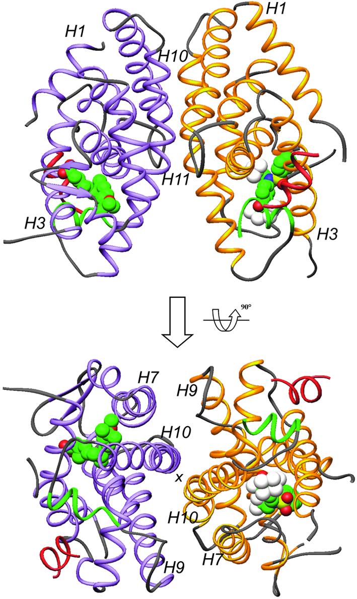
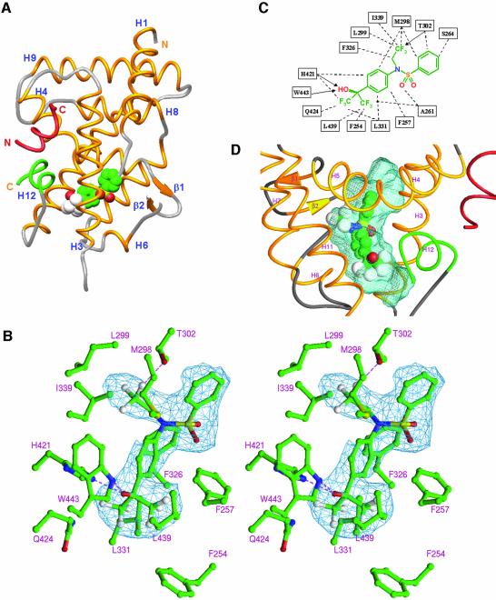
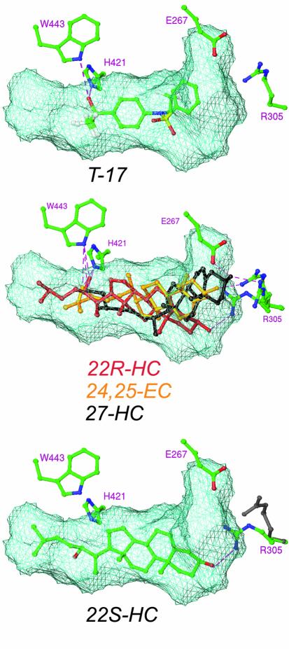
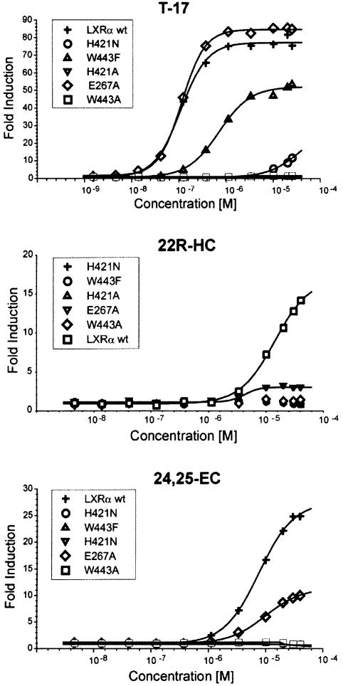
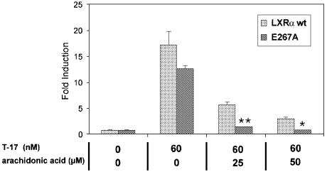
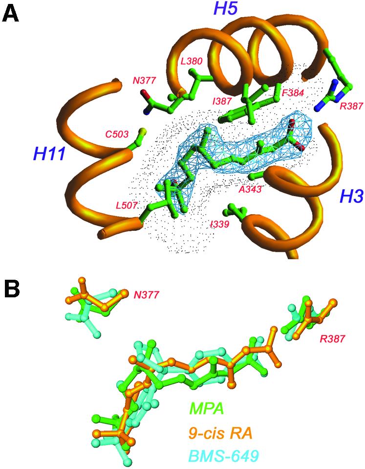
The nuclear receptor heterodimers of liver X receptor (LXR) and retinoid X receptor (RXR) are key transcriptional regulators of genes involved in lipid homeostasis and inflammation. We report the crystal structure of the ligand-binding domains (LBDs) of LXRalpha and RXRbeta complexed to the synthetic LXR agonist T-0901317 and the RXR agonist methoprene acid (Protein Data Base entry 1UHL). Both LBDs are in agonist conformation with GRIP-1 peptides bound at the coactivator binding sites. T-0901317 occupies the center of the LXR ligand-binding pocket and its hydroxyl head group interacts with H421 and W443, residues identified by mutational analysis as critical for ligand-induced transcriptional activation by T-0901317 and various endogenous oxysterols. The topography of the pocket suggests a common anchoring of these oxysterols via their 22-, 24- or 27-hydroxyl group to H421 and W443. Polyunsaturated fatty acids act as LXR antagonists and an E267A mutation was found to enhance their transcriptional inhibition. The present structure provides a powerful tool for the design of novel modulators that can be used to characterize further the physiological functions of the LXR-RXR heterodimer.


Figures

None
Fig. 1. Overall structure of the LXRα–RXRβ LBD-heterodimer presented in two views separated by 90°. The LXRα LBD is shown in yellow and the RXRβ LBD in purple, except for AF2 helices which are depicted in green. GRIP-1 peptides are colored red. T-17 and MPA are in space-filling representation, with carbon, oxygen, nitrogen, sulfur and fluoride atoms colored in green, red, blue, yellow and white, respectively. Selected secondary elements are annotated with numbers positioned at their N-terminal ends.
None
Fig. 2. Molecular basis of T-17 recognition by LXRα. T-17 bound in the core of the LBD with helix12 (AF2) capping the ligand-binding pocket and the GRIP-1-derived peptide bound at the coactivator-binding site. T-17 is shown in space-filling representation, and the coactivator peptide and AF2 helix are depicted in red and green, respectively. Secondary elements are annotated with numbers positioned at their N-terminal ends. Stereo diagram showing interactions between T-17 and LXRα. The difference electron density map used for modeling of T-17 is depicted in blue and contoured at 2.9 σ. Possible hydrogen bonds are indicated with broken lines (purple). Schematic representation of LXRα/T-17 interactions. Broken lines and arrows represent van der Waals contacts and hydrogen bonds, respectively. Topography of the ligand-binding pocket of LXR. T-17 is centrally positioned in the ligand-binding pocket. The ligand-accessible void is shown in blue.
None
Fig. 3. Docking of oxysterols into the ligand-binding pocket of LXRα. Docking of agonistic oxysterols suggests a common binding of their 22-, 24- or 27-hydroxyl group to H421 and W443 [exemplified by 22(R)-HC 24,25-EC and 27-HC], interactions that cannot be formed by the antagonistic oxysterol 22(S)-HC due to steric hindrance. By allowing conformational flexibility in the side chain of R305, all oxysterols docked with the 3-hydroxyl group hydrogen bonded to this residue. The original position of R305 is shown in gray. Possible hydrogen bonds are indicated with broken lines (purple).
None
Fig. 4. Transcriptional activation of LXRα by T-17, 22(R)-HC and 24,25-EC. CaCo-2/TC7 cells were transiently transfected with a 4× GAL4-RE luciferase reporter and a GAL4-LXR wild-type or mutant LBD fusion construct, and subsequently treated with T-17, 22(R)-HC or 24,25-EC in optimized serial dilutions, as indicated in the figures. Data are shown as fold induction of agonist-induced luciferase activity divided by luciferase activity of vehicle.
None
Fig. 5. Inhibition of T-17-induced transcriptional activation of the LXRα wild-type and E267A mutant by arachidonic acid. CaCo-2/TC7 cells were transiently transfected with a 4× GAL4-RE luciferase reporter and a GAL4-LXR wild-type or mutant LBD fusion construct, and subsequently treated with T-17 and arachidonic acid in the concentrations indicated. Data are shown as fold induction where luciferase activity was divided by reporter activity of pCMXGal4 lacking an insert. For statistical analysis, the E267A mutant was compared with wild-type LXR (Student’s t-test). *p ≤ 0.05; **p ≤ 0.01.
None
Fig. 6. Molecular basis of MPA recognition by RXRβ. In the RXR complex MPA adopts an l-shaped conformation resembling that of 9-cis RA. MPA interacts mainly with residues from helices 3, 5 and 11. The 2FoFc electron density map (blue) is contoured at 1.2 σ. The ligand-accessible void is shown as a dotted surface. Superimposition of three RXR–ligand complexes: MPA (green), 9-cis RA (yellow) and BMS-649 (blue). Differences in pocket size are mainly due to different conformations of N377 and R387.
None
Fig. 7. Comparison of the dimer interaction surfaces of RXRβ in the RXRβ homodimer (left; PDB accession code 1H9U) and in the LXRα–RXRβ heterodimer (right). The central hydrophobic core of the interface is conserved, whereas the polar interactions differ significantly between the two dimers. The area of the dimer interaction surface in the homodimer is 1443 Å2, while in the heterodimer it is 1115 Å2. Green, hydrophobic surface; blue, hydrogen bond donating groups; red, hydrogen bond accepting groups.

References

    1. Abagyan R.A., Totrov,M.M. and Kuznetsov,D.N. (1994) ICM—a new method for protein modeling and design. Applications to docking and structure prediction from the distorted native conformation. J. Comput. Chem., 15, 488–506.
    1. Anderson L.M., Choe,S.E., Yukhananov,R.Y., Hopfner,R.L., Church,G.M., Pratt,R.E. and Dzau,V.J. (2003) Identification of a novel set of genes regulated by a unique liver X receptor-α-mediated transcription mechanism. J. Biol. Chem., 278, 15252–15260. - PubMed
    1. Bourguet W., Ruff,M., Chambon,P., Gronemeyer,H. and Moras,D. (1995) Crystal structure of the ligand-binding domain of the human nuclear receptor RXR-α. Nature, 375, 377–382. - PubMed
    1. Bricogne G. (1993) Direct phase determination by entropy maximization and likelihood ranking: status report and perspectives. Acta Crystallogr. D, 49, 37–60. - PubMed
    1. Brzozowski A.M. et al. (1997) Molecular basis of agonism and antagonism in the oestrogen receptor. Nature, 389, 753–758. - PubMed

Publication types

Associated data