Comparative Study
. 2003 Sep 15;22(18):4846-55.
doi: 10.1093/emboj/cdg451.

VIVID is a flavoprotein and serves as a fungal blue light photoreceptor for photoadaptation

Affiliations
Comparative Study

VIVID is a flavoprotein and serves as a fungal blue light photoreceptor for photoadaptation

Carsten Schwerdtfeger et al. EMBO J. .

Abstract

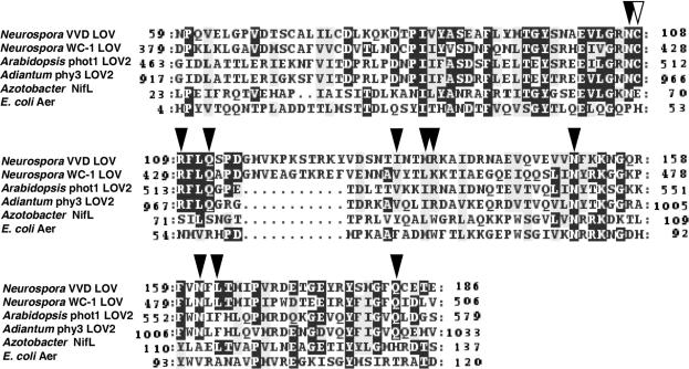
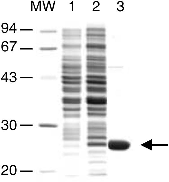
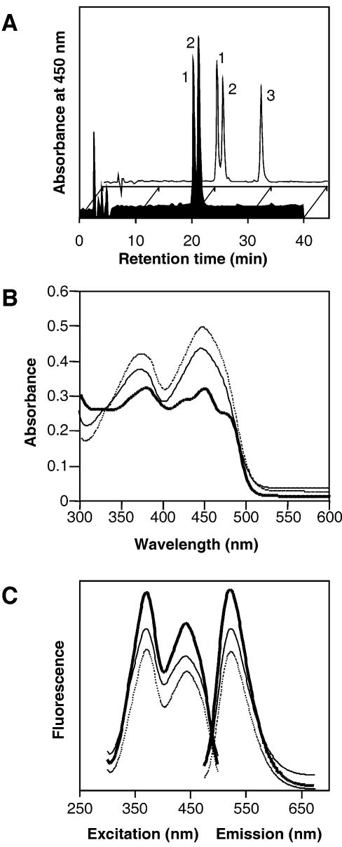
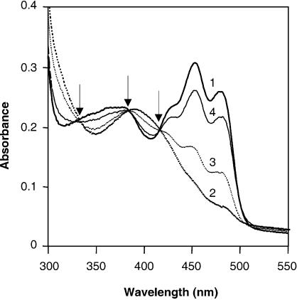
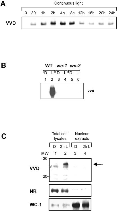
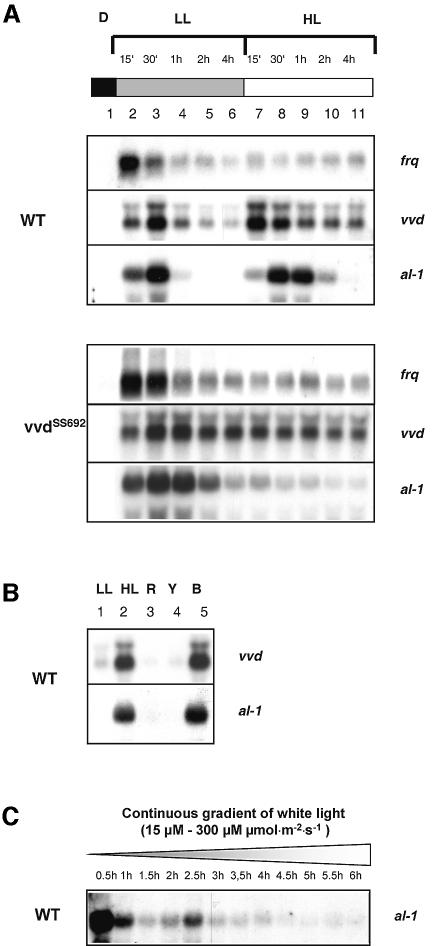
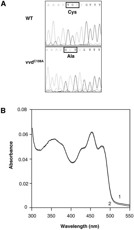
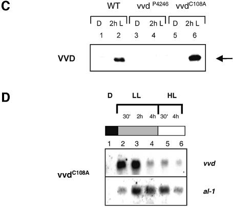
Blue light regulates many physiological and developmental processes in fungi. Most of the blue light responses in the ascomycete Neurospora crassa are dependent on the two blue light regulatory proteins White Collar (WC)-1 and -2. WC-1 has recently been shown to be the first fungal blue light photoreceptor. In the present study, we characterize the Neurospora protein VIVID. VIVID shows a partial sequence similarity with plant blue light photoreceptors. In addition, we found that VIVID non-covalently binds a flavin chromophore. Upon illumination with blue light, VIVID undergoes a photocycle indicative of the formation of a flavin-cysteinyl adduct. VVD is localized in the cytoplasm and is only present after light induction. A loss-of-function vvd mutant was insensitive to increases in light intensities. Furthermore, mutational analysis of the photoactive cysteine indicated that the formation of a flavin-cysteinyl adduct is essential for VIVID functions in vivo. Our results show that VIVID is a second fungal blue light photoreceptor which enables Neurospora to perceive and respond to daily changes in light intensity.


Figures

None
Fig. 1. Sequence alignment of VVD with the WC-1 LOV domain and FAD and FMN-binding domains of plant photoreceptors and redox sensing proteins. Identical residues are shown in black, similar residues are shaded in gray. Flavin-interacting residues as well as the photoactive cysteine of phy3 are marked by black and open arrowheads, respectively (DDBJ/EMBL/GenBank accession Nos: VVD, AAK08514; WC-1, Q01371; Arabidopsis phot1, AAC01753; Adiantum phy3, T30891; Azotobacter NifL, P30663, E.coli Aer, P50466).
None
Fig. 2. Expression in E.coli and purification of VVD. Proteins were separated on a 15% SDS–PAGE. Non-induced (lane 1) and induced E.coli cells expressing VVD as His-tagged fusion protein (lane 2), VVD protein after purification by affinity chromatography. The arrow on the right shows the expressed and purified VVD protein.
None
Fig. 3. VVD non-covalently binds FAD and FMN. (A) HPLC analysis of the VVD chromophores (black profile) and an aqueous mixtures (transparent profile) of FAD (peak 1), FMN (peak 2) and riboflavin (peak 3). (B) Absorbance spectra of the purified VVD protein before (solid line) and after SDS denaturation (dashed line) and of an aqueous FAD solution (dotted line). (C) Fluorescence excitation (left, 525 nm emission) and emission spectra (right, 370 nm excitation) of the VVD chromophores (solid line), FAD (dashed line) and FMN (dotted line).
None
Fig. 4. The VVD protein exhibits a reversible light-induced absorbance change. Absorbance spectra were recorded from purified VVD protein after incubation in the dark (spectrum 1), after a 30 s light induction (100 µmol photons/m2/s for 30 s, provided by universal white lamps Osram L65W/25S; spectrum 2) and after subsequent incubations in the dark for 10 min (spectrum 3), 2 h (spectrum 4) and 5 h (spectrum 1) at 4°C. The three isosbestic points are indicated by arrows.
None
Fig. 5. VVD is expressed only in the light and localized in the cytoplasm. (A) Expression of the VVD protein in N.crassa mycelia after growth in the dark and after different times of continuous light induction. (B) Northern blot analysis of the vvd gene expression in the Neurospora wild type and in wc-1 and wc-2 mutant background. Cultures were harvested either after growth in the dark (D) or after a low light induction for 30 min (L). (C) Western blot analysis of VVD, nitrate reductase (NR) and WC-1 in nuclear extracts and total cell lysates of Neurospora wild type. Total cell lysates (lanes 1 and 2) and nuclear extracts (lanes 3 and 4) were prepared after cultivation in the dark and after an additional incubation for 2 h in the light. The arrow on the right shows the VVD protein. A non-specific cross reaction of the VVD antiserum is indicated by an asterisk.
None
Fig. 6. The vvd mutant is insensitive to high light during growth under continuous illumination. (A) Cultures of Neurospora wild type and mutant vvdSS692 were grown in the dark and illuminated with low light (LL) for 4 h and subsequently with high light (HL) for an additional 4 h. Mycelia were harvested at the indicated time points. (B) Dark-grown wild-type mycelia were subjected to a 4 h low light induction (LL) and subsequently illuminated with either white (HL), red (R), yellow (Y) or blue light (B) for 1 h. (C) Cultures of Neurospora wild type were grown in the dark and illuminated using a continuous light gradient of white light. An initial light intensity of 15 µmol/m2/s was applied and light was gradually increased up to 300 µmol/m2/s after 6 h. Mycelia were harvested at the indicated time points. For northern blot analysis the carotenoid biosynthesis gene al-1, the circadian clock gene frq and vvd were used as specific probes.
None
Fig. 7. Cysteine108 is essential for the light-induced absorbance change in vitro and for the in vivo functions of VVD in photoadaptation. (A) Sequencing results of the vvd wild-type gene and of the vvdC108A gene (genomic DNA construct) following site-directed mutagenesis. (B) Absorbance spectra from purified VVD protein expressed in E.coli after incubation in the dark (spectrum 1) and after a 30 s light induction (100 µmol photons/m2/s, provided by universal white lamps Osram L65W/25S; spectrum 2). (C) Western blot analysis of VVD in Neurospora wild-type, vvdP4246 mutant and in a vvdp4246 mutant after transformation with the vvdC108A gene. Total cell lysates were prepared after cultivation in the dark (lanes 1, 3 and 5) and after an additional incubation for 2 h in the light (lanes 2, 4 and 6). The arrow on the right shows the VVD protein. (D) The vvdC108A mutant is defective in both photoadaptation and in the up-regulation of vvd and al-1 in response to higher light. Cultures of the vvdC108A strain were grown in the dark and illuminated with low light (LL) for 4 h and subsequently with high light (HL) for an additional 4 h. Mycelia were harvested at the indicated time points. For northern blot analysis the carotenoid biosynthesis gene al-1 and vvd were used as specific probes.

References

    1. Baima S., Macino,G. and Morelli,G. (1991) Photoregulation of the albino-3 gene in Neurospora crassa. J. Photochem. Photobiol. B, 11, 107–115. - PubMed
    1. Ballario P., Vittorioso,P., Magrelli,A., Talora,C., Cabibbo,A. and Macino,G. (1996) White collar-1, a central regulator of blue light responses in Neurospora, is a zinc finger protein. EMBO J., 15, 1650–1657. - PMC - PubMed
    1. Batschauer A. (1998) Photoreceptors of higher plants. Planta, 206, 479–492. - PubMed
    1. Baum J.A. and Giles,N.H. (1985) Genetic control of chromatin structure 5′ to the qa-x and qa-2 genes of Neurospora. J. Mol. Biol., 182, 79–89. - PubMed
    1. Briggs W.R. and Christie,J.M. (2002) Phototropins 1 and 2: versatile plant blue-light receptors. Trends Plant Sci., 7, 204–210. - PubMed

Publication types