Rational design of drugs that induce human immunodeficiency virus replication
- PMID: 12970407
- PMCID: PMC228450
- DOI: 10.1128/jvi.77.19.10227-10236.2003
Rational design of drugs that induce human immunodeficiency virus replication
Abstract
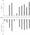
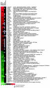
Drugs that induce human immunodeficiency virus type 1 (HIV-1) replication could be used in combination with highly active antiretroviral therapy (HAART) to reduce the size of the latent reservoir that is in part responsible for viral persistence. Protein kinase C (PKC) is a logical target for such drugs because it activates HIV-1 transcription through multiple mechanisms. Here we show that HIV-1 gene expression can be induced by potent synthetic analogues of the lipid second messenger diacylglycerol (DAG) synthesized on a five-member ring platform that reduces the entropy of binding relative to that of the more flexible DAG template. By varying the alkyl side chains of these synthetic DAG lactones, it was possible to maximize their potency and ability to render latently infected T cells sensitive to killing by an anti-HIV-1 immunotoxin while minimizing the side effects of CD4 and CXCR4 downregulation and tumor necrosis factor alpha upregulation. The two lead compounds, LMC03 and LMC07, regulated a series of PKC-sensitive genes involved in T-cell activation and induced viral gene expression in peripheral blood mononuclear cells from HIV-1-infected individuals. These studies demonstrate the potential for the rational design of agents that, in conjunction with HAART and HIV-specific toxins, can be used to decrease or eliminate the pool of latently infected reservoirs by forcing viral expression.
Figures


Similar articles
-
Dual role of novel ingenol derivatives from Euphorbia tirucalli in HIV replication: inhibition of de novo infection and activation of viral LTR.PLoS One. 2014 May 14;9(5):e97257. doi: 10.1371/journal.pone.0097257. eCollection 2014. PLoS One. 2014. PMID: 24827152 Free PMC article.
-
Induction of HIV-1 replication in latently infected CD4+ T cells using a combination of cytokines.J Exp Med. 1998 Jul 6;188(1):83-91. doi: 10.1084/jem.188.1.83. J Exp Med. 1998. PMID: 9653086 Free PMC article.
-
Drug-loaded red blood cell-mediated clearance of HIV-1 macrophage reservoir by selective inhibition of STAT1 expression.J Leukoc Biol. 2003 Nov;74(5):764-71. doi: 10.1189/jlb.0403156. Epub 2003 Aug 21. J Leukoc Biol. 2003. PMID: 12960277
-
Administration of HDAC inhibitors to reactivate HIV-1 expression in latent cellular reservoirs: implications for the development of therapeutic strategies.Biochem Pharmacol. 2004 Sep 15;68(6):1231-8. doi: 10.1016/j.bcp.2004.05.040. Biochem Pharmacol. 2004. PMID: 15313421 Review.
-
The challenge of viral reservoirs in HIV-1 infection.Annu Rev Med. 2002;53:557-93. doi: 10.1146/annurev.med.53.082901.104024. Annu Rev Med. 2002. PMID: 11818490 Review.
Cited by
-
Protein Kinase C: One Pathway towards the Eradication of Latent HIV-1 Reservoirs.Adv Virol. 2012;2012:805347. doi: 10.1155/2012/805347. Epub 2012 Mar 5. Adv Virol. 2012. PMID: 22500169 Free PMC article.
-
Human immunodeficiency virus reactivation by phorbol esters or T-cell receptor ligation requires both PKCalpha and PKCtheta.J Virol. 2005 Aug;79(15):9821-30. doi: 10.1128/JVI.79.15.9821-9830.2005. J Virol. 2005. PMID: 16014943 Free PMC article.
-
Eradication of Human Immunodeficiency Virus Type-1 (HIV-1)-Infected Cells.Pharmaceutics. 2019 Jun 1;11(6):255. doi: 10.3390/pharmaceutics11060255. Pharmaceutics. 2019. PMID: 31159417 Free PMC article. Review.
-
Molecular control of HIV-1 postintegration latency: implications for the development of new therapeutic strategies.Retrovirology. 2009 Dec 4;6:111. doi: 10.1186/1742-4690-6-111. Retrovirology. 2009. PMID: 19961595 Free PMC article. Review.
-
The ingenol-based protein kinase C agonist GSK445A is a potent inducer of HIV and SIV RNA transcription.PLoS Pathog. 2022 Jan 18;18(1):e1010245. doi: 10.1371/journal.ppat.1010245. eCollection 2022 Jan. PLoS Pathog. 2022. PMID: 35041707 Free PMC article.
References
-
- Altman, A., and M. Villalba. 2002. Protein kinase C-theta (PKCθ): a key enzyme in T cell life and death. J. Biochem. (Tokyo) 132:841-846. - PubMed
-
- Blankson, J. N., D. Persaud, and R. F. Siliciano. 2002. The challenge of viral reservoirs in HIV-1 infection. Annu. Rev. Med. 53:557-593. - PubMed
-
- Blumberg, P. M., P. Acs, D. K. Bhattacharyya, and P. S. Lorenzo. 2000. Inhibitors of protein kinase C and related receptors for the lipophilic second messenger sn-1,2-diacylglycerol, p. 347-364. In J. S. Gutkind (ed.), Signal transduction and cell cycle inhibitors. Humana Press, Totowa, N.J.
-
- Bocklandt, S., P. M. Blumberg, and D. H. Hamer. 2003. Activation of latent HIV-1 expression by the potent anti-tumor promoter 12-deoxyphorbol 13-phenylacetate. Antiviral Res. 59:89-98. - PubMed
-
- Brose, N., and C. Rosenmund. 2002. Move over protein kinase C, you've got company: alternative cellular effectors of diacylglycerol and phorbol esters. J. Cell Sci. 115:4399-4411. - PubMed
MeSH terms
Substances
LinkOut - more resources
Full Text Sources
Other Literature Sources
Research Materials

