. 2003 Oct;77(19):10327-38.
doi: 10.1128/jvi.77.19.10327-10338.2003.

Antigenic subclasses of polytropic murine leukemia virus (MLV) isolates reflect three distinct groups of endogenous polytropic MLV-related sequences in NFS/N mice

Affiliations

Antigenic subclasses of polytropic murine leukemia virus (MLV) isolates reflect three distinct groups of endogenous polytropic MLV-related sequences in NFS/N mice

Leonard H Evans et al. J Virol. 2003 Oct.

Abstract

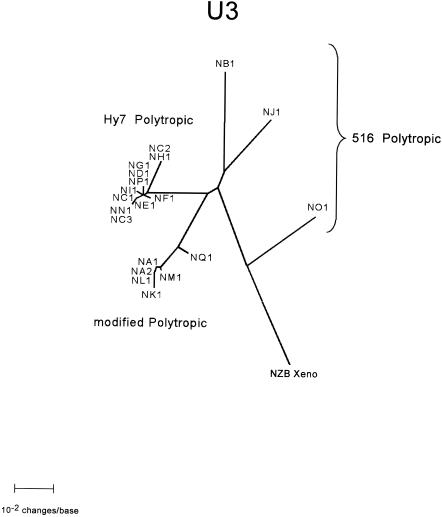
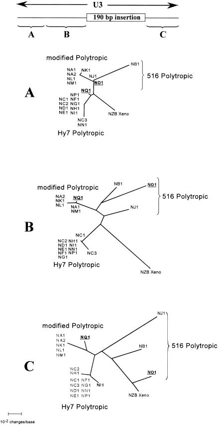
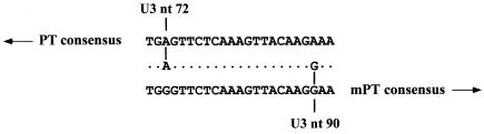
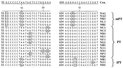
Polytropic murine leukemia viruses (MLVs) are generated by recombination of ecotropic MLVs with members of a family of endogenous proviruses in mice. Previous studies have indicated that polytropic MLV isolates comprise two mutually exclusive antigenic subclasses, each of which is reactive with one of two monoclonal antibodies termed MAb 516 and Hy 7. A major determinant of the epitopes distinguishing the subclasses mapped to a single amino acid difference in the SU protein. Furthermore, distinctly different populations of the polytropic MLV subclasses are generated upon inoculation of different ecotropic MLVs. Here we have characterized the majority of endogenous polytropic MLV-related proviruses of NFS/N mice. Most of the proviruses contain intact sequences encoding the receptor-binding region of the SU protein and could be distinguished by sequence heterogeneity within that region. We found that the endogenous proviruses comprise two major groups that encode the major determinant for Hy 7 or MAb 516 reactivity. The Hy 7-reactive proviruses correspond to previously identified polytropic proviruses, while the 516-reactive proviruses comprise the modified polytropic proviruses as well as a third group of polytropic MLV-related proviruses that exhibit distinct structural features. Phylogenetic analyses indicate that the latter proviruses reflect features of phylogenetic intermediates linking xenotropic MLVs to the polytropic and modified polytropic proviruses. These studies elucidate the relationships of the antigenic subclasses of polytropic MLVs to their endogenous counterparts, identify a new group of endogenous proviruses, and identify distinguishing characteristics of the proviruses that should facilitate a more precise description of their expression in mice and their participation in recombination to generate recombinant viruses.


Figures

FIG. 1.
FIG. 1.
Endogenous PT and mPT sequences of NFS/N mice. DNA from a prenatal NFS/N mouse (6 μg) was digested with BamHI and analyzed by electrophoresis on a 0.8% agarose gel. The gel was then blotted and hybridized to the DIG-labeled polytropic MLV env probe to detect homologous sequences. Nearly all polytropic MLVs have a BamHI site derived from the endogenous sequence near the 3′ end of the pol gene and no additional BamHI sites in the env gene or LTR. Thus, the bands correspond to fragments between the BamHI site in the endogenous pol gene and the nearest BamHI site after the 3′ end of the provirus.
FIG. 2.
FIG. 2.
Amplified region of endogenous proviruses encompassing the receptor-binding region of the SU protein. The diagram at the top represents an intact endogenous polytropic provirus (open rectangles) and the flanking sequences (solid rectangles). Approximate locations of the LTR, the gag, pol, and env genes, and sequences encoding the integrase (IN), the surface glycoprotein (SU), and the transmembrane protein (TM) are indicated. The expanded region at the bottom represents the PCR product of approximately 900 bp that was used for initial sequence comparisons among the endogenous proviruses. The product contains pol gene sequences encoding a portion of the IN protein, the env gene leader sequence (L), and the receptor-binding region (RBR) of the SU protein. The positions of the BamHI and EcoRI sites that define the polytropic MLV probe are also indicated.
FIG. 3.
FIG. 3.
Sequence properties of the ∼900-bp PCR products of lambda clones containing nondeleted receptor-binding regions. Diagrams depict the amplified region shown in Fig. 2. Vertical arrows indicate the positions at which additional sequences (given below) were located within the SU-encoding sequences. The approximate locations and natures of mutations rendering five of the proviruses defective are indicated. These include a mutation at nucleotide 251 in ND1 resulting in a tRNAIle as the initiator codon of env and a termination codon in pol, a frameshift mutation at nucleotide 785 in ND1, a premature termination codon in pol at nucleotide 252 in NF1 and NF2, a premature termination codon at nucleotide 385 in env in NF1 and NF2, and an identical mutation at nucleotide 385 in NH1 and NH2.
FIG. 4.
FIG. 4.
Phylogenetic relationships of NFS/N endogenous proviruses. An unrooted phylogram generated by NJ phylogenetic analysis of a sequence encompassing a 3′ pol gene sequence and approximately 850 bases of the env gene is presented. This sequence corresponds to the ∼900-bp PCR product initially sequenced (Fig. 3) plus an additional ∼200 bp obtained by sequence analyses of the PCR products flanking the 27-bp deletion (Fig. 4). The analyses included the 19 proviruses distinguishable by sequence in this region as well as the analogous sequence from the xenotropic MLV NZB-9-1 (XENO). Branch lengths are drawn to scale. The identity of each clone is indicated at the branch termini. The very closely related clades containing the Hy 7 PT proviruses and the mPT proviruses are expanded (within dashed circles) in order to present the branch termini of these clades clearly. Group 516 PT viruses (NB1, NO1, and NJ1) are indicated by a brace. The robustness of the map was assessed by bootstrap analysis repeated 1,000 times. Bootstrap values are shown as circled numbers on the major branches.
FIG. 5.
FIG. 5.
U3 regions of endogenous polytropic MLV-related proviruses. Schematic diagrams are shown depicting distinguishing structural features of the U3 regions of polytropic MLV-related proviruses described by Tomanaga and Coffin (52, 53). The diagrams are adapted from similar diagrams presented in their reports. U3 types P-I and P-II correspond to the U3 regions found in PT and mPT proviruses, respectively, identified in inbred mouse strains (46). Types P-III, P-IV, and P-V correspond to U3 regions of proviruses identified in wild mouse subspecies and may reflect features that are ancestral to the P-I and P-II U3 regions. The regions exhibiting the distinguishing features, numbered 1 through 6, are indicated by boxes and include direct repeats (indicated by a repeated number with an asterisk), unique sequences, and deletions exhibited by the proviruses. In addition, the 190-bp insertion is indicated. V's extending down from the horizontal axes of the diagrams indicate regions that are absent from the various U3 types and include deleted sequences identified in P-I and P-II. The diagrams do not indicate the precise locations or sizes of the various structural features.
FIG. 6.
FIG. 6.
Structural features of U3 regions of endogenous polytropic MLV-related proviruses identified in NFS/N mice. Structural features of the U3 regions of Hy 7 PT, mPT, and 516 PT proviruses identified in this report are presented. The features are indicated as described in the legend to Fig. 5. The structures of the U3 regions of two distinguishable xenotropic MLVs are also included in order to facilitate comparison to the structure of the 516 PT provirus NO1, which lacks the 190-bp insertion characteristic of all other polytropic MLV-related proviruses.
FIG. 7.
FIG. 7.
Phylogenetic relationships of the U3 regions of NFS/N endogenous proviruses. An unrooted phylogram generated by NJ phylogenetic analysis of alignments of the U3 regions of the proviruses is presented. U3 sequences extending from the 5′ end of the U3 up to, but not including, the TATA box were analyzed. The analyses included all 20 distinct proviruses identified in our analyses as well as the analogous sequence from the xenotropic MLV NZB-9-1. Branch lengths are drawn to scale.
FIG. 8.
FIG. 8.
Phylogenetic analysis of 5′ and 3′ segments of the U3 regions of NFS/N endogenous proviruses. Phylogenetic analyses were performed on alignments of two 5′ segments and a 3′ segment of the U3 region of the proviruses to assess shifts of putative recombinant provirus from one clade to another. The sequences included in the alignment are indicated by the braces in the diagram at the top of the figure, which is drawn to scale. Unrooted phylograms generated by NJ phylogenetic analysis of alignment of the segments of the U3 regions of the proviruses are presented. The analyses included all 20 distinct proviruses identified in our analyses as well as the analogous sequence from the xenotropic MLV NZB-9-1. The clones that are putative recombinant proviruses, NQ1 and NO1, are underlined and boldfaced for ease of identification. Branch lengths are drawn to scale.
FIG. 9.
FIG. 9.
Recombination region in a PT env-mPT U3 endogenous recombinant provirus. An alignment of the consensus sequence of PT proviruses (top), the corresponding sequence of the putative recombinant provirus NQ1 (center), and the consensus sequence of mPT proviruses (bottom) is presented. A 17-bp sequence in the alignment is flanked by a PT provirus-specific base on the 5′ side and an mPT provirus-specific base on the 3′ side. Positions in the NQ1 sequence that are identical in both the PT and mPT consensus sequences are indicated by dots, and positions that are specific to the PT or mPT consensus sequence and shared by NQ1 are indicated by a vertical line connecting the base of NQ1 to the base of the matching consensus sequence. NQ1 sequences 5′ of the 17 bp exhibit nearly complete agreement with the PT consensus sequence, whereas sequences 3′ of the 17 bp exhibit nearly complete agreement with the mPT consensus sequence. Arrows directed toward the 5′ (PT) or 3′ (mPT) sequences are intended to convey this observation.
FIG. 10.
FIG. 10.
Single-nucleotide polymorphisms characteristic of PT, mPT, and iPT proviruses and of specific proviruses. Two windows in the alignment of 19 distinct polytropic MLV-related proviruses with their consensus sequence (Con.) are shown and illustrate examples of nucleotide polymorphisms. Numbers on the left of each window indicate the nucleotide position starting from the beginning of the initiation codon for env. Nucleotides differing from the consensus sequence are boxed. A single-nucleotide polymorphism specific for PT proviruses is located at position 60. Single-nucleotide polymorphisms characteristic of mPT proviruses are located at positions 53 and 637, and a single-nucleotide polymorphism characteristic of iPT proviruses is located at position 638 in NB1. Single-nucleotide polymorphisms specific for individual proviruses are found at positions 70 (NN1) and 646 (NJ1).

Similar articles

Cited by

References

    1. Adachi, A., K. Sakai, N. Kitamura, S. Nakanishi, O. Niwa, M. Matsuyama, and A. Ishimoto. 1984. Characterization of the env gene and long terminal repeat of molecularly cloned Friend mink cell focus-inducing virus DNA. J. Virol. 50:813-821. - PMC - PubMed
    1. Bi, W., and P. J. Stambrook. 1997. CCR: a rapid and simple approach for mutation detection. Nucleic Acids Res. 25:2949-2951. - PMC - PubMed
    1. Bi, W., and P. J. Stambrook. 1998. Detection of known mutation by proof-reading PCR. Nucleic Acids Res. 26:3073-3075. - PMC - PubMed
    1. Bosselman, R. A., F. van Straaten, C. Van Beveren, I. M. Verma, and M. Vogt. 1982. Analysis of the env gene of a molecularly cloned and biologically active Moloney mink cell focus-forming proviral DNA. J. Virol. 44:19-31. - PMC - PubMed
    1. Britt, W. J., B. Chesebro, and J. L. Portis. 1984. Identification of a unique erythroleukemia-associated retroviral gp70 expressed during early stages of normal erythroid differentiation. J. Exp. Med. 159:1591-1603. - PMC - PubMed

Substances

LinkOut - more resources