. 2003 Oct;77(19):10394-403.
doi: 10.1128/jvi.77.19.10394-10403.2003.

An alphavirus replicon particle chimera derived from venezuelan equine encephalitis and sindbis viruses is a potent gene-based vaccine delivery vector

Affiliations

An alphavirus replicon particle chimera derived from venezuelan equine encephalitis and sindbis viruses is a potent gene-based vaccine delivery vector

Silvia Perri et al. J Virol. 2003 Oct.

Abstract

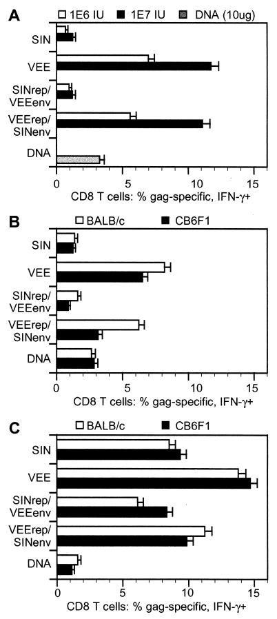
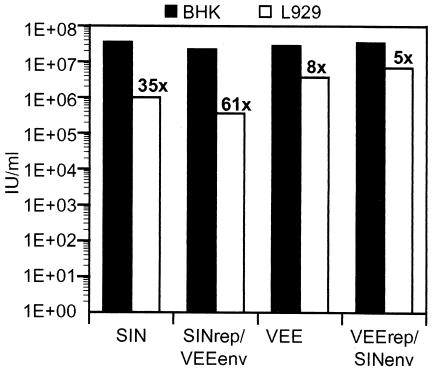
Alphavirus replicon particle-based vaccine vectors derived from Sindbis virus (SIN), Semliki Forest virus, and Venezuelan equine encephalitis virus (VEE) have been shown to induce robust antigen-specific cellular, humoral, and mucosal immune responses in many animal models of infectious disease and cancer. However, since little is known about the relative potencies among these different vectors, we compared the immunogenicity of replicon particle vectors derived from two very different parental alphaviruses, VEE and SIN, expressing a human immunodeficiency virus type 1 p55(Gag) antigen. Moreover, to explore the potential benefits of combining elements from different alphaviruses, we generated replicon particle chimeras of SIN and VEE. Two distinct strategies were used to produce particles with VEE-p55(gag) replicon RNA packaged within SIN envelope glycoproteins and SIN-p55(gag) replicon RNA within VEE envelope glycoproteins. Each replicon particle configuration induced Gag-specific CD8(+) T-cell responses in murine models when administered alone or after priming with DNA. However, Gag-specific responses varied dramatically, with the strongest responses to this particular antigen correlating with the VEE replicon RNA, irrespective of the source of envelope glycoproteins. Comparing the replicons with respect to heterologous gene expression levels and sensitivity to alpha/beta interferon in cultured cells indicated that each might contribute to potency differences. This work shows that combining desirable elements from VEE and SIN into a replicon particle chimera may be a valuable approach toward the goal of developing vaccine vectors with optimal in vivo potency, ease of production, and safety.


Figures

FIG. 1.
FIG. 1.
SINrep/VEEenv replicon particle chimeras. (A) Schematic illustration of the tripartite RNAs used in generating SINrep/VEEenv particle chimeras: SIN replicon expressing a gene-of-interest (G.O.I.), such as GFP or HIV-1 p55gag, and SIN-derived defective helpers expressing a SIN/VEE hybrid capsid protein and VEE envelope glycoproteins, respectively. SIN-derived sequences (white boxes), VEE-derived sequences (shaded boxes), and the subgenomic promoter (arrows) are indicated. (B) Alignment of capsid protein sequences in the RNA-binding domain. Sequences shown are from two SIN strains, DC+ (15) and HR (51), and two VEE strains, TRD (22) and 6119 (58). Identical residues (bold letters on shaded background), similar residues (residues shown boxed on white background), and the point where the amino terminus of the SIN capsid was fused to the carboxy terminus of VEE capsid (arrow) are indicated.
FIG. 2.
FIG. 2.
VEErep/SINenv replicon particle chimeras. (A) Construction of the VCR-Chim2.1 replicon. The packaging signal (PS) and 3′UTR (3′) from the SIN replicon were used to replace sequences in nsP3 and at the 3′ end of the VEE replicon, respectively. (B) VEE-derived defective helpers expressing SIN capsid and envelope glycoproteins. SIN-derived sequences (white boxes), VEE-derived sequences (shaded boxes), and the subgenomic promoter (arrows) are indicated.
FIG. 3.
FIG. 3.
RNA replication and packaging of alphavirus replicon particle chimeras. (A) Minus-strand RNA detection in cultured cells by semiquantitative RT-PCR. BHK-21 cells were electroporated with in vitro-transcribed replicon RNAs and harvested at different times, as indicated to the left of each gel panel. cDNA was synthesized from total RNA with an oligonucleotide primer complementary to minus-strand RNA, and a GFP fragment was amplified by PCR. PCR amplification mixtures were divided into six aliquots, and one aliquot per sample was removed after 0, 10, 15, 20, 25, and 30 amplification cycles as indicated above the gel lanes. (B) In vitro-transcribed replicon and defective helper RNAs were coelectroporated into BHK-21 cells to produce stocks of replicon particles. The titers of the replicon particle preparations were then determined by infecting naive BHK-21 cells with serial dilutions of the different particle preparations expressing GFP reporter and counting GFP-positive cells by flow cytometry at 16 h postinfection. The results are the averages of titers from three independent replicon particle preparations. 1E+0, 100; 2E+7, 2 × 10⁷; 4E+7, 4 × 10⁷.
FIG. 4.
FIG. 4.
Analysis of heterologous gene expression in cultured cells. (A) BHK or L929 cells were infected with parental and chimeric replicon particles encoding GFP at an MOI of 0.5 to 1. At the indicated postinfection times, cells were harvested and analyzed by flow cytometry to determine the mean fluorescence intensity (MFI) of the GFP-positive cell population. (B) BHK cells were infected with replicon particles expressing HIV p55gag at an MOI of 0.5 to 1. At the indicated times, supernatants and cell lysates were harvested. Levels of p55gag were determined with the Coulter HIV-1 p24 antigen assay. The percentage of cells infected with the particles was determined with intracellular staining at the 24-h time point.
FIG. 5.
FIG. 5.
Induction of HIV p55Gag-specific CD8+ T cells in mice by alphavirus replicon particles. (A) Groups of four BALB/c mice were immunized twice with either 106 (1E6) or 107 (1E7) IU of HIV p55gag-encoding replicon particles, or (B) groups of four BALB/c mice and four CB6F1 mice were immunized twice with 106 IU of HIV p55gag-encoding replicon particles. As a control, a DNA plasmid expressing HIV p55Gag was used. Two weeks after the final immunization, pooled spleen cell suspensions were prepared, and samples were analyzed for IFN-γ-secreting CD8+ T cells following stimulation with Gag peptides. (C) Groups of four BALB/c or CB6F1 mice primed with 10 μg of DNA plasmid expressing HIV p55gag were given a single booster immunization of 106 IU of replicon particles. Five days later, pooled spleen cell suspensions were prepared, and samples were analyzed for IFN-γ-secreting CD8+ T cells following stimulation with Gag peptides. The upper limit of the 95% confidence interval for the measured frequency is indicated by the error bar. Each panel represents the results of one of the two experiments done. In each experiment, the nonimmunized control group gave no background responses.
FIG. 6.
FIG. 6.
Replicon particle infection of different cell lines. Cells were infected with serial dilutions of the different replicon particle preparations expressing a GFP reporter. At 16 h postinfection, GFP-positive cells were counted by flow cytometry. Infectivity of the murine fibroblast L929 cell line was compared to the infectivity of BHK-21 cells as a ratio of the titer obtained in the BHK-21 to the titer obtained in the L929 cells. 1E+0, 100; 1E+01, 101; 1E+02, 102.
FIG. 7.
FIG. 7.
IFN-α/β sensitivity assay. Monolayers of cells were grown for 24 h in the presence of twofold dilutions of mouse-derived IFN-α/β, with concentrations ranging from 0 to 500 U/ml. Cells were then infected at an MOI of 0.5 to 1 (based on titers obtained on L929 cells). At 24 h postinfection, GFP-positive cells were counted by flow cytometry. Fifty percent inhibition (dotted lines) is indicated and represents the amount of IFN-α/β necessary to halve the number of cells expressing GFP [GFP(+)] compared to the samples that were not treated with IFN-α/β [(−)IFNα/β].

Similar articles

Cited by

References

    1. Borrow, P., H. Lewicki, B. H. Hahn, G. M. Shaw, and M. B. Oldstone. 1994. Virus-specific CD8+ cytotoxic T-lymphocyte activity associated with control of viremia in primary human immunodeficiency virus type 1 infection. J. Virol. 68:6103-6110. - PMC - PubMed
    1. Bredenbeek, P. J., I. Frolov, C. M. Rice, and S. Schlesinger. 1993. Sindbis virus expression vectors: packaging of RNA replicons by using defective helper RNAs. J. Virol. 67:6439-6446. - PMC - PubMed
    1. Byrnes, A. P., and D. E. Griffin. 2000. Large-plaque mutants of Sindbis virus show reduced binding to heparan sulfate, heightened viremia, and slower clearance from the circulation. J. Virol. 74:644-651. - PMC - PubMed
    1. Chastain, M., A. J. Simon, K. A. Soper, D. J. Holder, D. L. Montgomery, S. L. Sagar, D. R. Casimiro, and C. R. Middaugh. 2001. Antigen levels and antibody titers after DNA vaccination. J. Pharm. Sci. 90:474-484. - PubMed
    1. Choi, H. K., L. Tong, W. Minor, P. Dumas, U. Boege, M. G. Rossmann, and G. Wengler. 1991. Structure of Sindbis virus core protein reveals a chymotrypsin-like serine proteinase and the organization of the virion. Nature 354:37-43. - PubMed

Publication types

MeSH terms