. 2003 Oct;77(19):10515-27.
doi: 10.1128/jvi.77.19.10515-10527.2003.

Characterization of the expression, intracellular localization, and replication complex association of the putative mouse hepatitis virus RNA-dependent RNA polymerase

Affiliations

Characterization of the expression, intracellular localization, and replication complex association of the putative mouse hepatitis virus RNA-dependent RNA polymerase

Sarah M Brockway et al. J Virol. 2003 Oct.

Abstract

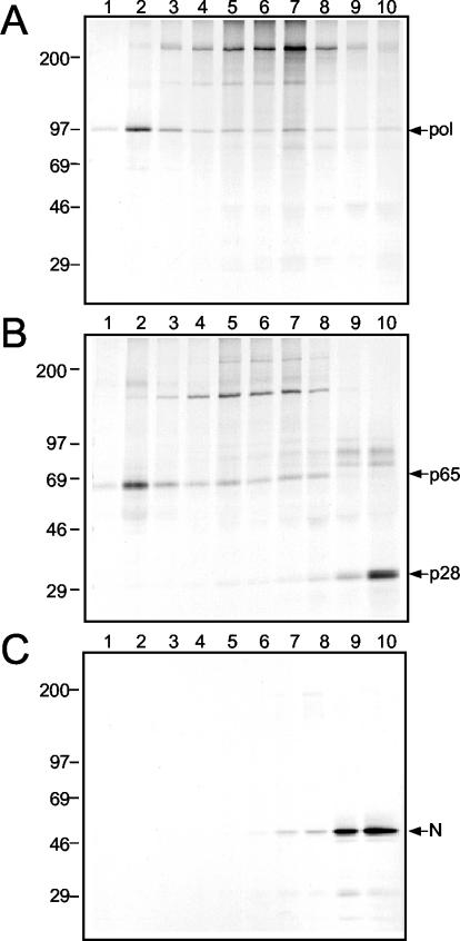
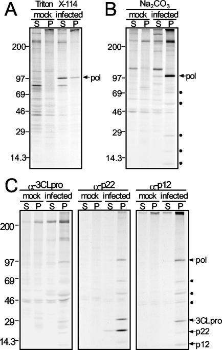
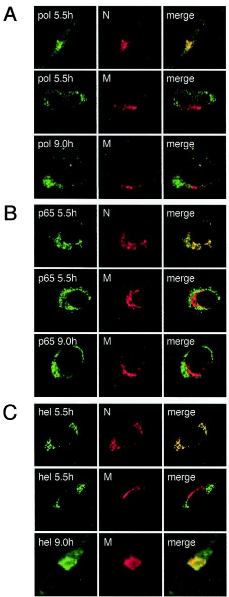
Mouse hepatitis virus (MHV) RNA synthesis is mediated by a viral RNA-dependent RNA polymerase (RdRp) on membrane-bound replication complexes in the host cell cytoplasm. However, it is not known how the putative MHV RdRp (Pol) is targeted to and retained on cellular membranes. In this report, we show that a 100-kDa protein was stably detected by an anti-Pol antiserum as a mature product throughout the virus life cycle. Gradient fractionation and biochemical extraction experiments demonstrated that Pol was not an integral membrane protein but was tightly associated with membranes and coimmunoprecipitated with the replicase proteins 3CLpro, p22, and p12. By immunofluorescence confocal microscopy, Pol colocalized with viral proteins at replication complexes, distinct from sites of virion assembly, over the entire course of infection. To determine if Pol associated with cellular membranes in the absence of other viral factors, the pol domain of gene 1 was cloned and expressed in cells as a fusion with green fluorescent protein, termed Gpol. In Gpol-expressing cells that were infected with MHV, but not in mock-infected cells, Gpol relocalized from a diffuse distribution in the cytoplasm to punctate foci that colocalized with markers for replication complexes. Expression of Gpol deletion mutants established that the conserved enzymatic domains of Pol were dispensable for replication complex association, but a 38-amino-acid domain in the RdRp unique region of Pol was required. This study demonstrates that viral or virus-induced factors are necessary for Pol to associate with membranes of replication complexes, and it identifies a defined region of Pol that may mediate its interactions with those factors.


Figures

FIG. 1.
FIG. 1.
MHV genome organization and Pol expression and stability. (A) MHV genome organization. Schematic shows the organization of the gene 1 polyprotein, as well as the location of genes 6 and 7, encoding structural proteins M and N, respectively. Confirmed or predicted mature gene 1 proteins are shown as boxes. Shaded boxes indicate replicase proteins of interest in the present study: the amino-terminal cleavage products (p28 and p65), the 3C-like proteinase (3CLpro), two carboxy-terminal ORF1a proteins (p22 and p12), the putative RdRp domain (Pol), and the putative helicase (Hel). The region of gene 1 used to generate the α-Pol rabbit polyclonal antiserum VU145 is indicated. (B and C) Pulse-label and pulse-chase translation of Pol. Cells were radiolabeled with [35S]Met-Cys at 5.5 h p.i., and either samples were withdrawn at the indicated times for pulse-labeling or, after 30 min of radiolabeling, cells were incubated in media containing excess unlabeled Met-Cys for 15 min to 16 h as described in Materials and Methods. Samples were immunoprecipitated with the α-Pol antiserum VU145, and proteins were analyzed by SDS-PAGE on a 5 to 18% acrylamide gradient gel followed by fluo-rography. For control lanes, cells were radiolabeled from 5.5 to 7.5 h after infection or mock infection. Lysates of mock-infected cells (M) were immunoprecipitated with VU145, and lysates of infected cells were also precipitated with preimmune sera (pre). Molecular mass standards (in kilodaltons) are shown to the left of the gel, and the locations of both Pol and a >150-kDa protein are shown on the right.
FIG. 2.
FIG. 2.
Detection of viral proteins after subcellular fractionation and differential centrifugation. MHV-infected or mock-infected cells were lysed by ball-bearing homogenization and subjected to differential centrifugation as described in Materials and Methods to obtain a nuclear pellet (NP), a large membrane pellet (P8), a small membrane pellet (P100), and a cytosolic fraction (S100) consisting of the supernatant after the 100,000-rpm spin. The fractions were subjected to SDS-PAGE on a 5 to 18% acrylamide gradient gel and then transferred to a nitrocellulose membrane for use in immunoblot assays with the α-Pol antiserum VU145 (A) or the α-p28-α-p65 antibody UP102 (B). Molecular mass standards (in kilodaltons) are indicated to the left of the blots, and the locations of Pol, p65, and p28 are indicated on the right.
FIG. 3.
FIG. 3.
Gradient fractionation of membranes from MHV-infected DBT cells. The P100 pellet from MHV-infected cells was resuspended and fractionated on an iodixanol gradient as described in Materials and Methods. Gradient fractions were immunoprecipitated, separated by SDS-PAGE on a 5 to 18% acrylamide gradient gel, and subjected to fluorography. Lane 1 indicates the top of the gradient (less dense), and lane 10 indicates the bottom of the gradient (denser). Molecular mass markers (in kilodaltons) are given to the left of the gels, and proteins of interest are indicated on the right. (A) VU145 (α-Pol); (B) UP102 (α-p28-α-p65); (C) J3.3 (α-N).
FIG. 4.
FIG. 4.
Triton X-114 and Na2CO3 extractions of DBT cells and detection of viral proteins. (A and B) The P100 pellet from radiolabeled MHV-infected or mock-infected cells was resuspended and treated with either a 1% Triton X-114 solution (A) or 200 mM Na2CO3 (B) at a pH of 11.0. The treated lysates were first separated into cytosolic, aqueous fractions (S) and membrane fractions (P) by centrifugation through sucrose cushions as described in Materials and Methods and then subjected to immunoprecipitation with the α-Pol antiserum VU145. Precipitated proteins were separated on a 5 to 18% acrylamide gel by electrophoresis and were visualized after fluorography. The location of Pol is shown to the right of the gels, and molecular size standards (in kilodaltons) are shown on the left. Coprecipitating proteins are indicated by solid circles on the right of the gel. (C) Na2CO3-treated lysates were immunoprecipitated with antisera against 3CLpro, p22, or p12 and analyzed as described above. The locations of Pol, 3CLpro, p22, and p12 are shown to the right of the gels. Coprecipitating proteins of unconfirmed identity are indicated by solid circles to the right of the gels. Molecular size standards (in kilodaltons) are shown on the left.
FIG. 5.
FIG. 5.
Time course of Pol localization during MHV infection. MHV-infected DBT cells were fixed at the indicated times p.i. prior to preparation for indirect immunofluorescence microscopy as described in Materials and Methods. Cells were imaged on a Zeiss LSM 510 confocal microscope. Images are single confocal slices taken by using a 40× objective and are representative of the cell population. (A) Dual-label imaging of Pol (green) and N (red) at 5.5 h p.i. or of Pol(green) and M (red) at 5.5 and 9 h p.i. (B) Dual-label imaging of p65 (green) and N (red) at 5.5 h p.i. or of p65 (green) and M (red) at 5.5 and 9 h p.i. (C) Dual-label imaging of Hel (green) and N (red) at 5.5 h p.i. or of Hel (green) and M (red) at 5.5 and 9 h p.i. Merged images are shown with areas of colocalization in yellow.
FIG. 6.
FIG. 6.
Targeting of Gpol to replication complexes during MHV infection. (A) Cloning of MHV Pol. The pol region of gene 1 was cloned by RT-PCR. Primer-generated substitutions were made to nucleotides in both the slippery sequence (boldfaced) and the pseudoknot in order to eliminate the potential for a −1 ribosomal frameshift while maintaining the coding sequence of pol. Mutated residues are shown in red, and the resulting amino acid sequences for both wild-type Pol and cloned Pol are given above nucleotide codons. pol cDNA was subcloned into pEGFP-C1 and expressed as a fusion to the carboxy terminus of GFP (Gpol). (B and C) DBT cells on glass coverslips were transfected with cDNA encoding either GFP or Gpol. Twenty-four hours posttransfection, cells were either infected with MHV or mock infected for 5.5 h, fixed, and either imaged for GFP fluorescence (B) or subjected to indirect immunofluorescence as described in Materials and Methods by using antibodies against the MHV structural proteins M and N (C). Multinucleated cells are a cytopathic effect of viral infection. In all images, GFP is shown as green and antibody staining is shown as red. Merged images are shown with areas of colocalization in yellow.
FIG.7.
FIG.7.
Replication complex association of Gpol deletion mutants. (A) Gpol deletion mutagenesis. Progressive carboxy-terminal truncations were made to Gpol cDNA (pEGFP-C1-pol) by digestion of the plasmid with BamHI and upstream restriction enzymes (listed above schematic) followed by blunt-end ligation after treatment with T4 DNA polymerase. Gene 1 nucleotide numbers of restriction cut sites are listed above the full-length Gpol diagram, and amino acid numbers within Pol are listed below. The predicted domains of Pol (RdRp unique, fingers, palm, and thumb) are indicated as differentially shaded regions on the Gpol schematic, and the location of the SDD core is shown above. GFP is shown as a green box fused to the amino terminus of the Pol protein and is not drawn to scale. The number of Pol amino acids remaining is given to the right of each mutant. The region between the dotted lines indicates a 178-aa domain within Pol (D373 to R551) that was required for replication complex association. (B) Immunofluorescence imaging of Gpol mutants. DBT cells on glass coverslips were transfected with cDNA encoding either Gpol or Gpol deletion mutants. Twenty-four hours posttransfection, cells were either infected with MHV or mock infected for 5.5 h and then subjected to indirect immunofluorescence with antibodies against N as described in Materials and Methods. In all images, GFP is shown as green and N staining is shown as red. Merged images are shown with areas of colocalization in yellow.
FIG.8.
FIG.8.
Identification of a domain required for Gpol replication complex association. (A) Gpol mutagenesis. The 178-aa region (D373 to R551) either was deleted from full-length Gpol or was cloned as a fusion to the carboxy terminus of GFP. Truncations of this region (D373 to R551) within GpolΔ2 were generated by PCR as described in Materials and Methods. The predicted domains of Pol (RdRp unique, fingers, palm, and thumb) are indicated as differentially shaded regions on the full-length Gpol schematic, and the location of the SDD core is shown above. GFP is shown as a green box fused to the amino terminus of the Pol protein and is not drawn to scale. The number of Pol amino acids remaining is indicated to the right of each mutant. The region between the dotted lines represents a 38-aa domain within Pol (F411 to D448) that was required for replication complex association. (B) Immunofluorescence imaging of Gpol mutants. DBT cells on glass coverslips were transfected with cDNA encoding either full-length Gpol or Gpol deletion mutants. Twenty-four hours posttransfection, cells were either infected with MHV or mock infected for 5.5 h and then subjected to indirect immunofluorescence with antibodies against N as described in Materials and Methods. In all images, GFP is shown as green and N staining is shown as red. Merged images are shown with areas of colocalization in yellow.
FIG. 9.
FIG. 9.
Amino acid alignment of the 38-aa Pol targeting domains from different coronavirus species. Shown is an alignment of amino acid residues 411 to 448 from MHV A59 (1) with the homologous sequences from the following coronaviruses: bovine coronavirus (BCoV) (46), severe acute respiratory syndrome coronavirus (SARS-CoV) (32), infectious bronchitis virus (IBV) (5), and human coronavirus 229E (HCoV229E) (21). Conserved residues are shaded, and the consensus sequence is given at the bottom.

Similar articles

Cited by

References

    1. Bonilla, P. J., A. E. Gorbalenya, and S. R. Weiss. 1994. Mouse hepatitis virus strain A59 RNA polymerase gene ORF 1a: heterogeneity among MHV strains. Virology 198:736-740. - PMC - PubMed
    1. Bordier, C. 1981. Phase separation of integral membrane proteins in Triton X-114 solution. J. Biol. Chem. 256:1604-1607. - PubMed
    1. Bost, A. G., R. H. Carnahan, X. T. Lu, and M. R. Denison. 2000. Four proteins processed from the replicase gene polyprotein of mouse hepatitis virus colocalize in the cell periphery and adjacent to sites of virion assembly. J. Virol. 74:3379-3387. - PMC - PubMed
    1. Bost, A. G., E. Prentice, and M. R. Denison. 2001. Mouse hepatitis virus replicase protein complexes are translocated to sites of M protein accumulation in the ERGIC at late times of infection. Virology 285:21-29. - PMC - PubMed
    1. Boursnell, M. E., T. D. Brown, I. J. Foulds, P. F. Green, F. M. Tomley, and M. M. Binns. 1987. Completion of the sequence of the genome of the coronavirus avian infectious bronchitis virus. J. Gen. Virol. 68:57-77. - PubMed

Publication types

Substances