. 2003 Oct;77(19):10528-36.
doi: 10.1128/jvi.77.19.10528-10536.2003.

Biochemical and genetic characterizations of a novel human immunodeficiency virus type 1 inhibitor that blocks gp120-CD4 interactions

Affiliations

Biochemical and genetic characterizations of a novel human immunodeficiency virus type 1 inhibitor that blocks gp120-CD4 interactions

Qi Guo et al. J Virol. 2003 Oct.

Abstract

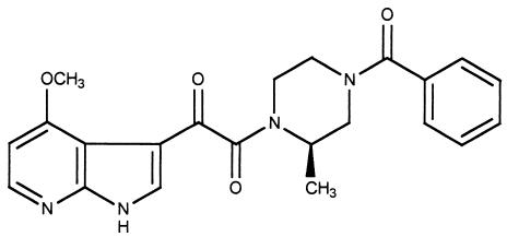
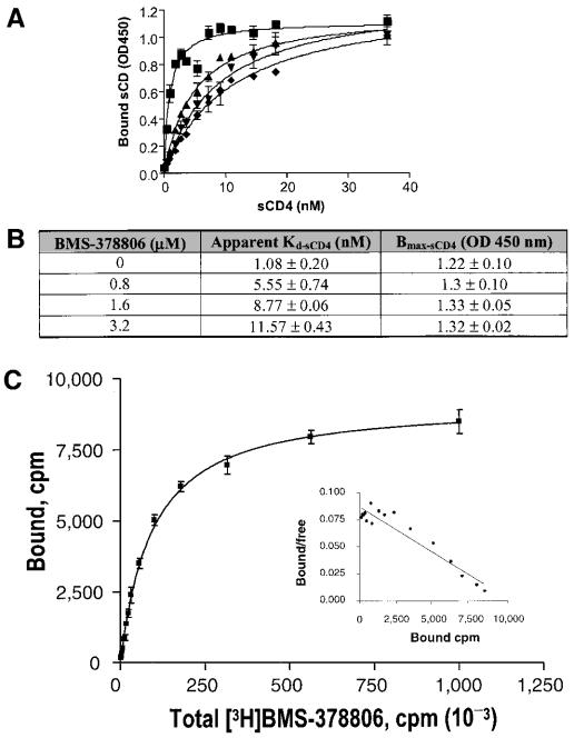
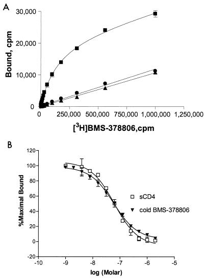
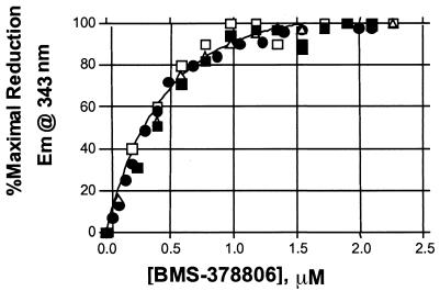
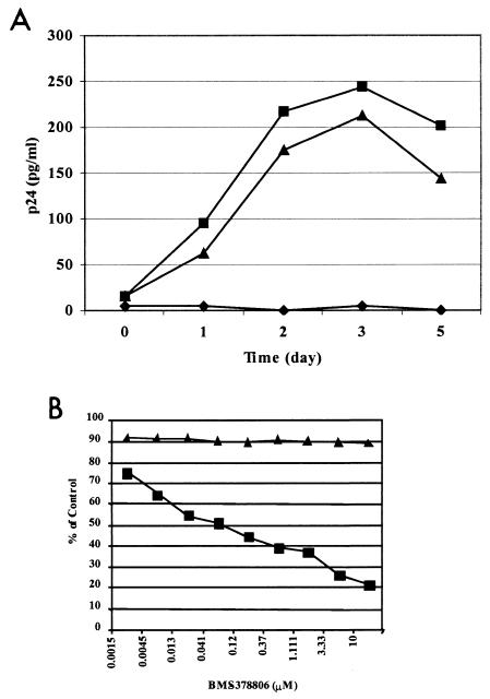
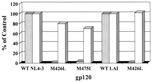
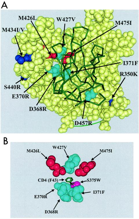
BMS-378806 is a recently discovered small-molecule human immunodeficiency virus type 1 (HIV-1) attachment inhibitor with good antiviral activity and pharmacokinetic properties. Here, we demonstrate that the compound targets viral entry by inhibiting the binding of the HIV-1 envelope gp120 protein to cellular CD4 receptors via a specific and competitive mechanism. BMS-378806 binds directly to gp120 at a stoichiometry of approximately 1:1, with a binding affinity similar to that of soluble CD4. The potential BMS-378806 target site was localized to a specific region within the CD4 binding pocket of gp120 by using HIV-1 gp120 variants carrying either compound-selected resistant substitutions or gp120-CD4 contact site mutations. Mapping of resistance substitutions to the HIV-1 envelope, and the lack of compound activity against a CD4-independent viral infection confirm the gp120-CD4 interactions as the target in infected cells. BMS-378806 therefore serves as a prototype for this new class of antiretroviral agents and validates gp120 as a viable target for small-molecule inhibitors.


Figures

FIG. 1.
FIG. 1.
The structure of BMS-378806.
FIG. 2.
FIG. 2.
Competitive inhibition of gp120-CD4 binding by BMS-378806 and binding affinity of BMS-378806 to gp120. (A) Saturation binding curve of BMS-378806. The levels of sCD4 bound to gp120 in the absence or presence of various concentrations of BMS-378806 and in escalating concentrations of sCD4 were measured by using the gp120-CD4 binding ELISA. BMS-378806 concentrations: ▪, 0 μM; ▴, 0.8 μM; ▾, 1.6 μM; ⧫, 3.2 μM. Data were analyzed according to the one-site binding model with GraphPad Prism. OD450, optical density at 450 nm. (B) Apparent binding constants for sCD4 in the absence and presence of BMS-378806. The Kd and Bmax values were derived by using nonlinear regression analysis with GraphPad Prism. Values were the means ± standard errors of the means, representing two independent experiments. (C) Binding affinity of BMS-378806 to gp120 by SPA. [3H]BMS-378806 binding to the gp120 protein was measured by employing gp120-coated SPA beads. The amount of gp120-bound [3H]BMS-378806 was determined by using a Packard TopCount scintillation counter. The results from Scatchard analysis of binding isotherm are shown in the insert. The x axis represents bound (corrected), and the y axis indicates bound/free.
FIG. 3.
FIG. 3.
Reversibility of BMS-378806-gp120 binding and Ki determination. (A) Competition of the gp120-bound [3H]BMS-378806 by cold compound. Binding of [3H]BMS-378806 to gp120JRFL (monomer) was performed with the gp120-bound SPA. After reaching equilibrium (at 4 h) (squares), specific binding was reversed to background levels (circles) by the addition of 60 μM unlabeled BMS-378806 (2,857-fold excess of Kd, at 15 h). Background binding was similar to that of control SPA beads lacking bound gp120 (triangles). (B) Ki determination for BMS-378806 and sCD4. The Ki values were determined by adding 106 cpm of [3H]BMS-378806 to SPA reaction mixtures containing various concentrations of unlabeled BMS-378806 or sCD4. The data reported here were expressed as percentages of the maximal counts per minute bound and represented the averages of at least four sets of replicates.
FIG. 4.
FIG. 4.
Binding stoichiometry of BMS-378806 to gp120 protein. gp120JRFL (1.0 μM) was titrated with BMS-378806, and the observed percent reduction in fluorescence from each of four independent experiments was plotted first as a function of BMS-378806 concentration (data not shown). Each of these data sets was then normalized to the percent maximal fluorescence reduction observed for each experiment in order to analyze the four sets of data together. The individual, normalized data sets are depicted as open squares, closed squares, closed diamonds, and open triangles. The curve represents the best fit to a saturation binding isotherm determined by using GraphPad Prism.
FIG. 5.
FIG. 5.
Lack of inhibition against a CD4-independent HIV-1 variant. (A) Replication kinetics of the ADA-S190R/N197S variant in Cf2Th cell lines. Virus titers in the supernatants of Cf2Th-CD4+ CCR5+ (square), CD4-negative Cf2Th-CCR5+ (triangle), and Cf2Th-CD4 CCR5 (diamond) cell cultures following infection with the recombinant virus ADA-S190R/N197S were monitored with a p24 ELISA. (B) Effect of BMS-378806 on HIV infection of CD4-negative and CD4-positive cells. The antiviral activity of BMS-378806 in Cf2Th-CD4+ CCR5+ (square) and CD4-negative Cf2Th-CCR5+ (triangle) cell cultures was determined 3 days following viral infection with ADA-S190R/N197S. The results are presented as percentages of inhibition relative to their respective untreated infected cell cultures. Results from a representative experiment are shown.
FIG. 6.
FIG. 6.
Binding of BMS-378806 to gp120-resistant variants is significantly reduced. The y axis denotes the percentage of BMS-378806 binding to variant gp120 proteins versus the envelope protein of corresponding wild-type HIV-1 strain (control). The x axis represents the various gp120 proteins tested. The data from the [3H]BMS-378806 binding experiment are shown in gray, and those from sCD4 are depicted in white.
FIG. 7.
FIG. 7.
Locations of BMS-378806-selected resistant substitutions and CD4 contact site mutations on gp120. (A) A space-filled model with atom spheres sized to the van der Waals radii was prepared with WebLab Viewerpro 4.0 (Accelrys, Inc., San Diego, Calif.). Yellow spheres, gp120 residues; red spheres, BMS-378806-selected substitutions located in the CD4 binding pocket (M426L and M475I); dark blue spheres, BMS-378806-selected substitutions (R350K, M434I/V, and S440R); turquoise spheres, CD4 contact site mutations (D368R, E370R, I371F, W427V, and D457R). The sCD4 protein is represented by a green diagram overlapping gp120. (B) Placement of the S375W residue. A simplified model shows only the relative locations of the CD4 contact site mutations (in turquoise) and the BMS-378806-selected substitutions (in red). The S375W residue (in pink) lies just below the Phe43 (F43) residue of CD4 (in green), at the base of the CD4 binding pocket.

Similar articles

Cited by

References

    1. Blair, W. S., P. F. Lin, N. A. Meanwell, and P. B. Wallace. 2000. HIV-1 entry-an expanding portal for drug discovery. Drug Discov. Today 5:183-194. - PubMed
    1. Campbell, I. D., and R. A. Dwek. 1984. Biological spectroscopy. Benjamin/Cummings, Menlo Park, Calif.
    1. Chan, D. C., and P. S. Kim. 1998. HIV entry and its inhibition. Cell 93:681-684. - PubMed
    1. Cohen, O. J., and A. S. Fauci. 2001. Current strategies in the treatment of HIV infection. Adv. Intern. Med. 46:207-246. - PubMed
    1. Copeland, R. A. 2000. A practical introduction to structure, mechanism, and data analysis, p. 104. In V. C. H. Wiley (ed.), Enzymes, 2nd ed. John Wiley & Sons, New York, N.Y.