. 2003 Oct;77(19):10663-9.
doi: 10.1128/jvi.77.19.10663-10669.2003.

Cell-derived sequences in the N-terminal region of the polyprotein of a cytopathogenic pestivirus

Affiliations

Cell-derived sequences in the N-terminal region of the polyprotein of a cytopathogenic pestivirus

A Müller et al. J Virol. 2003 Oct.

Abstract

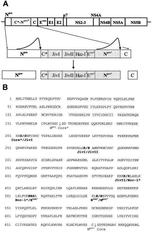
Efficient proteolytic release of nonstructural protein 3 (NS3) from the viral polyprotein is considered to be crucial for the cytopathogenicity of pestiviruses. Here we describe a novel cytopathogenic (cp) bovine viral diarrhea virus strain (BVDV CP8) with a complex insertion composed of viral and cell-derived sequences, including two fragments of the cellular J-domain protein Jiv (J-domain protein interacting with viral protein) located in the N-terminal region of the polyprotein. BVDV CP8 expresses a Jiv fusion protein of 513 amino acids in addition to a complete set of viral proteins. This protein has the capacity to induce NS2-3 cleavage in trans. Accordingly, CP8 is a representative of a novel type of cp pestivirus with a cp-specific mutation located outside of the NS2-3 gene.


Figures

FIG. 1.
FIG. 1.
Northern blot analysis. Total RNAs isolated 48 h postinfection from MDBK cells infected with BVDV strains NCP8, CP8, and NADL and from noninfected MDBK cells were blotted after separation by agarose gel electrophoresis and hybridized against [32P]-labeled probes specific for BVDV (left) or Jiv (right). The BVDV-specific probe is 83 or 95% identical to the genomes of BVDV NADL or CP8, respectively; the Jiv probe shows an identity of over 99% to the Jiv insertions in both genomes, explaining the differences in the signal intensities obtained with these probes. Due to its low abundance, the Jiv mRNA could not be detected in total cellular RNA (24). The size standards are shown on the left; the BVDV NADL genome has a length of about 12.6 kb (5) and served as an additional size marker to estimate the length of the CP8 genome.
FIG. 2.
FIG. 2.
(A) The top diagram is the genome organization of BVDV strain CP8 deduced from the analyzed cDNA clones (see also Fig. 3A). The N-terminal region of the CP8 polyprotein and the processing products are depicted below the diagram of the genome. The inserted region is marked by a grey bar. C*, truncated core protein; JivI and JivII, fragments of bovine Jiv; Hcc-1*, fragment of protein Hcc-1; Erns*, Erns-derived peptide; Npro*, truncated Npro. C*-Npro* represents a CP8-specific processing product consisting of 513 amino acids. (B) Amino acid sequence of the N-terminal region of the CP8 polyprotein. Proteolytic cleavage sites are indicated by vertical arrows. Slashes indicate the borders between the rearranged sequence blocks. C*, a 35-amino acid fragment of C (based on the nucleotide sequences, codon S203 is derived from C, while S204 originates from the Jiv mRNA); JivI, amino acids 151 to 226 of bovine Jiv; JivII, amino acids 527 to 692 or 693 of bovine Jiv (since codon K445 matches the corresponding codon of the Jiv mRNA, it was ascribed to JivII; however, to confirm that K445 is not derived from Hcc-1, the nucleotide sequence of the corresponding bovine mRNA would be required); Hcc-1*, amino acids 11 or 12 to 74 or 75 of protein Hcc-1 (to define the border between Hcc-1* and Erns*, the nucleotide sequences of the bovine Hcc-1 mRNA and the Erns gene of CP8 would be required); Erns*, amino acids 170 or 171 to 196 of Erns (see above). The part of Jiv representing Jiv90 is shown in italics.
FIG. 3.
FIG. 3.
(A) (Top) Scheme of the CP8 genome structure; black bars below the genome depict the cDNA clones and reverse transcription-PCR fragments used to analyze the genome structure. (Bottom) Scheme of expression constructs. Numbers below the bars denote the calculated molecular sizes (k, kilodaltons) of the expected processing products; numbers in brackets indicate the amino acids of the CP8 polyprotein which are encoded by their respective constructs. In the polypeptide encoded by pC8/E2-NS4A, S symbolizes the signal peptide preceding E2. (B) Processing of the N-terminal region of the CP8 polyprotein. Metabolically labeled proteins were isolated by radioimmunoprecipitation with anti-Jiv and anti-Npro sera and separated by sodium dodecyl sulfate-polyacrylamide gel electrophoresis on an 8% Tricine gel (28). The strong band at 55 kDa detected by the Jiv serum (lanes 1 to 5) was not recognized by a different Jiv-specific antiserum from another rabbit (data not shown), strongly suggesting that this protein is not related to cellular Jiv or putative Jiv homologues. Constructs used for protein expression are indicated above the lanes. The size standards are shown on the left. The positions of C*-Npro* and Npro are indicated by arrowheads. (C) Detection of C-His. For radioimmunoprecipitation a MAb directed against the His tag was used. Proteins were separated on a 12% Tricine gel, and the position of C-His is indicated by an arrowhead. All lanes were derived from the same gel. (D) Detection of the CP8-specific protein. Constructs used for transient expression in BHK cells are indicated above the lanes. MDBK cells infected with BVDV strain CP8 or NCP8 at a multiplicity of infection of 2 were analyzed at 48 h postinfection. Proteins were separated on an 8% Tricine gel; for Western blot analysis, the Jiv-specific serum was used. All lanes were derived from the same gel.
FIG. 3.
FIG. 3.
(A) (Top) Scheme of the CP8 genome structure; black bars below the genome depict the cDNA clones and reverse transcription-PCR fragments used to analyze the genome structure. (Bottom) Scheme of expression constructs. Numbers below the bars denote the calculated molecular sizes (k, kilodaltons) of the expected processing products; numbers in brackets indicate the amino acids of the CP8 polyprotein which are encoded by their respective constructs. In the polypeptide encoded by pC8/E2-NS4A, S symbolizes the signal peptide preceding E2. (B) Processing of the N-terminal region of the CP8 polyprotein. Metabolically labeled proteins were isolated by radioimmunoprecipitation with anti-Jiv and anti-Npro sera and separated by sodium dodecyl sulfate-polyacrylamide gel electrophoresis on an 8% Tricine gel (28). The strong band at 55 kDa detected by the Jiv serum (lanes 1 to 5) was not recognized by a different Jiv-specific antiserum from another rabbit (data not shown), strongly suggesting that this protein is not related to cellular Jiv or putative Jiv homologues. Constructs used for protein expression are indicated above the lanes. The size standards are shown on the left. The positions of C*-Npro* and Npro are indicated by arrowheads. (C) Detection of C-His. For radioimmunoprecipitation a MAb directed against the His tag was used. Proteins were separated on a 12% Tricine gel, and the position of C-His is indicated by an arrowhead. All lanes were derived from the same gel. (D) Detection of the CP8-specific protein. Constructs used for transient expression in BHK cells are indicated above the lanes. MDBK cells infected with BVDV strain CP8 or NCP8 at a multiplicity of infection of 2 were analyzed at 48 h postinfection. Proteins were separated on an 8% Tricine gel; for Western blot analysis, the Jiv-specific serum was used. All lanes were derived from the same gel.
FIG. 4.
FIG. 4.
(A) Expression of NS2-3 and NS3 in MDBK cells infected with NCP8 or CP8. Cell lysates prepared 48 h postinfection were separated on an 8% Tricine gel and subjected to Western blot analysis by using a MAb directed against NS3. Noninfected cells served as controls. Positions of NS2-3 and NS3 are marked by arrowheads. (B) Induction of NS2-3 cleavage in trans. Proteins were analyzed by Western blot with a MAb directed against NS3. Constructs used to drive protein expression are indicated above the lanes. Positions of NS2-3 and NS3 are marked by arrowheads. The molecular size standards are shown on the left; k, kilodaltons.

Similar articles

Cited by

References

    1. Baroth, M., M. Orlich, H.-J. Thiel, and P. Becher. 2000. Insertion of cellular NEDD8 coding sequences in a pestivirus. Virology 278:456-466. - PubMed
    1. Becher, P., G. Meyers, A. D. Shannon, and H.-J. Thiel. 1996. Cytopathogenicity of border disease virus is correlated with integration of cellular sequences into the viral genome. J. Virol. 70:2992-2998. - PMC - PubMed
    1. Becher, P., H.-J. Thiel, M. Collins, J. Brownlie, and M. Orlich. 2002. Cellular sequences in pestivirus genomes encoding gamma-aminobutyric acid (A) receptor-associated protein and Golgi-associated ATPase enhancer of 16 kilodaltons. J. Virol. 76:13069-13076. - PMC - PubMed
    1. Choong, M. L., L. K. Tan, S. L. Lo, E. C. Ren, K. Ou, S. E. Ong, R. C. Liang, T. K. Seow, and M. C. Chung. 2001. An integrated approach in the discovery and characterization of a novel nuclear protein over-expressed in liver and pancreatic tumors. FEBS Lett. 496:109-116. - PubMed
    1. Collett, M. S., R. Larson, C. Gold, D. Strick, D. K. Anderson, and A. F. Purchio. 1988. Molecular cloning and nucleotide sequence of the pestivirus bovine viral diarrhea virus. Virology 165:191-199. - PubMed

Publication types

MeSH terms

LinkOut - more resources