

doi: 10.1128/jvi.77.19.10700-10705.2003.
Lassa virus Z protein is a matrix protein and sufficient for the release of virus-like particles [corrected]
Affiliations
- PMID: 12970458
- PMCID: PMC228511
- DOI: 10.1128/jvi.77.19.10700-10705.2003
Item in Clipboard
Lassa virus Z protein is a matrix protein and sufficient for the release of virus-like particles [corrected]
J Virol.
2003 Oct.
Erratum in
- J Virol. 2003 Dec;77(23):12927
Abstract
Lassa virus is an enveloped virus with glycoprotein spikes on its surface. It contains an RNA ambisense genome that encodes the glycoprotein precursor GP-C, the nucleoprotein NP, the polymerase L, and the Z protein. Here we demonstrate that the Lassa virus Z protein (i). is abundant in viral particles, (ii). is strongly membrane associated, (iii). is sufficient in the absence of all other viral proteins to release enveloped particles, and (iv). contains two late domains, PTAP and PPXY, necessary for the release of virus-like particles. Our data provide evidence that Z is the Lassa virus matrix protein that is the driving force for virus particle release.
Figures
Quantification of Lassa virus proteins. Vero and SRD cells were infected with Lassa virus at a multiplicity of infection of 1 before cells were labeled with 800 μCi of [35S]methonine-cysteine (Premix; Amersham-Buchler) 24 h postinfection for 48 h. Virions of cell culture supernatants were precleared by centrifugation at 1,000 × g for 10 min and were then passed through a 20% sucrose cushion by UC at 20,000 rpm for 2 h (SW28 rotor; Beckman). Viral proteins were subjected to SDS-PAGE on 12% polyacrylamide gels and were quantified by using a FUJI BAS 1000 BioImaging analyzer system (raytest). Masses are given in kilodaltons on left.
Membrane association of Lassa virus Z protein. (A) Purified Lassa virus was left untreated (upper panel) or was treated with 1% Triton X-100, loaded on iodixanol (Sigma) gradients, and subjected to UC (1.5 h at 4°C, 41,000 rpm, SW41; Beckman). Aliquots of the fractions of both gradients were subjected to SDS-PAGE. Viral proteins were subsequently immunoblotted and were detected with the antisera anti-GP-2, anti-NP, and anti-Z (24). Antisera against NP and Z were raised in rabbits as described previously (24) by using peptides derived from Lassa virus strain Josiah NP (aa 53 to 76) and from the Z protein (aa 2 to 16). (B) Membrane association of solitarily expressed Z. Vero cells were transfected with the beta-actin promoter-driven pCAGGS vector (30) with insertions encoding Z, GP-C, and NP. Forty-eight hours posttransfection, cells were disrupted in 20 mM Tris-HCl, pH 7.4, by using a Dounce homogenizer and were centrifuged (10 min, 700 × g). Supernatant containing membranes and cytosol was adjusted with OptiPrep (Sigma) to a final concentration of 35% (wt/wt) and was overlaid with 30% OptiPrep in TNE and subsequently with TNE lacking OptiPrep. Samples were then centrifuged to equilibrium at 165,000 × g for 4 h at 4°C in an SW55 rotor (Beckman). Fractions of 0.5 ml were collected from the top of the gradient after centrifugation. Aliquots were subjected to SDS-PAGE followed by immunoblotting using specific antisera against GP-C, NP, and Z. (C) Detachment studies of membrane-bound Z protein. Aliquots of fraction 1 from the membrane flotation described in legend for panel B were either treated with 2 M KCl or with 50 mM EDTA for 1 h at room temperature or were treated for 1 h at 4°C with 1 M sodium bicarbonate (pH 10) that was neutralized with 1 M Tris-HCl (pH 6.8). Aliquots then were subjected to a second round of flotation analysis and were subsequently analyzed by SDS-PAGE followed by immunoblotting. Masses are given in kilodaltons on left of panels A through C.
Analysis of released Z-protein-containing particles. (A) Vero cell cultures were transfected with pCAGGS encoding Z and, as a control, NP. Cellular supernatants were collected, freed from cellular debris (3,000 × g, 10 min), and pelleted through a 20% sucrose cushion by UC. The UC pellet was dissolved in 100 μl of SDS-PAGE sample buffer, and transfected cells were dissolved in 500 μl of SDS-PAGE sample buffer. Twenty-microliter samples were subjected to SDS-PAGE followed by immunoblotting. NP and Z were visualized by specific immune reactions. C, cell lysate; S, pelleted material from the supernatant. (B) Protease protection assay for the identification of lipid-enveloped Z-containing particles. Aliquots of UC-pelleted Z were either treated with 0.1 μg of proteinase K alone per μl or proteinase K with 1% Triton X-100 for 30 min at 37°C or were left untreated. Samples then were analyzed by SDS-PAGE followed by immunoblotting. Masses are given in kilodaltons on left of both panels A and B.
Lassa virus wild-type Z protein and mutants. (A) The amino acid sequence of Lassa virus Z protein, strain Josiah, is shown in one-letter code (NCBI accession number NC_004296/004297). Amino acids associated with particular structures and possible functions are shown in boldfaced single letters. The cysteine and histidine residues that are proposed to form the RING domain are underlined, the two tetrapeptides postulated as late domains are doubly underlined, and the hydrophobic peptide (aa 50 to 61) is indicated with a dotted line. (B) Wild-type Z, deletion, and substitution mutants used in this study are shown schematically.
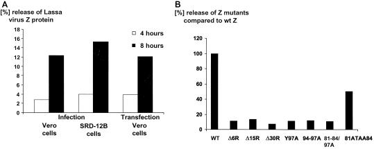
Release of Lassa virus, wild-type Z particles, and Z particles with mutated late domains. Vero and SRD-12B cells were infected with Lassa virus at a multiplicity of infection of 1. In addition, Vero cell cultures were transfected with pCAGGS encoding wild-type and mutated Z, respectively. Forty-eight hours postinfection and 24 h posttransfection, respectively, cells were metabolically labeled with [35S]methionine-cysteine for 4 and 8 h. Collected cells and cell supernatants were clarified from cell debris by centrifugation. Z protein was immunoprecipitated by using a precipitation buffer containing detergents, subjected to SDS-PAGE, and quantified by using a BioImaging analyzer system. Wild-type and mutated Z was tested in at least three independent experiments differing within 10%. Mean values are shown. (A) The release of Lassa virus and virus-like particles after infection of Vero and SRD-12B cells or with wild-type Z protein-transfected cells was quantified, and the ratio of Z protein detected in the supernatant to cellular Z protein was calculated (Tina software). (B) The ratio of released wild-type (wt) Z protein was set to 100%, and the ratio of released mutant Z protein is shown as the relative percentage compared to wild-type Z protein.
References
-
- Buchmeier, M. J. 2002. Arenaviruses: protein structure and function, p. 159-173. In M. B. A. Oldstone (ed.), Arenaviruses, vol. I. Springer, Berlin, Germany. - PubMed
Publication types
MeSH terms
Substances
LinkOut - more resources
Full Text Sources
Other Literature Sources
Miscellaneous
