. 2003 Oct;77(19):10725-9.
doi: 10.1128/jvi.77.19.10725-10729.2003.

A lipase isolated from the silkworm Bombyx mori shows antiviral activity against nucleopolyhedrovirus

Affiliations

A lipase isolated from the silkworm Bombyx mori shows antiviral activity against nucleopolyhedrovirus

Kangayam M Ponnuvel et al. J Virol. 2003 Oct.

Abstract

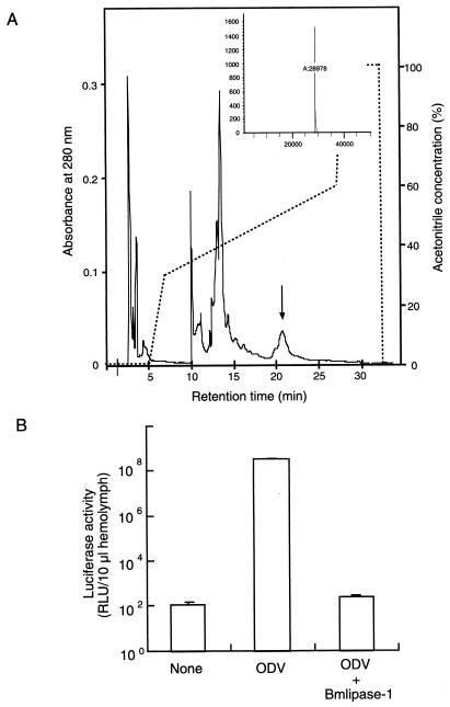
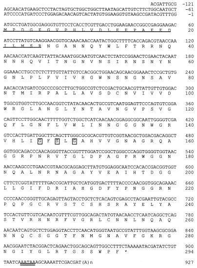
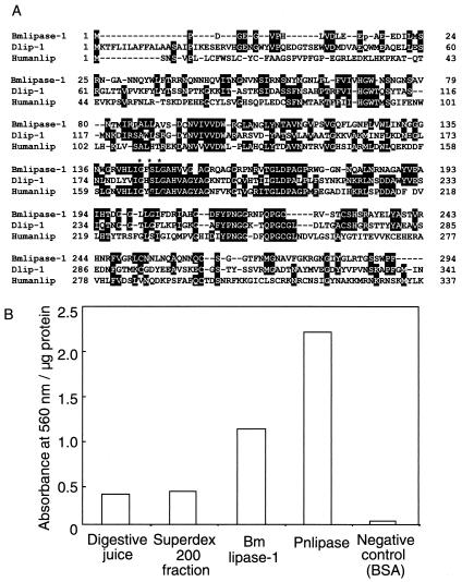
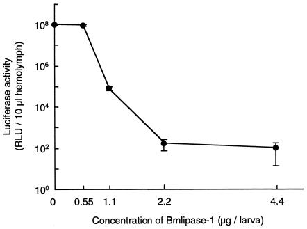
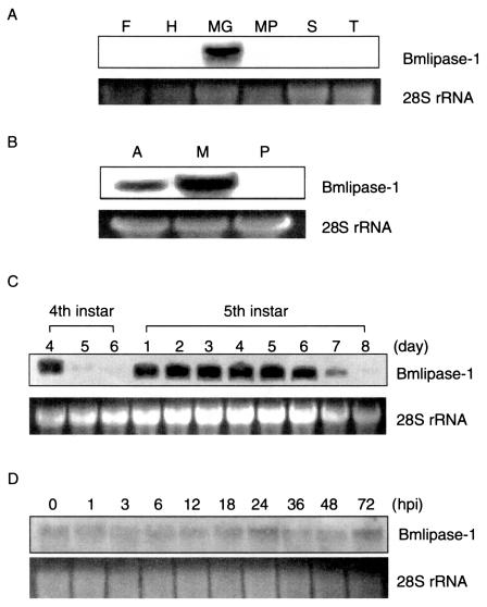
A protein showing strong antiviral activity against Bombyx mori nucleopolyhedrovirus (BmNPV) was purified from the digestive juice of B. mori larvae. A homology search of the deduced amino acid sequence of the protein cDNA revealed 56% homology with Drosophila melanogaster lipase and 21% homology with human lipase. As lipase activity of the protein was confirmed in vitro, this protein was designated Bmlipase-1. Northern blot analysis showed that the Bmlipase-1 gene is expressed in the midgut but not in other tissues, nor is it activated by BmNPV infection. In addition, the Bmlipase-1 gene was shown not to be expressed in the molting and wandering stages, indicating that the gene is hormonally regulated. Our results suggest that an insect digestive enzyme has potential as a physiological barrier against BmNPV at the initial site of viral infection.


Figures

FIG. 1.
FIG. 1.
Final HPLC purification profile and antiviral activity of Bmlipase-1. (A) Purification profile of Bmlipase-1. Bmlipase-1 was eluted with 49.5% acetonitrile-0.05% TFA by reverse-phase HPLC. The arrow indicates the Bmlipase-1 position. The inset shows the LC-MS spectrum with molecular mass of Bmlipase-1. B. mori larvae of the Daizo race were reared on mulberry leaves, whereas larvae of the Tokai × Asahi race were fed an artificial diet (Nihonnosanko). The silkworms were maintained in the silkworm rearing room under controlled environmental conditions at 27°C. The digestive juice was collected from B. mori (Daizo race) larvae in an ice-cold tube by mild electric shock, and ammonium sulfate was added to give a 40% saturation. The precipitate was suspended in 40 mM phosphate buffer (pH 7.4) and treated with 50% n-butanol (final concentration) overnight at 4°C, and the lower aqueous layer was collected after centrifugation at 10,000 × g for 30 min. Half-volume ice-cold acetone was added to this solution to precipitate proteins in the aqueous layer. The precipitated proteins were collected by centrifugation at 10,000 × g for 30 min, dried, and dissolved in 40 mM phosphate buffer (pH 7.4). The solution equilibrated with the same buffer was applied to a Superdex 200 HR 10/30 column attached to a fast-performance liquid chromatography system (Pharmacia). Each fraction was tested for antiviral activity. Antiviral fractions were further purified through a reverse-phase column, Sephasil C8 SC 2.1/10, attached to a SMART system (Pharmacia) with a linear gradient of acetonitrile-0.05% TFA. (B) Antiviral activity against BmNPV-ODV. Infectivity was expressed as relative light units/10 μl of hemolymph. The data shown are means ± standard deviations of results from five experiments. The infectivity of BmNPV treated with 2.2 μg of Bmlipase-1 per larva (indicated as ODV + Bmlipase-1) was compared to that of nontreated BmNPV (indicated as ODV). As a negative control, the same amount of hemolymph from nontreated B. mori larvae was used to measure the background levels of luciferase activity (indicated as None). The ODV concentration was22.5 ng/larva. Luciferase activity was measured at 136 hpi. The antiviral activity was assayed using BmNPVp10luc, which contained a luciferase reporter gene driven by the p10 promoter (15). The recombinant BmNPV was a gift from Shuichiro Tomita from our institute. The virus expresses a luciferase reporter gene at 15 hpi. The infection and mortality levels were quantified in the Tokai × Asahi race. The ODV was purified by ultracentrifugation on sucrose gradient (14). The ODV (22.5 ng/larva) suspension was mixed with the purified protein (2.2 μg/larva) and dissolved in 40 mM phosphate buffer (pH 8.0). This mixture was incubated at 30°C for 1 h. Fifth-instar larvae just after ecdysis were orally inoculated with 5 μl of ODV mixture. Thirty larvae were used for each treatment. The hemolymph was collected at various hpi and assayed for luciferase activity. Ten microliters of hemolymph was added to 50 μl of luciferase assay buffer (Promega), and luciferase activity was measured using a Luminocounter 700 (Microtech-Nition).
FIG.2.
FIG.2.
Nucleotide and deduced amino acid sequences of Bmlipase-1 cDNA. The putative signal sequence of purified Bmlipase-1 is underlined. Boxed amino acid residues denote the active site of the lipase family (10). An asterisk shows the termination codon (TAA). The polyadenylation signal is double-underlined. B. mori EST database (Silkbase; http://www.ab.a.u-tokyo.ac.jp/silkbase/) was screened based on the N-terminal amino acid sequence of the purified protein. A clone (mg809) isolated from the midgut cDNA library showed homology with this protein. The nucleotide sequence of the mg809 clone, kindly provided by Kazuei Mita from our institute, was determined to lack the sequence of the 5′ region. Complete nucleotide sequence was obtained by a First Choice RLM-RACE kit (Ambion) using mRNA extracted from the midgut and purified by a Quick Prep mRNA kit (Amersham Pharmacia Biotech). The nucleotide sequence was determined by dye-terminator cycle sequencing using a DNA sequencer (ABI 373A).
FIG. 3.
FIG. 3.
Structural and functional properties of Bmlipase-1. (A) Comparison of the amino acid sequence of Bmlipase-1 with that of D. melanogaster lipase (accession number AE003759-4) (1) and human lipase (6). The amino acid sequence from positions 338 to 500 of human lipase is omitted. Identical amino acid residues are boxed in black. The active site of the lipase family (GXSXG) is shown with asterisks. (B) Lipase activity measurement. The data shown are a typical result of three independent experiments. Lipase activity of both nonpurified digestive juice and Superdex 200 fraction is also shown. As positive and negative controls, Phycomyces nitens lipase (Pnlipase) and bovine serum albumin (BSA) were used, respectively. The protein concentration was determined using a Bio-Rad protein assay kit. Lipase activity was assayed according to the method of Arreguin-Espinosa et al. (2). P. nitens lipase was obtained from Wako. Lipase activity was measured at 560 nm and is expressed per milligram of protein.
FIG. 4.
FIG. 4.
Effect of Bmlipase-1 on BmNPV-ODV infectivity. ODV infectivity was examined after treatment of ODV (860 ng/larva) with different concentrations of Bmlipase-1. Data shown are means ± standard deviations of results from five experiments. Luciferase activity was measured at 136 hpi. Detailed experimental conditions are described in the legend to Fig. 1. Note that luciferase activity of hemolymph samples from nontreated B. mori larvae was also measured to determine the background level, and the level of relative light units (RLU) per 10 μl of hemolymph was 120 ± 3.4.
FIG. 5.
FIG. 5.
Northern blot analysis of Bmlipase-1 gene expression. (A) Bmlipase-1 gene expression in the fat body (F), hemocyte (H), midgut (MG), Malpighian tubule (MP), silk gland (S), and trachea (T). As an internal marker, 28S rRNA is shown in the lower panel. Tissues such as the midgut, hemocyte, silk gland, trachea, fat body, and Malpighian tubule were prepared from two fifth-instar larvae. The midgut was further separated into anterior, middle, and posterior portions. Total RNA from each tissue was isolated with ISOGEN (Nippon Gene). Northern blotting (11) was conducted with 20 μg of RNA and screened with a digoxygenin-labeled Bmlipase-1 probe prepared by PCR DIG labeling mix (Roche). (B) Bmlipase-1 gene expression in the anterior (A), middle (M), and posterior (P) portions of the midgut. (C) Bmlipase-1 gene expression in fourth and fifth instars. Fourth-instar days 5 and 6 are the molting stage. Fifth-instar day 8 is the wandering stage. (D) Effects of BmNPV infection on Bmlipase-1 gene expression. Bmlipase-1 gene expression was analyzed at different time intervals after BmNPV infection.

References

    1. Adams, M. D. A. 2000. The genome sequence of Drosophila melanogaster. Science 287:2185-2195. - PubMed
    1. Arreguin-Espinosa, R., B. Arreguin, and C. González. 2000. Purification and properties of a lipase from Cephaloleia presignis (Coleoptera, chrysomelidae). Biotechnol. Appl. Biochem. 31:239-244. - PubMed
    1. Barrett, J. W., A. J. Brownwright, M. J. Primavera, A. Retnakaran, and S. R. Palli. 1998. Concomitant primary infection of the midgut epithelial cells and the hemocytes of Trichoplusia ni by Autographa californica nucleopolyhedrovirus. Tissue Cell 30:602-616. - PubMed
    1. Boman, H. G., and D. Hultmark. 1987. Cell-free immunity in insects. Annu. Rev. Microbiol. 41:103-126. - PubMed
    1. Clem, R. J., M. Fechheimer, and L. K. Miller. 1991. Prevention of apoptosis by a baculovirus gene during infection of insect cells. Science 254:1388-1390. - PubMed

Publication types

Associated data

LinkOut - more resources