. 2003 Sep;133(1):47-62.
doi: 10.1104/pp.103.025676.

Differential regulation of glucose-6-phosphate dehydrogenase isoenzyme activities in potato

Affiliations

Differential regulation of glucose-6-phosphate dehydrogenase isoenzyme activities in potato

Rüdiger Hauschild et al. Plant Physiol. 2003 Sep.

Abstract

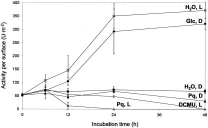
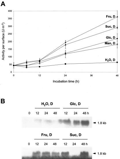
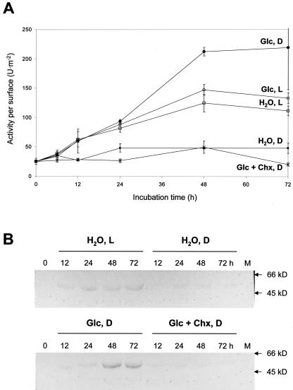
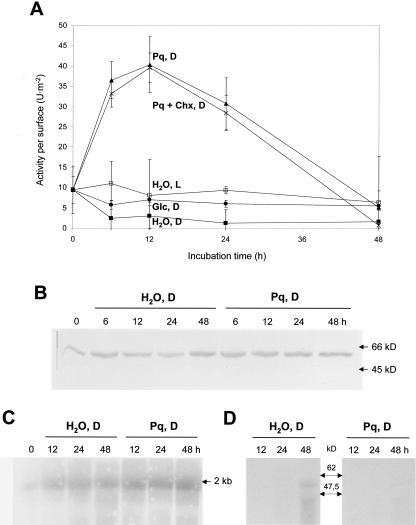
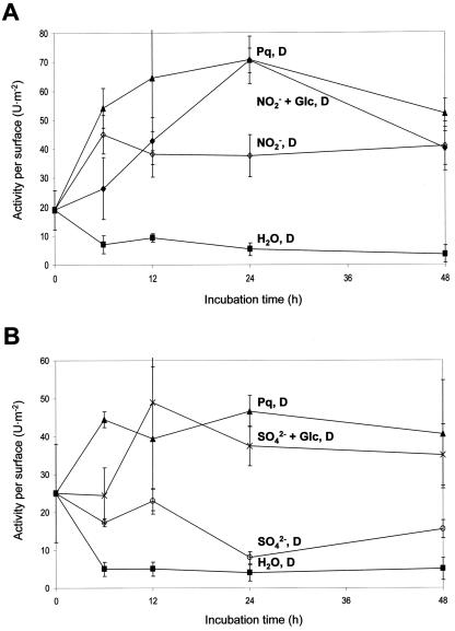
In plants, Glc-6-phosphate dehydrogenase (G6PDH) isoenzymes are present in the cytosol and in plastids. The plastidic enzymes (P1 and P2) are subject to redox regulation, but mechanisms that adjust cytosolic G6PDH activity are largely unknown. We adopted a leaf disc system for monitoring the effects of various conditions on G6PD isoform expression and enzyme activities in potato (Solanum tuberosum). Cytosolic G6PDH activity remained constant during water incubation in the dark. In continuous light or in the presence of metabolizable sugars in the dark, cytosolic G6PDH activity increased 6-fold within 24 h. Cycloheximide incubation demonstrated that enhanced cytosolic G6PDH activity depends on de novo protein synthesis. Osmotic change, phosphate sequestration, or oxidative stress did not affect cytosolic G6PDH activity. Furthermore, enzyme activity and protein contents closely followed the corresponding mRNA levels. Together with the fact that multiple SURE elements are present in the promoter region of the gene, these results suggest that cytosolic G6PDH activity is regulated by sugar availability at the transcriptional level. Plastidic G6PDH activity stayed constant during water incubation in the light and dropped to minimal levels within 6 h in the dark. Conversely, plastidic G6PDH activity of leaf discs incubated on Paraquat rose to 10-fold higher levels, which was not prevented by cycloheximide. Similar increases were found with nitrite, nitrate, or sulfate. No major changes in protein or mRNA contents of the plastidic P1 and P2 isoforms were registered. K(m) (Glc-6-phosphate) values of plastidic G6PDH activity differed between samples incubated on water or Paraquat, suggesting posttranslational modification of the plastidic enzyme(s). Immunoprecipitation of (32)P-labeled samples with P1 isoform-specific antibodies showed that the chloroplast enzyme is subject to protein phosphorylation. Obviously, in extended dark periods, G6PDH activity in the stroma is restricted but can be stimulated in response to high demands for NADPH.


Figures

Figure 1.
Figure 1.
Cytosolic G6PDH activities per surface (units per meter squared) in leaf discs incubated for 48 h on water in the dark (H2O, D), in continuous light (H2O, L), or on 50 mm Glc in the dark (Glc, D). In addition, 5 μm Paraquat in the dark (Pq, D) or in the light (Pq, L) and incubation on 100 μm 3-(3,4-dichlorophenyl)-1,1′-dimethyl urea (DCMU), which uncouples photosynthetic electron transport in the light (DCMU, L), was tested.
Figure 2.
Figure 2.
A, cytosolic G6PDH activities per surface (units per meter squared) in leaf discs incubated for 48 h in the dark on either water (H2O, D), 50 mm Man (Man, D), 50 mm Glc (Glc, D), 50 mm Fru (Fru, D), or 25 mm Suc (Suc, D), respectively. B, Northern-blot analyses conducted with total RNA (15 μg each) isolated from potato leaf discs incubated on different sugars in the dark. Samples were separated in denaturing agarose gels. After northern-blot transfer, membranes were hybridized with radiolabeled cDNA fragments of the cytosolic isoform, washed under stringent conditions (three times at 68°C with 0.1× SSC and 0.1% [w/v] SDS), and exposed to x-ray film. Numbers above the lanes represent incubation time in hours. Note that an RNA sample is missing (in the lane labeled 24 h Suc, D).
Figure 3.
Figure 3.
A, Cytosolic G6PDH activities per surface (units per meter squared) in leaf discs incubated for 72 h on water in the light (H2O, L) or in the dark (H2O, D), on 50 mm Glc in the light (Glc, L) or in the dark (Glc, D), and in additional presence of 1 mm cycloheximide (Chx; Glc + Chx, D). Note that water incubation in the light was not always lower compared with Glc incubation in the dark (compare graphs H2O, L and Glc, D in Figs. 1 and 3). B, Immunolabeling of cytosolic G6PDH protein on western blots of cleared extracts prepared from leaf discs incubated under conditions indicated. M, Molecular mass standard; arrows indicate sizes of apparent kilodaltons. Numbers above the lanes represent incubation times in hours.
Figure 4.
Figure 4.
Plastidic (DTTred-sensitive) G6PDH activity of leaf discs incubated for 48 h. A, Leaf discs were incubated on water in the light (H2O, L) or in the dark (H2O, D), on 5 μm Paraquat in the dark (Pq, D), and in additional presence of 1 mm Chx (Pq + Chx, D). Decreases of plastidic G6PDH activity after 24 h were observed repeatedly but not in all experiments (compare Fig. 5). B, Immunodetection of plastidic G6PDH (P1 isoform) on western blots of leaf discs incubated on either water or Paraquat in the dark for the hours indicated. C, Northern-blot analysis of total RNA (20 μg per lane) isolated from leaf discs incubated on either water or Paraquat in the dark for the hours indicated. The blot was hybridized with radiolabeled cDNA fragments of the P1 isoform (von Schaewen et al., 1995; Wendt et al., 1999) and washed under stringent conditions (three times at 68°C with 0.1× SSC and 0.1% [w/v] SDS). D, Autoradiogram of immunoprecipitated P1 protein after radiolabeling of leaf discs with 500 μCi 32P in the dark incubated on either water or Paraquat. kD, Molecular mass standards. Numbers above the lanes refer to incubation time in hours.
Figure 5.
Figure 5.
Plastidic (DTTred-sensitive) G6PDH activities in leaf discs incubated in the dark for 48 h. A, Incubations were conducted either on water (H2O, D), 5 μm Paraquat (Pq, D), 20 mm NO2- (D), or 20 mm NO2- plus 50 mm Glc (NO2- + Glc, D). B, Incubations were conducted either on water (H2O, D), 5 μm Paraquat (Pq, D), 100 mm SO42- (D), or 10 mm SO42- plus 50 mm Glc (SO42- + Glc, D).
Figure 6.
Figure 6.
Sketch of the promoter region of the gene coding for cytosolic G6PDH. Motifs similar to sugar regulatory elements in other plant genes are shown above and below the sequenced 2-kb region of the promoter (gray box) and are indicated as follows: CHS, conserved region in chalcone synthase genes of Petunia hybrida and Arabidopsis (Tsukaya et al., 1991); IMH2 and IMH5, homologous motifs in isocitrate lyase and malate synthase promoters from Cucumis sativus (Sarah et al., 1996); R2, repetitive sequence of patatin class I promoters from potato (Mignery et al., 1988); SP8a and SP8b, recognition sequences of a DNA-binding protein in sugar-regulated genes of sporamin and β-amylase from Ipomoea batatas (Ishiguro and Nakamura, 1994); SporA1, repetitive sugar regulatory region of Sporamin from I. batatas (Kim et al., 1991); and SURE1 and SURE2, sugar-responsive elements 1 and 2 identified in several genes isolated from potato (Fu et al., 1995). CAAT and TATA boxes are also indicated. Position numbers refer to the translation start (+1ATG).
Figure 7.
Figure 7.
Model of G6PDH regulation in the cytosol and in chloroplasts of potato leaf tissue. We chose the more general terms “sugar” and “sugar sensor,” although our results indicate that sugar sensing occurs at the level of hexoses (with Hexokinase as possible sensor). SURE stands for all regulatory promoter elements in the cytosolic G6PD gene involved in sugar-mediated signaling to the nucleus (compare with Fig. 6). For clarity, redox regulation of the chloroplast enzyme was omitted. ATG, Translation start; Chx, inhibitor of cytosolic protein translation; DCMU (inhibitor of photosynthetic electron transport); C5P, C5-sugar phosphates; E4P, erythrose-4-phosphate; 6PG, 6-phosphogluconate. For further explanations, see “Conclusions.”

References

    1. Anderson DJ, Blobel G (1983) Immunoprecipitation of proteins from cell-free translations. Methods Enzymol 96: 111-120 - PubMed
    1. Ashton AR, Burnell JN, Hatch MD (1984) Regulation of C4 photosynthesis: inactivation of pyruvate, Pi dikinase by ADP-dependent phosphorylation and activation by phosphorolysis. Arch Biochem Biophys 230: 492-503 - PubMed
    1. Aubert S, Gout E, Bligny R, Douce R (1994) Multiple effects of glycerol on plant cell metabolism. J Biol Chem 269: 21420-21427 - PubMed
    1. Baginsky S, Tiller K, Link G (1997) Transcription factor phosphorylation by a protein kinase associated with chloroplast RNA polymerase from mustard (Sinapis alba). Plant Mol Biol 34: 181-189 - PubMed
    1. Batz O, Logemann E, Reinold S, Hahlbrock K (1998) Extensive reprogramming of primary and secondary metabolism by fungal elicitor or infection in parsley cells. Biol Chem 379: 1127-1135 - PubMed

Publication types

LinkOut - more resources