. 2003 Sep;133(1):100-12.
doi: 10.1104/pp.103.023358.

The ARG1-LIKE2 gene of Arabidopsis functions in a gravity signal transduction pathway that is genetically distinct from the PGM pathway

Collaborators, Affiliations

The ARG1-LIKE2 gene of Arabidopsis functions in a gravity signal transduction pathway that is genetically distinct from the PGM pathway

Changhui Guan et al. Plant Physiol. 2003 Sep.

Abstract

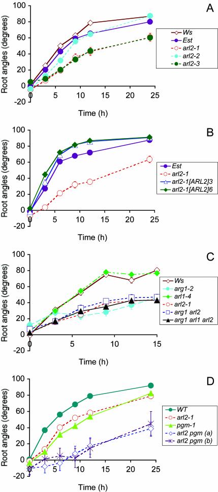
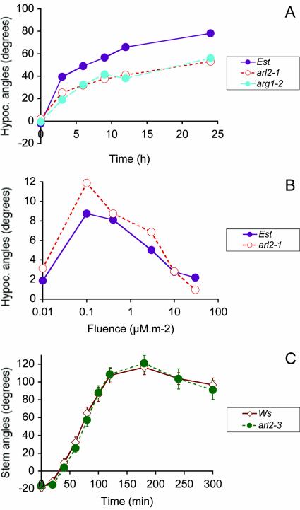
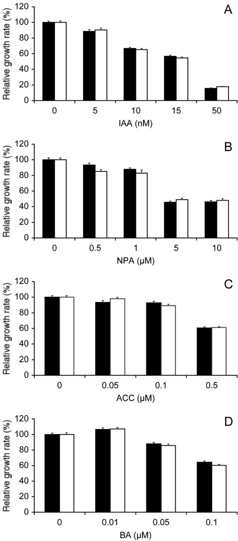
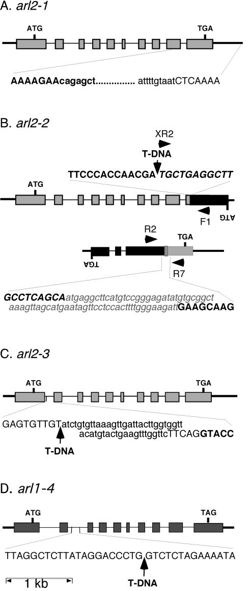
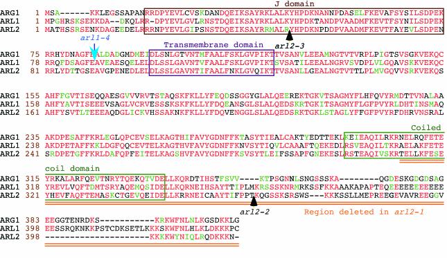
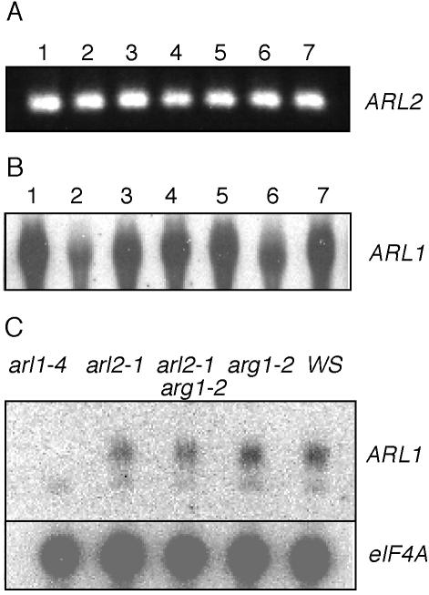
The arl2 mutants of Arabidopsis display altered root and hypocotyl gravitropism, whereas their inflorescence stems are fully gravitropic. Interestingly, mutant roots respond like the wild type to phytohormones and an inhibitor of polar auxin transport. Also, their cap columella cells accumulate starch similarly to wild-type cells, and mutant hypocotyls display strong phototropic responses to lateral light stimulation. The ARL2 gene encodes a DnaJ-like protein similar to ARG1, another protein previously implicated in gravity signal transduction in Arabidopsis seedlings. ARL2 is expressed at low levels in all organs of seedlings and plants. arl2-1 arg1-2 double mutant roots display kinetics of gravitropism similar to those of single mutants. However, double mutants carrying both arl2-1 and pgm-1 (a mutation in the starch-biosynthetic gene PHOSPHOGLUCOMUTASE) at the homozygous state display a more pronounced root gravitropic defect than the single mutants. On the other hand, seedlings with a null mutation in ARL1, a paralog of ARG1 and ARL2, behave similarly to the wild type in gravitropism and other related assays. Taken together, the results suggest that ARG1 and ARL2 function in the same gravity signal transduction pathway in the hypocotyl and root of Arabidopsis seedlings, distinct from the pathway involving PGM.


Figures

Figure 1.
Figure 1.
Gravitropic phenotype of wild-type, arl2, and arl1 mutant seedlings in darkness (A and C) or in light (B and D). Average root tip angles from the horizontal are shown at each time point. In A to D, genotypes of tested seedlings are indicated in a legend box. A, Root gravitropism of wild-type Est, Wassilewskija (Ws), and arl2-1, arl2-2, and arl2-3 mutant seedlings. This graph shows the results of two independent experiments, one involving Est and arl2-1, the other one involving Ws, arl2-2, and arl2-3. Wild-type Ws seedlings were included in both experiments, where they showed almost identical kinetics of root gravitropism (data not shown). Hence, we show only the Ws response for the second experiment. n = 33 to 84 for the first experiment and 38 to 120 for the second experiment. B, Root gravitropism of wild-type Est, untransformed arl2-1 mutant seedlings, and progeny of two independent arl2-1 transformants carrying the p35S-His6::ARL2 construct (arl2-1[ARL2]3 and arl2-1[ARL2]6, respectively; n = 41-53). C, Root gravitropism of wild-type Ws, single mutants arl1-4, arg1-2, and arl2-1, double mutant arg1-2 arl2-1, and triple mutant arg1-2 arl2-1 arl1-4 (n = 28-61). D, Root gravitropism of wild type (WT), single mutants arl2-1 and pgm-1, and double mutant arl2-1 pgm-1 seedlings. All wild-type and mutant lines tested in this experiment were derived from individual segregating F2 progeny from a cross between arl2-1 and pgm-1. Two independent arl2-1 pgm-1 double-mutant lines (a and b) were analyzed (n = 14-40). In A to D, vertical bars representing ses are shown at each time point. However, they are often masked by the curve symbols.
Figure 2.
Figure 2.
Tropic phenotypes of arl2 shoots. A, Kinetics of hypocotyl gravitropism in darkness for wild-type Est, arg1-2, and arl2-1 mutant seedlings (n = 111-168). B, Hypocotyl phototropism of 42-h-old seedlings exposed to a single pulse of blue light (450 nm) at a fluence rate of 0.15 μm m-2 s-1. Hypocotyl curvatures were measured 30 min after phototropic stimulation (n = 58-104). C, Kinetics of primary inflorescence stem gravitropism in darkness for 3- to 3.5-week-old wild-type and arl2-3 mutant plants (n = 15-16). As in Figure 1, ses are shown by vertical bars that are often masked by the curve symbols.
Figure 3.
Figure 3.
Root growth sensitivity to phytohormones and a polar auxin transport inhibitor. A to D, Relative root growth rate of wild-type Est (black bars) and arl2-1 mutant seedlings (white bars) in the presence of varying concentrations of IAA (A), naphthylphthalamic acid (NPA; B), 1-aminocyclopropane-1-carboxylic acid (ACC; C), or N6-benzyladenine (BA; D). Four-day-old seedlings were transferred onto fresh germination medium (GM) containing 0.1% (v/v) ethanol (A and D), 0.05% (v/v) dimethyl sulfoxide (B), or 0.05% (v/v) isopropanol (C) with the indicated concentrations of IAA, NPA, ACC, or BA. Root growth was measured over a period of 2 d. Average root growth rates were determined for each compound concentration and divided by the corresponding growth rate in absence of the compound (control). ses are represented by vertical bars. The numbers of seedlings tested in these experiments were 36 to 52 (A), 27 to 43 (B), 44 to 91 (C), and 81 to 95 (D). Average growth rates in the absence of added compounds were: A, 5.2 ± 0.09 (Est) and 4.5 ± 0.08 (arl2-1) mm d-1; B, 4.6 ± 0.12 (Est) and 4.5 ± 0.12 (arl2-1) mm d-1; C, 4.4 ± 0.09 (Est) and 5.09 ± 0.1 (arl2-1) mm d-1; and D, 5.2 ± 0.09 (Est) and 5.0 ± 0.1 (arl2-1) mm d-1.
Figure 4.
Figure 4.
Genomic structure of arl2-1 (A), arl2-2 (B), arl2-3 (C), and arl1-4 (D). Exons are represented by rectangles, introns by thin bars, and intergenic chromosomal regions by thick bars. ATG, Translation initiation codons; TGA or TAG, stop codons, ARL2 and At2g20050 or ARL1. Initiation and stop codons for lower strand open reading frames (ORFs; B) are indicated by inverted letters. The nucleotide sequence of a segment of DNA flanking the mutation site in each allele is indicated at the bottom or top of the corresponding diagram. The nucleotide sequence of an ARL2 or ARL1 exon fragment is represented in bold and uppercase characters, whereas that of an intronic or intergenic region is represented by uppercase characters. The sequence of At2g20050 exon fragments (B) is represented by italic uppercase characters. Sequences deleted within a specific allele are represented by lowercase characters, whereas new sequences added at a specific site (translocation breakpoint between the ARL2 and At2g20050 3′end fragments in arl2-2) are represented by a gray box, and written in bold, italicized, lowercase gray characters. T-DNA inserts are represented by arrows. For arl2-2 (B), the two ARL2 sequence-containing loci derived from a reciprocal translocation between chromosomes 1 and 2 are shown, along with the position of primers (black arrowheads) used in diagnostic PCR reactions (Fig. 5).
Figure 5.
Figure 5.
arl2-2/At2g20050 recombinant DNA fragments can be PCR amplified from arl2-2 genomic DNA but not from wild-type DNA. Primers used to PCR amplify wild-type ARL2 or recombinant arl2-2/At2g20050 fragments from Ws or arl2-2 DNAs are shown by horizontal arrowheads on the diagram shown in Figure 4B and marked F1 (PP2C-F1), R2 (PP2C-R2), XR2, and R7 (ARL2-R7) on that diagram and at the top of the gel. Their respective sequences are provided in Table I. The DNA used as a template in diagnostic PCR reactions is indicated above each electrophoretic lane. The sizes of amplified fragments, as determined by comparison with Mr markers loaded on the same gel, are shown on the right of the gel.
Figure 6.
Figure 6.
Amino acid sequence alignments of the ARG1 (top), ARL1 (middle), and ARL2 (bottom) proteins (Ws ecotype). Amino acid positions within each protein are shown at the left of each sequence (brown numbers). Protein names are indicated at the left of these numbers. Amino acids that are identical or conserved between at least two of the three proteins are shown in red and green characters, respectively. Gaps introduced within a protein to allow better alignment of flanking sequences are represented by dashes. The J domain, putative transmembrane domain, and predicted coiled-coil region are boxed. The arl2-1 mutation deletes the doubly underlined (orange) carboxyl end of the protein, whereas positions of T-DNA inserts within ARL2 and ARL1 alleles are indicated by black and blue arrows, respectively. Alignments were obtained by the Boxshade server (Institut Pasteur, Paris; http://bioweb.pasteur.fr/seqanal/interfaces/boxshade.html).
Figure 7.
Figure 7.
The ARL2 and ARL1 genes are expressed ubiquitously in Arabidopsis seedlings and plants. A, 3′ ARL2 cDNA fragments can be reverse transcription-PCR amplified from mRNAs extracted from siliques (1), cauline leaves (3), rosette leaves (4), stems (5), and flowers (6) of mature plants, roots of 3-week-old liquid-grown plants (7), and cotyledon and leaves of 5-d-old seedlings (2). B, Northern-blot analysis of total RNAs extracted from the plant organs defined in A, using ARL1 cDNA as a probe. Twenty micrograms of total RNA was loaded in each lane. C, Northern-blot analysis of total RNAs extracted from arl1-4, arl2-1, arg1-2 arl2-1, arg1-2, and wild-type Ws seedlings, using ARL1 (upper) or eIF4A (lower; loading control) cDNA sequences as probes. In this experiment, the ARL1 probe detected a low-intensity, nonspecific signal in all RNAs tested (including arl1-4). However, the specific ARL1 signal was not detectable in RNAs extracted from arl1-4 seedlings. In each panel, the source of mRNA is indicated above each lane, whereas the probe is identified at the right of the panel.

Similar articles

Cited by

References

    1. Arabidopsis Genome Initiative (2000) Analysis of the genome sequence of the flowering plant Arabidopsis thaliana. Nature 408: 796-814 - PubMed
    1. Ausubel FM, Brent R, Kingston RE, Moore DD, Seidman JG, Smith JA, Struhl K (1994) Current Protocols in Molecular Biology: Updates 1994-2002. John Wiley and Sons, New York.
    1. Bell CJ, Ecker JR (1994) Assignment of 30 microsatellite loci to the linkage map of Arabidopsis. Genomics 19: 137-144 - PubMed
    1. Bennett MJ, Marchant A, Green HG, May ST, Ward SP, Millner PA, Walker AR, Schulz B, Feldmann KA (1996) Arabidopsis AUX1 gene: a permease-like regulator of root gravitropism. Science 273: 948-950 - PubMed
    1. Bent AF (2000) Arabidopsis in planta transformation: uses, mechanisms, and prospects for transformation of other species. Plant Physiol 124: 1540-1547 - PMC - PubMed

Publication types

MeSH terms

Associated data