Multicenter Study
. 2003 Oct;73(4):835-48.
doi: 10.1086/378566. Epub 2003 Sep 10.

Confirmation of the type 2 myotonic dystrophy (CCTG)n expansion mutation in patients with proximal myotonic myopathy/proximal myotonic dystrophy of different European origins: a single shared haplotype indicates an ancestral founder effect

Affiliations
Multicenter Study

Confirmation of the type 2 myotonic dystrophy (CCTG)n expansion mutation in patients with proximal myotonic myopathy/proximal myotonic dystrophy of different European origins: a single shared haplotype indicates an ancestral founder effect

Linda L Bachinski et al. Am J Hum Genet. 2003 Oct.

Abstract

Myotonic dystrophy (DM), the most common form of muscular dystrophy in adults, is a clinically and genetically heterogeneous neuromuscular disorder. DM is characterized by autosomal dominant inheritance, muscular dystrophy, myotonia, and multisystem involvement. Type 1 DM (DM1) is caused by a (CTG)(n) expansion in the 3' untranslated region of DMPK in 19q13.3. Multiple families, predominantly of German descent and with clinically variable presentation that included proximal myotonic myopathy (PROMM) and type 2 DM (DM2) but without the DM1 mutation, showed linkage to the 3q21 region and were recently shown to segregate a (CCTG)(n) expansion mutation in intron 1 of ZNF9. Here, we present linkage to 3q21 and mutational confirmation in 17 kindreds of European origin with PROMM and proximal myotonic dystrophy, from geographically distinct populations. All patients have the DM2 (CCTG)(n) expansion. To study the evolution of this mutation, we constructed a comprehensive physical map of the DM2 region around ZNF9. High-resolution haplotype analysis of disease chromosomes with five microsatellite and 22 single-nucleotide polymorphism markers around the DM2 mutation identified extensive linkage disequilibrium and a single shared haplotype of at least 132 kb among patients from the different populations. With the exception of the (CCTG)(n) expansion, the available markers indicate that the DM2 haplotype is identical to the most common haplotype in normal individuals. This situation is reminiscent of that seen in DM1. Taken together, these data suggest a single founding mutation in DM2 patients of European origin. We estimate the age of the founding haplotype and of the DM2 (CCTG) expansion mutation to be approximately 200-540 generations.


Figures

Figure  1
Figure 1
Physical and genetic map of the DM2 region in 3q21. A, Locations of the 27 microsatellite markers used. Marker order was determined initially through use of the deCODE map (Kong et al. 2002) and then was refined using physical locations and by minimizing recombinations in the families tested. Locations of recombinations observed are indicated by arrowheads. B, BAC clones used to map markers close to ZNF9.
Figure  2
Figure 2
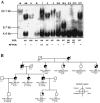
Molecular diagnosis of the DM2 (CCTG)n expansion mutation in an Italian family (I01) with PROMM and other selected cases. A, FIGE Southern blot showing expanded (CCTG)n alleles at the DM2 locus. Shown beneath each lane are the PCR allele sizes across the DM2 repeat and the results (+/−) of the RP-PCR assay. Pedigree numbers in the lane headers refer to those shown in the pedigree (B). A and B are haploid hybrids for the normal and mutant chromosome 3, respectively; AB is the donor patient from which the hybrid cell lines were established; M is the size marker lane; and cases 1–3 are individual, unrelated patients. B, Pedigree of PROMM kindred I01 (Meola et al. 1996), illustrating commonly encountered diagnostic problems and the importance of using multiple molecular diagnostic tests. Individual II:4 exemplifies a false-positive diagnosis. He had myotonia but had two normal alleles and showed no expansion by either RP-PCR or Southern analysis (“S”). For individual II:6, who has myotonia and muscle weakness, Southern analysis gave an unclear molecular diagnosis, whereas RP-PCR clearly identified him as a mutation carrier. Individual IV:1 has a false-negative diagnosis. Although phenotypically normal, he has an expansion of +16.5 kb. Individual IV:3 is a true homozygote. Both RP-PCR and Southern analysis confirm the absence of the expansion, in spite of the presence of only a single allele.
Figure  3
Figure 3
Chromosomal location and physical map of the BAC and PAC clones from the ZNF9 region determined by FISH. A, RPC1-3_436B3 was assigned to 3q21 by metaphase FISH (red). B, Ideogram of human chromosome 3. C, Mutual order of RP11-814L21, RPC1-3 436B3, and RP11-529F4, and the size of the gap in the physical map determined by fiber-FISH. Cohybridization of the three clones shows the order RP11-814L21–RPC1-3_436B3–RP11-529F4. A schematic representation of the genomic clones is shown below the image. The approximate size of the gap between RP11-814L21 and RPC1-3 426B3 was estimated at 227 kb (SD 59 kb).
Figure  4
Figure 4
Shared microsatellite marker haplotypes in families with DM2 over 5 Mb flanking the DM2 mutation. The country of origin of samples is indicated (CH = Switzerland; D = Germany; E = Spain; F = France; FIN = Finland; I = Italy; NY = United States; UK = United Kingdom). The 154-bp allele at 814L21-GT1 and 183-bp allele at 221E20-GT1 are seen predominantly on DM2 chromosomes of northern European origin (Finland, Germany, United Kingdom, and France). The 148-bp and 176-bp alleles at these respective loci are seen predominantly on DM2 chromosomes of southern European origin (Spain, Italy, and France). Alleles in common are indicated by shading.
Figure  5
Figure 5
Physical map of the ZNF9 region in 3q21 with the DM2 (CCTG)n expansion mutation (not drawn to scale). Bins are determined by overlap with the three other BAC clones available. Alleles found on DM2 and normal chromosomes are indicated, along with their frequencies. Approximately 30 kb of unordered fragments could be located either in intron 1 of ZNF9 or telomeric. Thus, the genomic size of ZNF9 can be estimated as at least 13 kb but could be as large as 44 kb. Consequently, the invariant haplotypes on DM2 chromosomes (boxed region) could be as small as 132 kb or as large as 163 kb.
Figure  6
Figure 6
A, Haplotypes observed across the DM2 region. Ten normal haplotypes (A–J) are shown, along with the DM2 haplotype, which is identical to haplotype A except for the presence of the DM2 (CCTG)n expansion. B, Haplotype evolutionary network based on parsimony. The areas of circles representing each haplotype are roughly proportional to the frequency of the haplotype being represented. Each haplotype is one mutational step from all of the haplotypes to which it is connected by a line. Haplotypes G and H are the only two haplotypes for which relationships are unclear. Haplotype G is either the result of a recombination or gene conversion event between the common haplotypes A and B (indicated by X) or the result of a repeat mutation (homoplasy), in which haplotype A changed by acquiring the mutation that is diagnostic for B or vice versa.
Figure  7
Figure 7
Evidence for LD with disease status for markers near the DM2 (CCTG)n expansion. The Y-axis shows nominal P values (on a formula image scale) for the moving-window haplotype contingency-table test. The evidence when marker 814L21-GT1 is included (unblackened circles) peaks centromeric to the expansion and is much stronger than when the marker is excluded (blackened circles). Permutation-based P values for the entire set of markers gave P<.001 for both curves.

References

Electronic-Database Information

    1. dbSNP Home Page, http://www.ncbi.nlm.nih.gov/SNP/
    1. GenBank, http://www.ncbi.nlm.nih.gov/Genbank/ (for RP11-814L21 [accession number AC022944], RP11-309B5 [accession number AC135587], RP11-723O4 [accession number AC112484], and RP11-434H6 [accession number AC108673], and chromosome 3 contig [accession number NT_006025])
    1. Gene Viewer, Cancer Genome Anatomy Project (CGAP), http://gai.nci.nih.gov/cgi-bin/GeneViewer.cgi (for CGAP SNPs)
    1. NCBI Reference Sequence (RefSeq), http://www.ncbi.nlm.nih.gov/RefSeq/
    1. Online Mendelian Inheritance in Man (OMIM), http://www.ncbi.nlm.nih.gov/Omim/ (for DM1, DM2, and PROMM)

References

    1. Abbruzzese C, Krahe R, Liguori M, Tessarolo D, Siciliano MJ, Ashizawa T, Giacanelli M (1996) Myotonic dystrophy phenotype without expansion of (CTG)n repeat: an entity distinct from proximal myotonic myopathy (PROMM)? J Neurol 243:715–721 - PubMed
    1. Ashizawa T, Epstein HF (1991) Ethnic distribution of myotonic dystrophy gene. Lancet 338:642–643 - PubMed
    1. Bassez G, Attarian S, Laforet P, Azulay JP, Rouche A, Ferrer X, Urtizberea JA, Pellissier JF, Duboc D, Fardeau M, Pouget J, Eymard B (2001) Proximal myotonial myopathy (PROMM): clinical and histology study. Rev Neurol 157:209–218 - PubMed
    1. Borrego S, Wright FA, Fernandez RM, Williams N, Lopez-Alonso M, Davuluri R, Antinolo G, Eng C (2003) A founding locus within the RET proto-oncogene may account for a large proportion of apparently sporadic Hirschsprung disease and a subset of cases of sporadic medullary thyroid carcinoma. Am J Hum Genet 72:88–100 - PMC - PubMed
    1. Brock GJ, Anderson NH, Monckton DG (1999) Cis-acting modifiers of expanded CAG/CTG triplet repeat expandability: associations with flanking GC content and proximity to CpG islands. Hum Mol Genet 8:1061–1067 - PubMed

Publication types