. 2003 Sep;14(9):3519-28.
doi: 10.1091/mbc.e03-04-0239. Epub 2003 May 29.

The association between integrin-associated protein and SHPS-1 regulates insulin-like growth factor-I receptor signaling in vascular smooth muscle cells

Affiliations

The association between integrin-associated protein and SHPS-1 regulates insulin-like growth factor-I receptor signaling in vascular smooth muscle cells

Laura A Maile et al. Mol Biol Cell. 2003 Sep.

Abstract

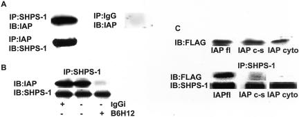
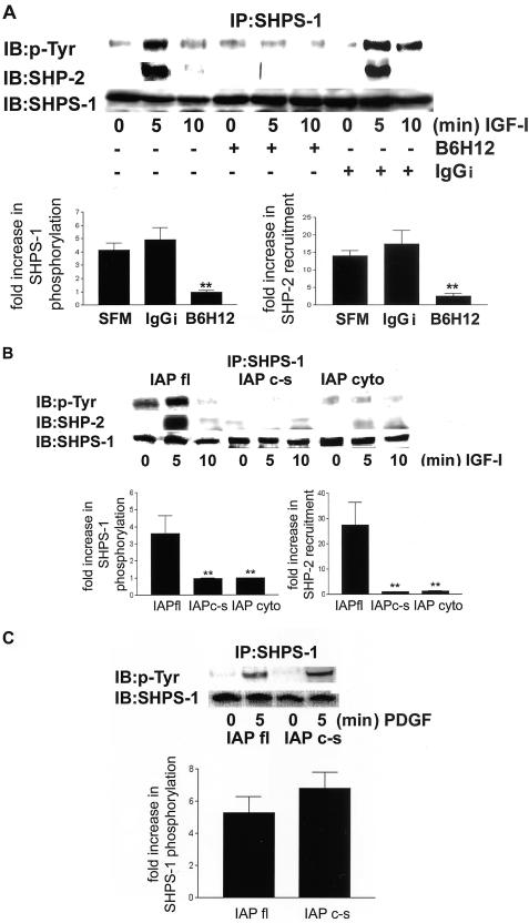
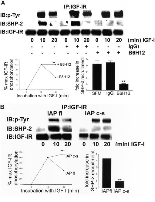
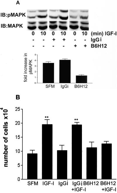
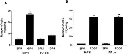
Growth factor signaling is usually analyzed in isolation without considering the effect of ligand occupancy of transmembrane proteins other than the growth factor receptors themselves. In smooth muscle cells, the transmembrane protein Src homology 2 domain containing protein tyrosine phosphatase substrate-1 (SHPS-1) has been shown to be an important regulator of insulin-like growth factor-I (IGF-I) signaling. SHPS-1 is phosphorylated in response to IGF-I, leading to recruitment of Src homology 2 domain tyrosine phosphatase (SHP-2). Subsequently, SHP-2 is transferred to IGF-I receptor and regulates the duration of IGF-I receptor phosphorylation. Whether ligand occupancy of SHPS-1 influences SHPS-1 phosphorylation or SHP-2 recruitment, thereby altering growth factor signaling, is unknown. Previous studies have shown that integrin associated protein (IAP) associates with SHPS-1. We undertook these studies to determine whether this interaction controlled SHPS-1 phosphorylation and/or SHP-2 recruitment and thereby regulated IGF-I signaling. Disruption of IAP-SHPS-1 binding, by using an IAP monoclonal antibody or cells expressing mutant forms of IAP that did not bind to SHPS-1, inhibited IGF-I-stimulated SHPS-1 phosphorylation and SHP-2 recruitment. This was associated with a lack of SHP-2 transfer to IGF-I receptor and sustained receptor phosphorylation. This resulted in an inability of IGF-I to stimulate sustained mitogen-activated protein kinase activation, cell proliferation, and cell migration. The effect was specific for IGF-I because disruption of the IAP-SHPS-1 interaction had no effect on platelet-derived growth factor-stimulated SHPS-1 phosphorylation or cell migration. In summary, our results show that 1) ligand occupancy of SHPS-1 is a key determinant of its ability to be phosphorylated after IGF-I stimulation, and 2) the interaction between IAP and SHPS-1 is an important regulator of IGF-I signaling because disruption of the results in impaired SHP-2 recruitment and subsequent inhibition of IGF-I-stimulated cell proliferation and migration.


Figures

Figure 1.
Figure 1.
Coprecipitation of IAP with SHPS-1 and disruption with anti IAP antibody. (A) Cell lysates were either immunoprecipitated with an anti-IAP antibody and coprecipitation of SHPS-1 determined by immunoblotting with anti-SHPS-1 antiserum or they were immunoprecipitated with anti SHPS-1 antibody and coprecipitation of IAP determined by immunoblotting with an anti-IAP antibody. As a control cell lysates were also immunoprecipitated with nonimmune rabbit serum (IgG) and immunoblotted with an anti-IAP antibody. (B) Quiescent pSMCs were incubated for 2 h ± the addition of the anti-IAP mAb, B6H12 or IgGi (both at 4 μg/ml). Coprecipitation of IAP with SHPS-1 was then determined by immunoprecipitating with an SHPS-1 antibody and immunoblotting with an anti-IAP antibody. The amount of SHPS-1 protein in each lane is shown in the lower row. (C) Expression of FLAG-labeled IAP and association with SHPS-1. Top, expression of FLAG-labeled IAP was determined by immunoblotting whole-cell lysates from cells transfected with each of the IAP cDNA constructs by using an anti-FLAG antibody. The results as scanning units are lane 1, 38,018; lane 2, 39,274; and lane 3, 46,779. Bottom, cell lysates were immunoprecipitated with an anti-SHPS-1 antibody and then coprecipitation of FLAG-labeled IAP was determined by immunoblotting with an anti-FLAG antibody. The amount of SHPS-1 that was immunoprecipitated in each lane is shown in the lower row. The results shown in this figure are representative of at least three independent experiments with similar results.
Figure 2.
Figure 2.
(A) SHPS-1 phosphorylation and SHP-2 recruitment to SHPS-1 in response to IGF-I after disruption of the association between IAP and SHPS-1 by the anti-IAP antibody B6H12. Quiescent cells were incubated for2h ± B6H12 antibody or IgGi (both at 4 μg/ml) and then exposed to IGF-I (100 ng/ml) as indicated. Cell lysates were immunoprecipitated with an anti-SHPS-1 antibody and then SHPS-1 phosphorylation was determined by immunoblotting with a pTyr. The association of SHP-2 with SHPS-1 was visualized by immunoblotting by using an anti SHP-2 antibody. The amount of SHPS-1 protein in each lane is shown in the lower row. The fold increase in SHPS-1 phosphorylation and SHP-2 recruitment after IGF-I stimulation as determined by scanning densitometric analysis of Western immunoblots from three separate experiments is shown. **p < 0.05 when the response to IGF-I of cells preincubated with B6H12 is compared with the IGF-I response in cells preincubated in SFM or control IgGi. (B) SHPS-1 phosphorylation and SHP-2 recruitment in response to IGF-I after disruption of the association between IAP and SHPS-1 in cells expressing mutated forms of IAP. Cells were exposed to IGF-I (100 ng/ml) for various periods. Cell lysates were immunoprecipitated with an anti-SHPS-1 antibody and SHPS-1 phosphorylation was determined by immunoblotting with an anti-phosphotyrosine antibody (pTyr). The association of SHP-2 was visualized by immunoblotting using an anti SHP-2 antibody. The amount of SHPS-1 protein in each lane is shown in the lower row. The increase in SHPS-1 phosphorylation and SHP-2 recruitment after IGF-I stimulation as determined by scanning densitometric analysis of Western immunoblots from three separate experiments is shown. **p < 0.05 when the response of cells expressing either of the mutant forms of IAP to IGF-I is compared with the IGF-I response in cells expressing IAPfl. (C) SHPS-1 phosphorylation in response to PDGF. Cells were exposed to PDGF (10 ng/ml) for 5 min. After cell lysis and immunoprecipitation with an anti-SHPS-1 antibody, SHPS-1 phosphorylation was determined by immunoblotting with a pTyr.
Figure 3.
Figure 3.
IGF-IR phosphorylation time course and SHP-2 recruitment after disruption of the interaction between IAP and SHPS-1. (A) Quiescent cells were incubated ± B6H12 or IgGi (4 μg/ml) and then exposed to IGF-I (100 ng/ml) for various lengths of time. After lysis and immunoprecipitation with an anti-IGF-IR antibody phosphorylation of the receptor was determined by immunoblotting with a pTyr. The association of SHP-2 was determined by immunoblotting with an anti SHP-2 antibody. The amount of IGF-IR protein in each lane is shown in the lower row. The level of tyrosine phosphorylation of IGF-IR as a percentage of maximum phosphorylation detected as determined by scanning densitometric analysis of Western immunoblots from three separate experiments is shown. The increase in SHP-2 recruitment after IGF-I stimulation as determined by scanning densitometry analysis of Western immunoblots from three separate experiments is also shown. **p < 0.05 when the response of cells preincubated with B6H12 to IGF-I is compared with cells preincubated in SFM alone or containing control IgGi. (B) Cells were incubated with IGF-I (100 ng/ml) for various times. After lysis and immunoprecipitation with an anti-IGF-IR antibody, phosphorylation of the receptor was determined by immunoblotting with a pTyr. The association of SHP-2 was determined by immunoblotting with an anti-SHP-2 antibody. The amount of IGF-IR protein in each lane is shown in the lower row. The changes IGF-IR phosphorylation and SHP-2 recruitment after IGF-I stimulation as determined by scanning densitometric analysis of Western immunoblots from three separate experiments is shown. **p < 0.05 when the response of cells expressing IAPc-s that were stimulated with IGF-I is compared with the response of cells expressing IAPfl.
Figure 4.
Figure 4.
(A) Phosphorylation of MAPK and cell proliferation in response to IGF-I. Cells were plated and grown as described in MATERIALS AND METHODS before a 2-h incubation ± B6H12 or IgGi (both at 4 μg/ml) and then treated with IGF-I (100 ng/ml) for 10 min. The level of p42/44 MAPK phosphorylation was determined by immunoblotting with a phosphospecific MAPK antibody. The total amount of MAPK in each sample was determined by immunoblotting with a MAPK antibody. (B) Cells were plated and grown as described in MATERIALS AND METHODS before a 2-h incubation ± B6H12 or IgGi (both at a concentration of 4 μg/ml) and then treated with IGF-I (100 ng/ml) for 48 h. Cell number in each well was then determined. Each data points represent the mean of three independent experiments. **p < 0.05 when the response of cells incubated in the presence of B6H12 is compared with cells incubated with control IgGi or in the absence of antibody.
Figure 5.
Figure 5.
IGF-I- and PDGF-stimulated cell migration in cells expressing full-length IAP and IAPc-s. Confluent cells were wounded then incubated ± IGF-I (100 ng/ml) (A) or PDGF (10 ng/ml) (B) for 48 h. The number of cells migrating across the wound edge in at least five preselected regions was counted. Each data point represents the mean ± SEM of three independent experiments. **p < 0.05 when migration of cells expressing IAPfl in the presence of IGF-I or PDGF is compared with incubation in SFM alone or when cells expressing IAPc-s stimulated with PDGF are compared with SFM alone.

References

    1. Andre, F., Rigot, V., Thimonier, J., Montixi, C., Parat, F., Pommier, G., Marvaldi, J., and Luis, J. (1999). Integrins and E-cadherin cooperate with IGF-I to induce migration of epithelial colonic cells. Int. J. Cancer 83, 497–505. - PubMed
    1. Babic, I., Schallhorn, A., Lindberg, F.P., and Jirik, F.R. (2000). SHPS-1 induces aggregation of Ba/F3 pro-B cells via an interaction with CD47. J. Immunol. 164, 3652–3658. - PubMed
    1. Brown, E., Hooper, L., Ho, T., and Gresham, H. (1990). Integrin-associated protein: a 50-kD plasma membrane antigen physically and functionally associated with integrins. J. Cell Biol. 111, 2785–2794. - PMC - PubMed
    1. Fujioka, Y., Matozaki, T., Noguchi, T., Iwamatsu, A., Yamao, T., Takahashi, N., Tsuda, M., Takada, T., and Kasuga, M. (1996). A novel membrane glycoprotein, SHPS-1, that binds the SH2-domain-containing protein tyrosine phosphatase SHP-2 in response to mitogens and cell adhesion. Mol. Cell. Biol. 16, 6887–6899. - PMC - PubMed
    1. Gockerman, A., Prevette, T., Jones, J.I., and Clemmons, D.R. (1995). Insulin-like growth factor (IGF)-binding proteins inhibit the smooth muscle cell migration responses to IGF-I and IGF-II. Endocrinology 136, 4168–4173. - PubMed

Publication types

MeSH terms

Substances