. 2003 Sep;14(9):3782-803.
doi: 10.1091/mbc.e03-01-0018. Epub 2003 May 29.

RAM: a conserved signaling network that regulates Ace2p transcriptional activity and polarized morphogenesis

Affiliations

RAM: a conserved signaling network that regulates Ace2p transcriptional activity and polarized morphogenesis

Bryce Nelson et al. Mol Biol Cell. 2003 Sep.

Abstract

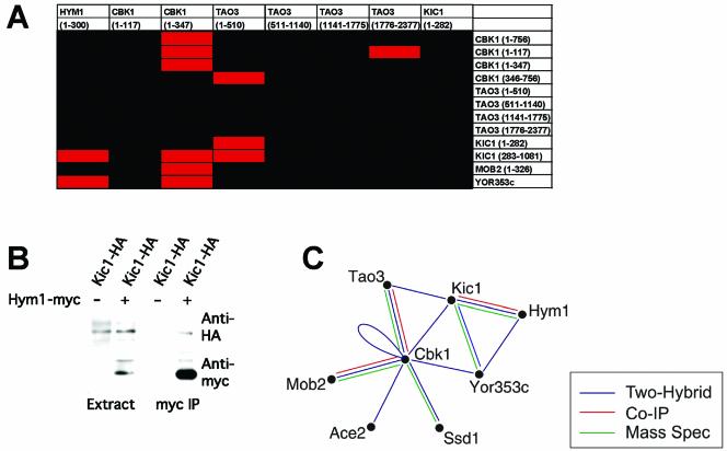
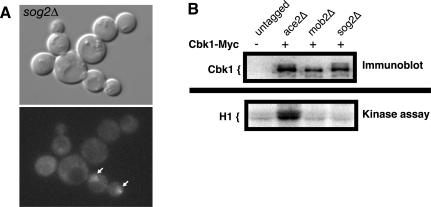
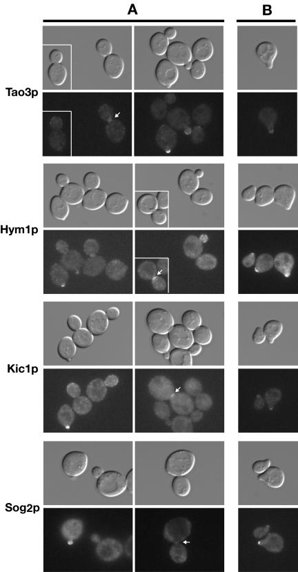
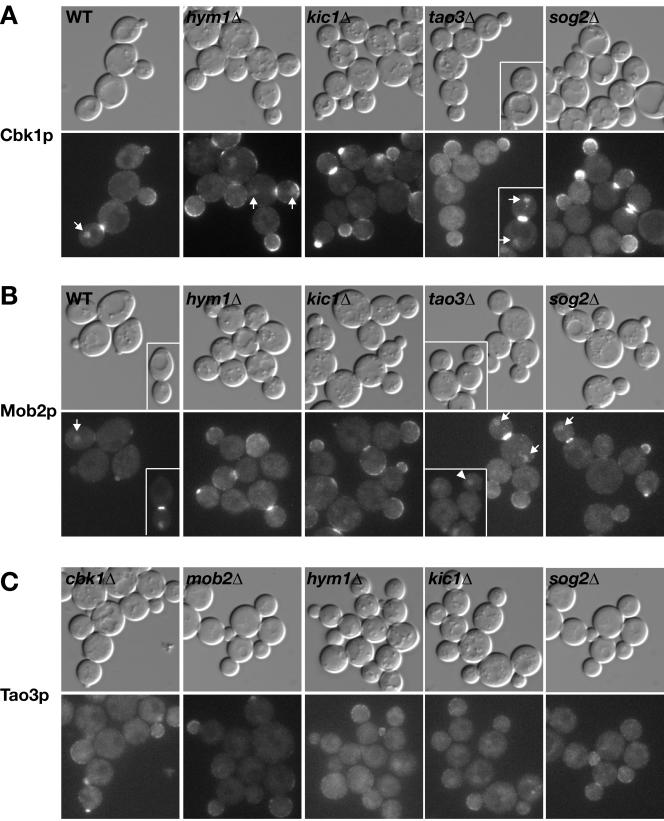
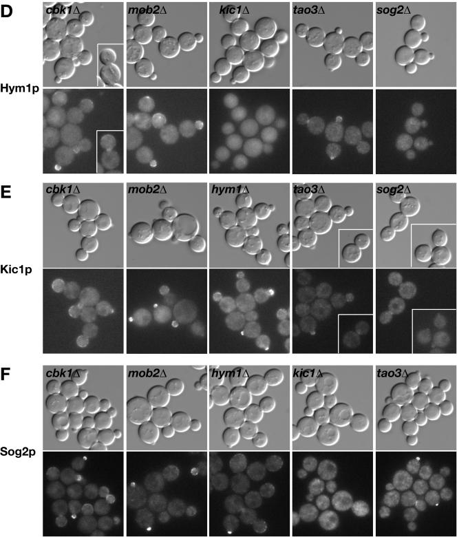
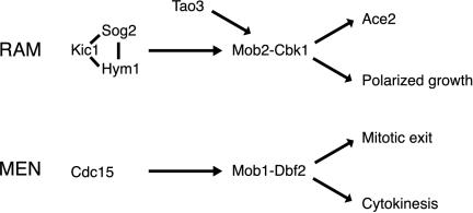
In Saccharomyces cerevisiae, polarized morphogenesis is critical for bud site selection, bud development, and cell separation. The latter is mediated by Ace2p transcription factor, which controls the daughter cell-specific expression of cell separation genes. Recently, a set of proteins that include Cbk1p kinase, its binding partner Mob2p, Tao3p (Pag1p), and Hym1p were shown to regulate both Ace2p activity and cellular morphogenesis. These proteins seem to form a signaling network, which we designate RAM for regulation of Ace2p activity and cellular morphogenesis. To find additional RAM components, we conducted genetic screens for bilateral mating and cell separation mutants and identified alleles of the PAK-related kinase Kic1p in addition to Cbk1p, Mob2p, Tao3p, and Hym1p. Deletion of each RAM gene resulted in a loss of Ace2p function and caused cell polarity defects that were distinct from formin or polarisome mutants. Two-hybrid and coimmunoprecipitation experiments reveal a complex network of interactions among the RAM proteins, including Cbk1p-Cbk1p, Cbk1p-Kic1p, Kic1p-Tao3p, and Kic1p-Hym1p interactions, in addition to the previously documented Cbk1p-Mob2p and Cbk1p-Tao3p interactions. We also identified a novel leucine-rich repeat-containing protein Sog2p that interacts with Hym1p and Kic1p. Cells lacking Sog2p exhibited the characteristic cell separation and cell morphology defects associated with perturbation in RAM signaling. Each RAM protein localized to cortical sites of growth during both budding and mating pheromone response. Hym1p was Kic1p- and Sog2p-dependent and Sog2p and Kic1p were interdependent for localization, indicating a close functional relationship between these proteins. Only Mob2p and Cbk1p were detectable in the daughter cell nucleus at the end of mitosis. The nuclear localization and kinase activity of the Mob2p-Cbk1p complex were dependent on all other RAM proteins, suggesting that Mob2p-Cbk1p functions late in the RAM network. Our data suggest that the functional architecture of RAM signaling is similar to the S. cerevisiae mitotic exit network and Schizosaccharomyces pombe septation initiation network and is likely conserved among eukaryotes.


Figures

Figure 1.
Figure 1.
RAM deletion mutants are round in morphology and are defective for cell separation and Ace2p localization. Logarithmically growing wild-type (FLY811), hym1Δ (FLY1081), cbk1Δ (FLY853), tao3Δ (FLY1124), kic1Δ (FLY1134), and mob2Δ (FLY849) cells expressing Ace2-GFP were visualized by differential interference contrast microscopy (top) and fluorescence microscopy (bottom). Arrows point to Ace2-GFP in the nuclei of representative cells. Note that the relative fluorescence of Ace2-GFP in the nuclei of RAM mutants is weaker than in the daughter cell nuclei of wild-type cells. Morphological and cell separation phenotypes were not affected by Ace2-GFP expression (our unpublished data). All images were captured and processed identically.
Figure 2.
Figure 2.
DNA microarray analysis of RAM deletion mutants. DNA microarray analysis of hym1Δ (Y1614), cbk1Δ (Y1726), tao3Δ (Y3152), kic1Δ (Y3323), and mob2Δ (Y3424) cells show overlapping expression profiles. Logarithmically growing cells were harvested or treated with 50 nM α-factor for 2 h before harvesting. Cells growing for vegetative profiling were grown in synthetic media containing all amino acids, cells treated with α-factor were grown in rich media. Two-dimensional clustering of DNA microarray profiles is presented. Gene expression is measured on a color scale (log10) with increased gene induction (red) or repression (green) corresponding to increased intensity. A gene cluster corresponding to Ace2p regulated genes is enlarged from the two-dimensional clustering for detailed examination (middle).
Figure 3.
Figure 3.
HYM1, CBK1, TAO3, KIC1, and MOB2 are necessary for formation of mating projections. Logarithmically growing cells were treated with 50 nM α-factor for 2 h and stained with rhodamine-phalloidin to visualize filamentous actin. (A) Representative micrographs are shown of cells that successfully formed mating projections. Cells were treated with rhodamine-phalloidin for detecting F-actin. Top, differential interference contrast microscopy; bottom, fluorescence microscopy. (B) Wild-type, hym1Δ, cbk1Δ, tao3Δ, kic1Δ, and mob2Δ and bni1Δ cells were scored for their ability to form mating projections. (C) RAM double mutants were scored for their ability to form mating projections. (D) Cells harboring RAM gene deletions in combination with bni1Δ were scored for their ability to form mating projections. More than 100 cells were scored in three separate counts. Strains used were wild type (SY2625), hym1Δ (Y1744), cbk1Δ (1726), tao3Δ (3481), kic1Δ (3487), mob2Δ (3482), bni1Δ (Y587), hym1Δcbk1Δ (Y1749), hym1Δtao3Δ (Y3609), hym1Δkic1Δ (Y3628), hym1Δmob2Δ (Y3608), cbk1Δtao3Δ (Y3627), cbk1Δkic1Δ (Y3606), cbk1Δmob2Δ (Y3626), tao3Δkic1Δ (Y3644), tao3Δmob2Δ (Y3645), and kic1Δmob2Δ (Y3625).
Figure 4.
Figure 4.
HYM1, CBK1, TAO3, KIC1, and MOB2 are important for apical growth. (A) Cells overexpressing GAL1pr-CLN1 are shown. Top, differential interference contrast microscopy; bottom, fluorescence microscopy. Strains containing GAL1-CLN1 were induced by addition of galactose and stained with rhodamine-phalloidin to visualize filamentous actin. (B) Cells were scored for bud hyperelongation over time. Scoring consisted of counting >100 cells three independent times. The strains used were wild type (W3031A), bni1Δ (Y581), bud6Δ (Y837), spa2Δ (Y580), hym1Δ (Y1603), cbk1Δ (Y1747), tao3Δ (Y3152), kic1Δ (Y3323), and mob2Δ (Y3424). pMT485, containing GAL1pr-CLN1 with the epitope tag HAx3 was provided by Mike Tyers (Samuel Lunenfeld Research Institute, Toronto, Ontario, Canada).
Figure 5.
Figure 5.
Cbk1p kinase activity is dependent on Mob2p, Hym1p, Kic1p, and Tao3p. Top, immunoblot of Cbk1-Myc immunoprecipitated from mating pheromone-treated wild-type (Y3280), mob2Δ (Y4035), hym1Δ (Y4032), kic1Δ (Y4034), tao3Δ (Y4033), ace2Δ (FLY1268), or untagged cells (W303-1A). The immunoblot was probed with anti-Myc antibody (9E10). Middle, an autoradiograph of the corresponding histone H1 kinase assays. The bottom panel shows graph of relative kinase activity. The Cbk1p-dependent histone H1 kinase activity from untagged cells was normalized to zero. The data from one of five experiments are presented; each experiment showed similar results. The maximal Cbk1p kinase activities in wild-type and ace2Δ cells are variable from experiment to experiment; thus, the apparent difference between kinase activities of wild-type and ace2Δ cells in the presented experiment is not significant.
Figure 6.
Figure 6.
RAM protein interaction network. (A) Pairwise two-hybrid qualitative LacZ assays were performed among the various RAM proteins. The corresponding amino acid regions of each protein are noted in parentheses. Red boxes indicate a positive interaction; black boxes indicate no interaction. (B) Coimmunoprecipitation of Kic1-HA with Hym1-Myc. Extracts prepared from cells expressing Hym1-Myc or untagged Hym1p were immunoprecipitated with anti-Myc. Immunoprecipitated proteins were detected by immunoblot analysis with antibodies directed against HA (top) or Myc (bottom). Strains used were Kic1-HA Hym1-Myc (Y4089) and Kic1-HA (Y3615). (C) Summary of the RAM protein interaction network based on our data and that of others (Racki et al., 2000; Ito et al., 2001; Du and Novick, 2002; Ho et al., 2002; Weiss et al., 2002). YOR353c = SOG2.
Figure 7.
Figure 7.
Sog2p is required for proper Ace2p localization, cell morphogenesis, and Cbk1p kinase activity. (A) Logarithmically growing sog2Δ (FLY1470) cells expressing Ace2-GFP were visualized by differential interference contrast microscopy (top) and fluorescence microscopy (bottom). Arrows point to Ace2-GFP in the nuclei of representative cells. (B) Top, immunoblot of Cbk1-Myc immunoprecipitated from mating pheromone-treated untagged cells (W303-1A), ace2Δ (FLY1268), mob2Δ (Y4035), and sog2Δ cells (FLY1492). The immunoblot was probed with anti-Myc antibody (9E10). Bottom, an autoradiograph of the corresponding histone H1 kinase assays. The relative kinase activity of immunoprecipitated Cbk1p from sog2Δ and mob2Δ cells was as low as immunoprecipitated kinase activity from untagged cells and was ∼10-fold less than from ace2Δ cells. Similar data was obtained from asynchronous cells (our unpublished data).
Figure 8.
Figure 8.
Tao3p, Hym1p, Kic1p, and Sog2p localize to sites of polarized growth. (A) Logarithmically growing homozygous diploid strains expressing Tao3-GFP (FLY1267), Hym1-GFP (FLY1244), Kic1-GFP (FLY1258), and Sog2-GFP (FLY1382) were visualized by differential interference contrast or fluorescence microscopy. Arrows point to bud neck localizations. (B) Tao3-GFP, Hym1-GFP, Kic1-GFP, and Sog2-GFP proteins localize to the tips of the mating projections upon pheromone treatment. MATa cells (FLY1263, FLY891, FLY947, and FLY1347) were treated with 5 μM mating pheromone for 2 h before visualization. All images were captured and processed identically.
Figure 9.
Figure 9.
Relationship between Cbk1p, Mob2p, Tao3p, Hym1p, Kic1p, and Sog2p for protein localization. Logarithmically growing cells expressing GFP-tagged proteins were visualized by differential interference contrast and fluorescence microscopy. (A) Localization of Cbk1-GFP in wild-type (FLY895), hym1Δ (FLY1383), kic1Δ (FLY1344), tao3Δ (FLY1259), and sog2Δ cells (FLY1515). Arrows point to nuclear localization of Cbk1-GFP in late mitotic cells. (B) Localization of Mob2-GFP in wild-type (FLY893), hym1Δ (FLY1342), kic1Δ (FLY1343), tao3Δ (FLY1260) and sog2Δ (FLY1499) cells. Arrows point to nuclear localization of Mob2-GFP. The arrowhead points to a G1 tao3Δ cell (inset) with Mob2-GFP in the nucleus. (C) Localization of Tao3-GFP in cbk1Δ (FLY1324), mob2Δ (FLY1386), hym1Δ (FLY1326), kic1Δ (FLY1325), and sog2Δ (FLY1500) cells. (D) Localization of Hym1-GFP in cbk1Δ (FLY1246), mob2Δ (FLY1245), kic1Δ (FLY1261), tao3Δ (FLY1262), and sog2Δ (FLY1514) cells. (E) Localization of Kic1-GFP in cbk1Δ (FLY1248), mob2Δ (FLY1247), hym1Δ (FLY1278), tao3Δ (FLY1249), and sog2Δ (FLY1465) cells. (F) Localization of Sog2-GFP in cbk1Δ (FLY1475), mob2Δ (FLY1474), hym1Δ (FLY1472), kic1Δ (FLY1471), and tao3Δ (FLY1473) cells. Immunoblots of the deletion strains reveal no significant differences in RAM protein levels (our unpublished data). Each image was captured and processed identically.
Figure 9.
Figure 9.
Relationship between Cbk1p, Mob2p, Tao3p, Hym1p, Kic1p, and Sog2p for protein localization. Logarithmically growing cells expressing GFP-tagged proteins were visualized by differential interference contrast and fluorescence microscopy. (A) Localization of Cbk1-GFP in wild-type (FLY895), hym1Δ (FLY1383), kic1Δ (FLY1344), tao3Δ (FLY1259), and sog2Δ cells (FLY1515). Arrows point to nuclear localization of Cbk1-GFP in late mitotic cells. (B) Localization of Mob2-GFP in wild-type (FLY893), hym1Δ (FLY1342), kic1Δ (FLY1343), tao3Δ (FLY1260) and sog2Δ (FLY1499) cells. Arrows point to nuclear localization of Mob2-GFP. The arrowhead points to a G1 tao3Δ cell (inset) with Mob2-GFP in the nucleus. (C) Localization of Tao3-GFP in cbk1Δ (FLY1324), mob2Δ (FLY1386), hym1Δ (FLY1326), kic1Δ (FLY1325), and sog2Δ (FLY1500) cells. (D) Localization of Hym1-GFP in cbk1Δ (FLY1246), mob2Δ (FLY1245), kic1Δ (FLY1261), tao3Δ (FLY1262), and sog2Δ (FLY1514) cells. (E) Localization of Kic1-GFP in cbk1Δ (FLY1248), mob2Δ (FLY1247), hym1Δ (FLY1278), tao3Δ (FLY1249), and sog2Δ (FLY1465) cells. (F) Localization of Sog2-GFP in cbk1Δ (FLY1475), mob2Δ (FLY1474), hym1Δ (FLY1472), kic1Δ (FLY1471), and tao3Δ (FLY1473) cells. Immunoblots of the deletion strains reveal no significant differences in RAM protein levels (our unpublished data). Each image was captured and processed identically.
Figure 10.
Figure 10.
Models of conserved regulatory MEN and RAM networks in yeast. The Mob2p–Cbk1p kinase complex likely functions late in the RAM network because 1) Cbk1p kinase activity is dependent on all known RAM proteins, and 2) Mob2p and Cbk1p are the only RAM proteins detectable in the daughter cell nucleus at the end of mitosis. Kic1p, Sog2p, and Hym1p may function together as a complex because Hym1p binds Sog2p and Kic1p and is Sog2p- and Kic1p-dependent for localization. By analogy to the MEN protein Cdc15p, which activates the Mob1p–Dbf2p kinase complex in vitro, Kic1p may phosphorylate and activate the Mob2p–Cbk1p complex. Tao3p is a 270-kDa protein of unknown function that interacts with Cbk1p and Kic1p. Thus, Tao3p may serve as a scaffold that facilitates activation of Mob2p–Cbk1p kinase by Kic1p.

Similar articles

Cited by

References

    1. Amberg, D.C., Zahner, J.E., Mulholland, J.W., Pringle, J.R., and Botstein, D. (1997). Aip3p/Bud6p, a yeast actin-interacting protein that is involved in morphogenesis and the selection of bipolar budding sites. Mol. Biol. Cell 8, 729–753. - PMC - PubMed
    1. Andersen, S.S., and Bi, G.Q. (2000). Axon formation: a molecular model for the generation of neuronal polarity. Bioessays 22, 172–179. - PubMed
    1. Baillat, G., Moqrich, A., Castets, F., Baude, A., Bailly, Y., Benmerah, A., and Monneron, A. (2001). Molecular cloning and characterization of phocein, a protein found from the Golgi complex to dendritic spines. Mol. Biol. Cell 12, 663–673. - PMC - PubMed
    1. Bardin, A.J., and Amon, A. (2001). Men and sin: what's the difference? Nat. Rev. Mol. Cell. Biol. 2, 815–826. - PubMed
    1. Bidlingmaier, S., Weiss, E.L., Seidel, C., Drubin, D.G., and Snyder, M. (2001). The Cbk1p pathway is important for polarized cell growth and cell separation in Saccharomyces cerevisiae. Mol. Cell. Biol. 21, 2449–2462. - PMC - PubMed

Publication types

MeSH terms