. 2003 Sep;14(9):3929-41.
doi: 10.1091/mbc.e03-01-0854. Epub 2003 May 29.

The yeast hnRNP-like protein Hrp1/Nab4 sccumulates in the cytoplasm after hyperosmotic stress: a novel Fps1-dependent response

Affiliations

The yeast hnRNP-like protein Hrp1/Nab4 sccumulates in the cytoplasm after hyperosmotic stress: a novel Fps1-dependent response

Michael F Henry et al. Mol Biol Cell. 2003 Sep.

Abstract

The Hrp1/Nab4 shuttling protein belongs to a family of RNA binding proteins that bind to nascent RNA polymerase II transcripts and form hnRNP complexes. Members of this family function in a staggering array of cellular activities, ranging from transcription and pre-mRNA processing in the nucleus to cytoplasmic mRNA translation and turnover. It has recently been recognized that the yeast stress response can include alterations in hnRNP-mediated mRNA export. We now report that the steady-state localization of Hrp1p rapidly shifts from the nucleus to the cytoplasm in response to osmotic stress. In contrast to a general stress response resulting in a transient relocation, Hrp1p redistribution is specific to hyperosmotic stress and is only reversed after stress removal. Hrp1p relocalization requires both the CRM1/XPO1 exportin and the FPS1 glycerol transporter genes but is independent of ongoing RNA transcription and protein arginine methylation. However, mutations in the high osmolarity glycerol and protein kinase C osmosensing pathways do not impact the Hrp1p hyperosmotic response. We present a working model for the cytoplasmic accumulation of Hrp1 and discuss the implications of this relocalization on Hrp1p function.


Figures

Figure 1.
Figure 1.
Hrp1p exhibits a steady-state nuclear localization. (A) Localization of Hrp1p at 25–36°C. Wild-type cells were grown to 1 × 10⁷ cells/ml in SC medium at 25°C (top row), 30°C (middle row), or 36°C (bottom row). Cells were then fixed and processed as described for immunofluorescence. Cells were photographed by use of Nomarski optics (right column) and DNA was stained with 4,6-diamidino-2-phenylindole (middle column). Hrp1p was visualized with anti-Hrp1 antisera (left column). (B) Schematic diagram of Hrp1p shuttling and steady-state localization during standard growth conditions. In this diagram, Hrp1p seems to localize to the nucleus because its rate of nuclear import is faster than its rate of export.
Figure 2.
Figure 2.
Hrp1p rapidly relocates to the cytoplasm in response to hyperosmolarity. (A) Time course of Hrp1p cytoplasmic accumulation after hyperosmotic exposure. Cells were incubated at 30°C, transferred to hyperosmotic conditions (0.7 M NaCl), and then incubated at either 30°C (top two panels) or 4°C (bottom two panels). Hrp1p was assessed 0, 5, 15, 30, and 60 min after hyperosmotic exposure. Hrp1p was also localized in cells grown continuously (>10 generations) in media containing 0.7 M NaCl. Cells from each time point were subjected to immunofluorescence microscopy with anti-Hrp1 antisera. Hrp1p was visualized with fluorescein isothiocyanate (FITC)-labeled anti-rabbit antibody (top). Cells were photographed by use of Nomarski optics (bottom). (B) Hypotonic shock does not induce Hrp1 cytoplasmic accumulation. For hypotonic shock, cultures grown at 30°C in YEPD were diluted with 4 volumes of water. The Hrp1p protein was assessed at 0, 5, 15, 30, and 60 min after hypotonic shock. Cells from each time point were subjected to immunofluorescence microscopy with anti-Hrp1 antisera. Hrp1p was visualized with FITC labeled anti-rabbit antibody (top). (C) Hrp1p remains intact after hyperosmotic exposure. Yeast extracts were prepared after they were grown to log phase and shifted to hyperosmotic conditions without (–, left gel) or with (+, right gel) cycloheximide addition. The lysates were separated on a 10% SDS-polyacrylamide gel and analyzed on a Western blot with antibodies against Hrp1p. (D) The cytoplasmic accumulation of Hrp1p after hyperosmotic shock is at least partially due to the nuclear export of preexisting Hrp1p. Wild-type cells expressing myc-Hrp1 were grown, radiolabeled, lysed, and immunoprecipitates were prepared. Immune complexes were analyzed by Western blot and autoradiography. Cells were labeled in medium containing glucose (lane 1) followed by a 1-h chase in the same medium containing 0.7 M NaCl (lanes 2–4).
Figure 3.
Figure 3.
Cytoplasmic Hrp1p is imported into the nucleus after a return to standard osmolarity. Wild-type cells were grown to log phase and stressed with 0.7 M NaCl for 30 min (first row). The NaCl was removed and Hrp1p localization was examined at 15- (second row), 30- (third row), and 60 (fourth row)-min time points. Cells were prepared for immunofluorescence with anti-Hrp1p antisera and then with fluorescein isothiocyanate-labeled anti-rabbit antibody to visualize Hrp1p (top row). Cells were photographed by use of Nomarski optics (bottom row).
Figure 4.
Figure 4.
Hyperosmotic-mediated Hrp1p export is independent of both RNA synthesis and arginine methylation. Wild-type (B, E, and H), rpb1-1 (A, D, and G), or ΔHMT1 (C, F, and I) cells were grown to log phase in SC medium at 30°C. The rpb1-1 cells were shifted to 36°C for 30 min (A) and then stressed with 0.7 M NaCl for 15 (D) and 30 (G) min. The wild-type cells were incubated at 30°C in the presence of thiolutin for 15 min (B) and then transferred to medium containing 0.7 M NaCl for 15 (E) and 30 (H) min. The ΔHMT1 cells were incubated at 30°C (C) and then transferred to medium containing 0.7 M NaCl for 15 (F) and 30 (I) min. Cells were prepared for immunofluorescence with anti-Hrp1p antisera and then with fluorescein isothiocyanate-labeled anti-rabbit antibody to visualize Hrp1p.
Figure 5.
Figure 5.
Schematic representation of the main components of the S. cerevisiae Hog1p and Pkc1p pathways. (A) The HOG pathway. Two osmosensors, Sho1p and Sln1p, stimulate the HOG MAP kinase cascade by different mechanisms. Activation of this MAP kinase cascade leads to the phosphorylation of the Hog1 MAP kinase, which is then imported into the nucleus. Four putative nuclear targets of Hog1p (Msn1p, Msn2p, Msn3p, and Hot1p) are also shown. (B) The Pkc1p pathway. Dotted arrows indicate activation of the Pkc1 pathway in which the detailed mechanisms of signal transduction are not yet known.
Figure 6.
Figure 6.
The Hog1p and Pkc1 kinase cascades are not required for the hyperosmotic localization of Hrp1p. (A) Localization of Hrp1 in strains deleted for components of the PKC1 pathway. Hrp1p was localized 0 (first row), 15 (second row), and 60 min (third row) after exposure to hyperosmolarity, as well as 10 min (fourth row) after the return to standard osmolarity. (B) Localization of Hrp1p in wild-type and Δpkc1 cells. Wild-type (MHY745) and Δpkc1 (MHY746) cells were collected at 0 (first row), 30 (second row), 60 (third row), and 120 min (fourth row) after the sorbitol level was increased from 0.5 to 1.4 M. (C) Localization of Hrp1p in wild-type and Δpkc1 BCK1–20 cells. Hrp1p was localized 0 (first row), 15 (second row), 60 (third row), and 120 min (fourth row) after exposure to 0.7 M NaCl. All cells were prepared for immunofluorescence with anti-Hrp1p antisera and then with fluorescein isothiocyanate-labeled anti-rabbit antibody to visualize Hrp1p.
Figure 7.
Figure 7.
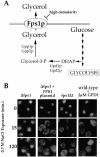
Hyperosmotic localization of Hrp1p requires the glycerol export protein, Fps1, but is independent of intracellular glycerol levels. (A) Glycerol efflux is controlled by the activity of the Fps1p channel protein. Glycerol is produced as the product of a side branch of glycolysis. DHAP is converted into glycerol-3-P by the actions of Gdp1p and Gdp2p. The glycerol-3-P is subsequently dephosphorylated by Gpp1p and Gpp2p. Under conditions of increased osmolarity, the Fps1p glycerol facilitator is closed enabling the cells to accumulate intracellular glycerol. When glycerol is produced during normal growth conditions, however, glycerol is leaked through the Fps1 channel to the outside medium. (B) Localization of Hrp1p in Δfps1 cells (first column), Δfps1 + FPS1 plasmid (second column), fps1Δ1 (third column), and wild-type + 2 μ GDP1 (fourth column) after hyperosmotic shock for 0, 15, or 120 min. Cells were prepared for immunofluorescence with anti-Hrp1p antisera and then with fluorescein isothiocyanate-labeled anti-rabbit antibody to visualize Hrp1p.
Figure 8.
Figure 8.
Hrp1p export after hypertonic stress is CRM1/XPO1 dependent. Localization of Hrp1p in xpo1ts cells at 36°C (first column) and 25°C (second column), Δlos1 (third column), Δmsn5 (fourth column) after hyperosmotic shock for 0, 15, and 120 min. Cells were prepared for immunofluorescence as described previously.
Figure 9.
Figure 9.
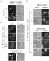
The Kap104 importin protein is a likely candidate to mediate hyperosmotic Hrp1 cytoplasmic accumulation. (A) Hrp1p accumulates in the cytoplasm of a kap104-16 mutant strain after a shift to the restrictive temperature. Cells were incubated at 25°C (top two panels) and then shifted to 36°C (top two panels) for 60 min. Cells from both time points were subjected to immunofluorescence microscopy with anti-Hrp1 antisera. Hrp1p was visualized with fluorescein isothiocyanate (FITC)-labeled anti-rabbit antibody (left). Cells were photographed by use of Nomarski optics (right). (B) Time course of Nab2p cytoplasmic accumulation after hyperosmotic exposure. Cells were incubated at 30°C and then transferred to hyperosmotic conditions (0.7 M NaCl). Nab2p was localized at 0, 30 min, and 10 generations after hyperosmotic exposure. Hrp1p was also localized 5 min after the removal of hyperosmotic conditions. Cells from each time point were subjected to immunofluorescence microscopy with anti-Nab2p antisera. Nab2p was visualized with fluorescein isothiocyanate (FITC)-labeled anti-mouse antibody (left) and cells were photographed by use of Nomarski optics (right). (C) Cytoplasmic Hrp1p does not accumulate in the nuclei of crm1 (xpo1-1) cells at the restrictive temperature. Cells incubated at the permissive temperatue of 25°C were transferred to hyperosmotic conditions (0.7 M NaCl) and Hrp1p was localized at 0 and 30 min after hyperosmotic exposure (top two panels). The cells were then shifted to 36°C and Hrp1p was localized at 15, 60, and 120 min (bottom three panels). Cells from each time point were subjected to immunofluorescence microscopy with anti-Hrp1p antisera. Hrp1p was visualized with FITC-labeled anti-rabbit antibody (right) and cells were photographed by use of Nomarski optics (left).

Similar articles

Cited by

References

    1. Aitchison, J.D., Blobel, G., and Rout, M.P. (1996). Kap104p: a karyopherin involved in the nuclear transport of messenger RNA binding proteins. Science 274, 624–627. - PubMed
    1. Arts, G.J., Fornerod, M., and Mattaj, I.W. (1998). Identification of a nuclear export receptor for tRNA. Curr. Biol. 8, 305–314. - PubMed
    1. Blondel, M., Alepuz, P.M., Huang, L.S., Shaham, S., Ammerer, G., and Peter, M. (1999). Nuclear export of Far1p in response to pheromones requires the export receptor Msn5p/Ste21p. Genes Dev. 13, 2284–2300. - PMC - PubMed
    1. Boeke, J.D., LaCroute, F., and Fink, G.R. (1984). A positive selection for mutants lacking orotidine-5′-phosphate decarboxylase activity in yeast: 5-fluoro-orotic acid resistance. PG. Mol. Gen. Genet. 197, 345–347. - PubMed
    1. Bossie, M.A., DeHoratius, C., Barcelo, G., and Silver, P. (1992). A mutant nuclear protein with similarity to RNA binding proteins interferes with nuclear import in yeast. Mol. Biol. Cell 3, 875–893. - PMC - PubMed

Publication types

MeSH terms

LinkOut - more resources