Comparative Study
. 2003 Oct;23(19):6909-21.
doi: 10.1128/MCB.23.19.6909-6921.2003.

Functions of the ectodomain and cytoplasmic tyrosine phosphatase domains of receptor protein tyrosine phosphatase Dlar in vivo

Affiliations
Comparative Study

Functions of the ectodomain and cytoplasmic tyrosine phosphatase domains of receptor protein tyrosine phosphatase Dlar in vivo

Neil X Krueger et al. Mol Cell Biol. 2003 Oct.

Abstract

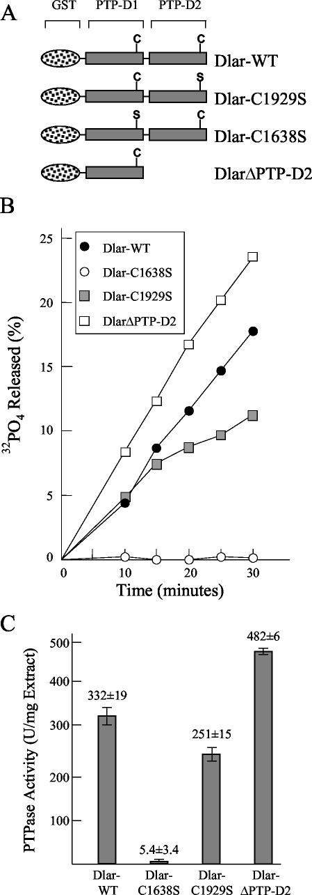
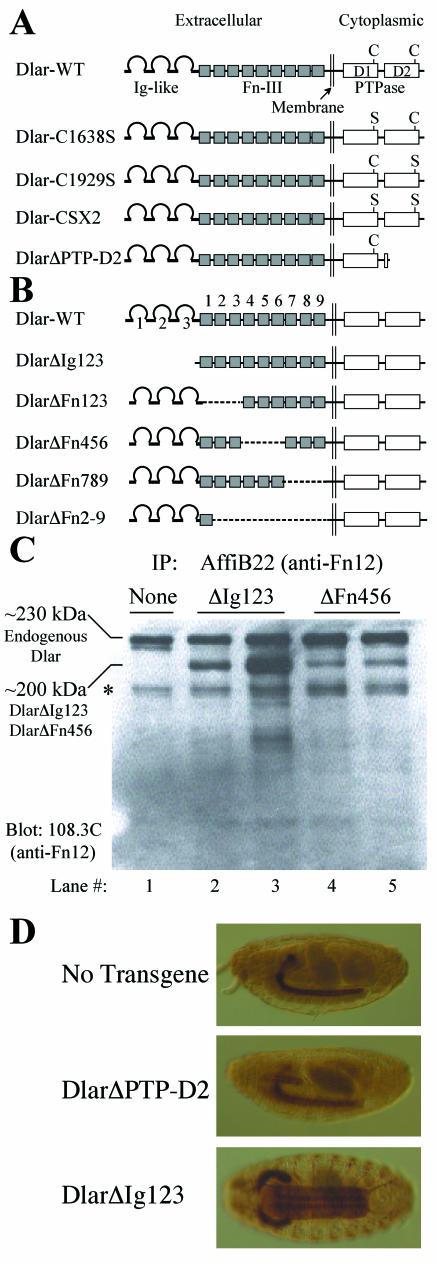
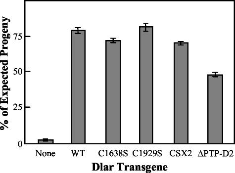
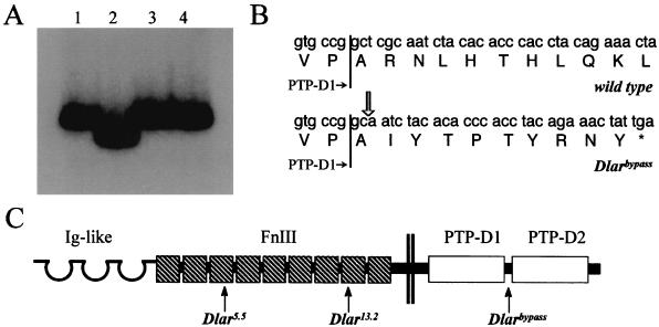
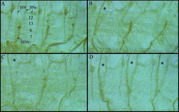
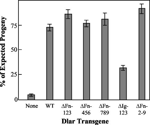
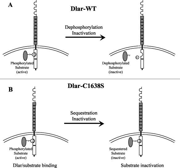
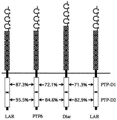
The receptor protein tyrosine phosphatase (PTPase) Dlar has an ectodomain consisting of three immunoglobulin (Ig)-like domains and nine fibronectin type III (FnIII) repeats and a cytoplasmic domain consisting of two PTPase domains, membrane-proximal PTP-D1 and C-terminal PTP-D2. A series of mutant Dlar transgenes were introduced into the Drosophila genome via P-element transformation and were then assayed for their capacity to rescue phenotypes caused by homozygous loss-of-function genotypes. The Ig-like domains, but not the FnIII domains, are essential for survival. Conversely, the FnIII domains, but not the Ig-like domains, are required during oogenesis, suggesting that different domains of the Dlar ectodomain are involved in distinct functions during Drosophila development. All detectable PTPase activity maps to PTP-D1 in vitro. The catalytically inactive mutants of Dlar were able to rescue Dlar(-/-) lethality nearly as efficiently as wild-type Dlar transgenes, while this ability was impaired in the PTP-D2 deletion mutants DlarDeltaPTP-D2 and Dlar(bypass). Dlar-C1929S, in which PTP-D2 has been inactivated, increases the frequency of bypass phenotype observed in Dlar(-/-) genotypes, but only if PTP-D1 is catalytically active in the transgene. These results indicate multiple roles for PTP-D2, perhaps by acting as a docking domain for downstream elements and as a regulator of PTP-D1.


Figures

FIG. 1.
FIG. 1.
In vitro PTPase assay of GST-Dlar fusion proteins. (A) Constructs used for PTPase assay. (B) Time course of Dlar phosphorylation by GST-Dlar fusion proteins. The fusion proteins were purified to apparent homogeneity as assessed by bromophenol blue staining of SDS-polyacrylamide gels, and the purified proteins were assayed at various time points for their ability to dephosphorylate the [32P]tyrosine-phosphorylated peptide substrate Raytide. (C) The time course data from panel B were used to calculate specific enzyme activity. The data are represented as means ± standard errors of the means.
FIG. 2.
FIG. 2.
Dlar transgenes and their expression in vivo. (A) Dlar cytoplasmic domain transgenes. Dlar-WT is shown at the top, and the Cys-to-Ser point mutants are diagrammed below. The DlarΔPTP-D2 truncation mutant is truncated at the beginning of PTP-D2. (B) Dlar ectodomain truncation mutant transgenes. The top line shows wild-type Dlar. The Ig-like domains and FnIII domains are numbered from the amino terminus (left). The missing domains in these mutants are shown by dotted lines. (C) Expression of Dlar transgenes. Extracts from wild-type embryos or embryos expressing Dlar transgenes under control of the C155 driver were immunoprecipitated (IP) with affinity-purified anti-Dlar polyclonal antibodies directed toward FnIII repeats 1 + 2 and immunoblotted with anti-Dlar monoclonal antibody (108.3C), also directed toward FnIII repeats 1 + 2. Lane 1, nontransgenic wild-type embryo (yw); lanes 2 and 3, extracts from two independent DlarΔIg123 transgenic lines; lanes 4 and 5, extracts from two independent DlarΔFn456 transgenic lines. The asterisk marks a protein isoform likely to be the proteolytically processed extracellular domain of Dlar. All transgenic embryos shown express Dlar transgenes in a wild type background. The bands corresponding to endogenous Dlar and transgenic Dlar are indicated at the left. (D) Embryo staining with anti-Dlar antibodies. (Top panel) Lateral view of a wild-type (yw) embryo stained with anti-Dlar antibodies. (Middle panel) Lateral view of a transgenic embryo expressing DlarΔPTP-D2 under control of the C155 driver. Note the characteristic salivary gland expression induced by C155. (Bottom panel) Ventral view of a transgenic embryo expressing DlarΔIg123 under C155 control. In this view, the cell bodies of ISN motoneurons can be easily visualized in the neuropils.
FIG. 3.
FIG. 3.
Bar graph of rescue of Dlar−/− lethality by Dlar cytoplasmic domain mutant transgenes. The bars represent the mean percentage of expected progeny observed ± standard deviation when the indicated Dlar transgene is expressed under control of the C155-GAL4 driver in a loss-of-function background. The results are pooled from rescue of several different Dlar−/− genotypes that were generated by using multiple combinations of Dlar loss-of-function alleles. At least two independently inserted Dlar transgenes were tested for each mutant shown.
FIG. 4.
FIG. 4.
The Dlarbypass allele encodes a protein truncated after PTP-D1. (A) Autoradiograph of 32P-labled PCR products subjected to SSCP electrophoresis. Lane 1, Wild-type parental genotype (A49/A49); Lane 2, Dlarbypass/Dlarbypass; lane 3, Dlar5.5/+; lane 4, Dlar13.2/+. (B) Dlarbypass/Dlarbypass genomic DNA from the region recognized by the primers in panel A was subcloned and sequenced. Dlarbypass has a 4-bp deletion relative to the wild-type Dlar sequence. The location of the deletion is indicated by the open arrow. The asterisk indicates a stop codon. The end of the coding sequence for PTP-D1 is indicated by vertical lines. (C) Proteins encoded by Dlarbypass, Dlar5.5, and Dlar13.2. Arrows indicate the locations where the alleles are truncated by mutation.
FIG. 5.
FIG. 5.
The frequency of bypass phenotypes is increased in Dlar/Dlar embryos by overexpression of Dlar-C1929S. Embryonic motoneuron pathways were visualized in stage 16 embryos by staining with anti-fasciclin II monoclonal antibody. The numbers in panel A indicate specific muscles within each hemisegment; ISN, ISNa, and ISNb are indicated by arrows. In all panels, asterisks indicate hemisegments with a bypass phenotype. The frequencies of bypass phenotype observed in this experiment are 32.3% ± 3.0% (n = 369 hemisegments scored) for Dlar5.5/13.2 embryos expressing no transgene, 34.6% ± 3.1% (n = 350) for Dlar5.5/13.2 embryos expressing the Dlar-WT transgene, 38.9% ± 4.9% (n = 162) for Dlar5.5/13.2 embryos expressing Dlar-C1638S, 54.8% ± 3.6% (n = 429) for Dlar5.5/13.2 embryos expressing Dlar-C1929S, and 25.3% ± 3.2% (n = 249) for Dlar5.5/13.2 embryos expressing Dlar-CSX2. Expression of Dlar-WT does not generate statistically significant rescue of the bypass phenotype, but expression of Dlar-C1929S significantly increases bypass frequency. (A) A wild-type embryo. Note the wild-type pattern of innervation of muscles 6, 7, 12, and 13 by ISNb. (B) A Dlar5.5/13.2 embryo expressing no transgenes. One hemisegment is bypass and two are wild type in this photograph. (C) A Dlar5.5/13.2 embryo expressing Dlar-WT. (D) A Dlar5.5/13.2 embryo expressing Dlar-C1929S. All three hemisegments shown exhibit bypass phenotypes.
FIG. 6.
FIG. 6.
Rescue of Dlar−/− lethality by Dlar ectodomain deletion mutant transgenes. The ability of Dlar transgenes to rescue the lethality of the genotype Dlar13.2/Dlar5.5 was assessed. The represent the mean percentage of expected progeny observed ± standard deviation when the indicated Dlar transgene is expressed under control of the C155-GAL4 driver in a Dlar13.2/Dlar5.5 background. The results shown are pooled from multiple independent transgenic lines for each transgene tested.
FIG. 7.
FIG. 7.
Speculative model of the mechanism of rescue of lethality by Dlar-C1638S. The ability of Dlar-C1638S to rescue lethality nearly as well as Dlar-WT does not necessarily mean that PTPase catalytic activity is not required for Dlar function. While they are unable to catalyze dephosphorylation, it is possible for Cys-to-Ser PTPase domain mutants to stably bind to substrates (1, 18). If the in vivo function of Dlar is to functionally inactivate its substrate via dephosphorylation, as diagrammed in panel A, then panel B shows that by binding and sequestering substrate, Dlar-1638S could mimic the function of Dlar-WT without being catalytically active.
FIG. 8.
FIG. 8.
Amino acid sequence comparison in the LAR subfamily of RPTPs. The percentages of amino acid identity between the indicated PTPase domains are shown. Within the LAR subfamily, catalytically inactive PTP-D2 has a greater degree of sequence conservation than catalytically active PTP-D1.

Similar articles

Cited by

References

    1. Allard, J. D., R. Herbst, P. M. Carroll, and M. A. Simon. 1998. Mutational analysis of the SRC homology 2 domain protein-tyrosine phosphatase Corkscrew. J. Biol. Chem. 273:13129-13135. - PubMed
    1. Aricescu, A. R., I. W. McKinnell, W. Halfter, and A. W. Stoker. 2002. Heparan sulfate proteoglycans are ligands for receptor protein tyrosine phosphatase σ. Mol. Cell. Biol. 22:1881-1892. - PMC - PubMed
    1. Bateman, J., R. S. Reddy, H. Saito, and D. Van Vactor. 2001. The receptor tyrosine phosphatase Dlar and integrins organize actin filaments in the Drosophila follicular epithelium. Curr. Biol. 11:1317-1327. - PubMed
    1. Bateman, J., H. Shu, and D. Van Vactor. 2000. The guanine nucleotide exchange factor trio mediates axonal development in the Drosophila embryo. Neuron 26:93-106. - PubMed
    1. Bilwes, A. M., J. den Hertog, T. Hunter, and J. P. Noel. 1996. Structural basis for inhibition of receptor protein-tyrosine phosphatase-α by dimerization. Nature 382:555-559. - PubMed

Publication types

MeSH terms

LinkOut - more resources