Comparative Study
. 2003 Oct;23(19):6944-57.
doi: 10.1128/MCB.23.19.6944-6957.2003.

A novel human Ada2 homologue functions with Gcn5 or Brg1 to coactivate transcription

Affiliations
Comparative Study

A novel human Ada2 homologue functions with Gcn5 or Brg1 to coactivate transcription

Nickolai A Barlev et al. Mol Cell Biol. 2003 Oct.

Abstract

In yeast, the transcriptional adaptor yeast Ada2 (yAda2) is a part of the multicomponent SAGA complex, which possesses histone acetyltransferase activity through action of the yGcn5 catalytic enzyme. yAda2, among several SAGA proteins, serves to recruit SAGA to genes via interactions with promoter-bound transcription factors. Here we report identification of a new human Ada2 homologue, hAda2beta. Ada2beta differs both biochemically and functionally from the previously characterized hAda2alpha, which is a stable component of the human PCAF (human Gcn5 homologue) acetylase complex. Ada2beta, relative to Ada2alpha, interacted selectively, although not stably, with the Gcn5-containing histone acetylation complex TFTC/STAGA. In addition, Ada2beta interacted with Baf57 (a component of the human Swi/Snf complex) in a yeast two-hybrid screen and associated with human Swi/Snf in vitro. In functional assays, hAda2beta (but not Ada2alpha), working in concert with Gcn5 (but not PCAF) or Brg1 (the catalytic component of hSwi/Snf complex), increased transcription via the B-cell-specific transcription factor Pax5/BSAP. These findings support the view that Gcn5 and PCAF have distinct roles in vivo and suggest a new mechanism of coactivator function, in which a single adaptor protein (Ada2beta) can coordinate targeting of both histone acetylation and chromatin remodeling activities.


Figures
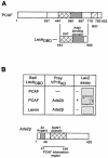
FIG. 1.
FIG. 1.
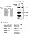
LexADBD-PCAF two-hybrid screen. (A) PCAF domain structure. Nucleosomal recognition, aa 1 to 357; HAT domain, aa 440 to 580; Ada2-binding domain, aa 580 to 667; bromodomain, aa 718 to 780. The region of PCAF (aa 444 to 695) used as bait in the two-hybrid screen is shown below. (B) Two-hybrid interaction between PCAF and Ada2β. In the upper part of the panel, LexADBD-PCAF444-695 or LexADBD-Lamin were cotransformed into yeast with the amino terminus of Ada2β fused to the VP16 activation domain. Interaction was tested by using a LacZ assay of colonies transferred to filters. Positively interacting blue colonies are shown after 30 min of color development at 30°C. In the lower part of the panel is a diagram of Ada2β. Domains conserved between different Ada2 species are indicated. The region of interaction with PCAF is indicated. (C) Sequence alignment of hAda2β, mAda2β, and hAda2α. Alignment was performed by using CLUSTAL W. Identical amino acids are shown in black boxes, and similar amino acids are shown in gray boxes.Continued.
FIG. 1.
FIG. 1.
LexADBD-PCAF two-hybrid screen. (A) PCAF domain structure. Nucleosomal recognition, aa 1 to 357; HAT domain, aa 440 to 580; Ada2-binding domain, aa 580 to 667; bromodomain, aa 718 to 780. The region of PCAF (aa 444 to 695) used as bait in the two-hybrid screen is shown below. (B) Two-hybrid interaction between PCAF and Ada2β. In the upper part of the panel, LexADBD-PCAF444-695 or LexADBD-Lamin were cotransformed into yeast with the amino terminus of Ada2β fused to the VP16 activation domain. Interaction was tested by using a LacZ assay of colonies transferred to filters. Positively interacting blue colonies are shown after 30 min of color development at 30°C. In the lower part of the panel is a diagram of Ada2β. Domains conserved between different Ada2 species are indicated. The region of interaction with PCAF is indicated. (C) Sequence alignment of hAda2β, mAda2β, and hAda2α. Alignment was performed by using CLUSTAL W. Identical amino acids are shown in black boxes, and similar amino acids are shown in gray boxes.Continued.
FIG. 2.
FIG. 2.
Examination of Ada2β association with PCAF. (A) Ada2β peptide antibodies. HeLa nuclear extract was fractionated on phosphocellulose p11 column, and bound material was step-eluted with increasing KCl (lanes 1 and 3 are a 0.3 M KCl elution, and lanes 2 and 4 are a 0.5 M KCl elution). The fractions were probed with anti-Ada2β sera either mock treated (left panel) or preincubated with the corresponding peptide (right panel). The position of the Ada2β-specific signal is shown. (B) Test of Ada2β association with PCAF in vivo. Flag-PCAF expression was induced in HeLa Tet-off cells by withdrawal of tetracycline (+). Cells treated with tetracycline served as negative control (−). Flag-agarose was used to immunoprecipitate FLAG-PCAF. Bound material was eluted with excess Flag-peptide. Immunoblotting was done with indicated antibodies. (C) Test of Ada2β interaction with PCAF or Gcn5 in the yeast two-hybrid assay. Yeast were cotransformed with PCAF, Gcn5, or Rho fused to LexADBD or LexADBD alone, along with the PCAF interacting region of Ada2β fused to VP16 activation domain. The LacZ reporter contained LexA binding sites. β-Galactosidase activity was measured in units per milligram of protein. The numbers are averages of three independent experiments.
FIG. 3.
FIG. 3.
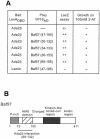
LexADBD-Ada2β two-hybrid screen. (A) Test of LexADBD-Ada2β interaction with Baf57. Either LexADBD-Ada2β or LexADBD-Lamin was cotransformed into yeast with deletion mutants of Baf57 (dimensions are indicated in parentheses) fused to the VP16 activation domain. The strength and specificity of interactions were assessed by two means: (i) color LacZ assay on filters and (ii) ability to grow on 100 mM 3-AT. (B) Schematic of Baf57. Functional domains of Baf57 are indicated. The Ada2β-interacting region is shown: P-rich (proline-rich), HMG domain (high-mobility-group domain), Charged, Kinesin-like coiled-coil region, and Acidic region.
FIG. 4.
FIG. 4.
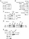
Analysis of physical interactions between Ada2β, Gcn5, and Baf57 proteins. (A) p11 column elution profiles of Gcn5, Ada2β, and Baf57. HeLa cell nuclear extract was fractionated by p11 chromatography. Flowthrough and salt elutions of bound material were analyzed by immunoblotting for Gcn5, Ada2β, and Baf57 with specific antisera. (B) GST-Ada2β interaction with Gcn5 and Baf57. In the upper panel, glutathione-beads bearing GST-Ada2α, GST-Ada2β, or GST proteins were incubated with HeLa 0.5 M fraction after p11 column chromatography. The bead-bound material was analyzed by Western blotting for indicated proteins. In the lower panel, a Coomassie blue stain of GST fusion proteins (20% of beads) used in the binding experiments is shown. (C) Coimmunoprecipitation of Brg1 and Gcn5 with Ada2β. HEK 293 cells were transiently transfected with Flag-Ada2β and Flag-Gcn5 constructs. At 48 h posttransfection, cell extracts were prepared, and Ada2β was immunoprecipitated with anti-Ada2β sera. The IgG fraction of an irrelevant polyclonal sera was used as a control for specificity. The Ada2β immunocomplexes were analyzed for the presence of Flag-Ada2β, Flag-Gcn5, and Brg1 by Western blotting with the indicated antibodies. (D) Endogenous Ada2β coimmunoprecipitates with Brg1 and Gcn5 in vivo. Endogenous Gcn5 and Brg1 were immunoprecipitated from dialyzed HeLa nuclear extract with Gcn5- or Brg1-specific polyclonal antibody. SDS-eluted material was analyzed for the presence of Brg1, Gcn5, and Ada2β with the indicated sera. The IgG fraction of an irrelevant polyclonal sera was used in mock immunoprecipitation to determine specificity of the interactions. (E) Analysis of Ada2β in purified TFTC and human Swi/Snf. The purified TFTC and hSwi/Snf (43) complexes (indicated as TFTC and hSwi/Snf, respectively) were analyzed for the presence of Ada2β. Input lane (INP) represents the starting HeLa nuclear extract material used in purification of the complexes. The positions of the TFTC- and hSwi/Snf-specific components are indicated.
FIG. 5.
FIG. 5.
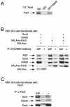
Pax5 physical interactions with Ada2β, Gcn5, and Brg1. (A) Physical interaction of Pax5 with GST-Ada2β in vitro. In vitro-translated Pax5 was incubated with beads bearing GST or GST-Ada2β. After several washes, bound material was eluted and resolved by SDS-PAGE. The gel was enhanced and exposed to X-ray film for 24 h to detect S35-labeled Pax5. (B) Coimmunoprecipitations of GAL4DBD-Pax5 with Gcn5, Ada2β, and Brg1. Whole-cell extract from HEK 293 cells transfected with GAL4DBD, GAL4DBD-Pax5, Gcn5, and Ada2β (as indicated) were subjected to immunoprecipitations with anti-GAL4 monoclonal antibody. The immunoprecipitated samples were analyzed by Western blotting for the presence of the indicated proteins with specific antisera. The input lanes (INP) represent 10% of the starting material. (C) Pax5 immunoprecipitation from HEK 293 cells. Whole-cell extract from cells transfected with Pax5 was immunoprecipitated with anti-Pax5 monoclonal antibody or mock precipitated with the IgG fraction of unrelated monoclonal antibodies. Immunoblotting was done for Gcn5, Ada2β and Pax-5.
FIG. 6.
FIG. 6.
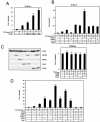
Pax5 functional interactions with Ada2α/Ada2β, Gcn5/PCAF, and Brg1. (A) Effect of increasing amounts of Ada2β on Gal4DBD-Pax5. HEK 293 cells were transfected with Gal4DBD-Pax5 expression vector and various amounts (0.2, 0.5, and 1 μg) of pCDNA3-Ada2β vector. Cells were cotransfected with a reporter bearing the luciferase gene under control of Gal4-binding sites integrated into the E1B promoter region (E1Bluc). The basal activity of E1Bluc was ∼1,000 light units. Error bars indicate the standard deviation. (B) Effect of coactivators on Gal4DBD-Pax5-dependent transcription. For the upper part of panel B, HEK 293 cells were transiently transfected with 200 ng of the Gal4DBD-Pax5 mammalian expression vector in various combinations with the indicated expression vectors, together with the Gal4-responsive luciferase reporter, E1Bluc. Typically, 0.5 μg of Ada2β or Ada2α expression vectors and 1 μg of Gcn5 or PCAF expression vectors were used in transfections. As shown in the lower section of panel B, Gal4DBD alone, or in any combination with the indicated coactivators (Ada2β, Gcn5, or PCAF), did not activate transcription of the E1Bluc reporter. (C) Western analysis of transfected proteins. Immunoblot analysis of ectopically expressed proteins in HEK 293 cells was done. Cells were transfected as for the upper panel and then immunoblotted. The protein expression levels were determined with specific antibodies: anti-Pax5 (Pharmingen) and anti-Flag (Sigma) for detection of Ada2β and PCAF, anti-HA (Babco) for detection of Ada2α, and anti-Gcn5 custom-made polyclonal serum. (D) Pax5 cotransfections with coactivators in H1299 cells. Pax5 DNA was transfected in different combinations with the indicated DNAs. Cells were cotransfected with the luciferase reporter pGL3 (no Pax5-binding sites) or pGL3 (CD19-2), bearing two Pax5-binding sites derived from the CD-19 promoter, a gift of James Hagman. The basal activity of pGL3 was ∼10,000 light units. Error bars are shown.
FIG. 7.
FIG. 7.
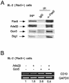
Interaction of Pax5 with Ada2β, Gcn5, and Brg1 in Burkitt lymphoma cells. (A) Coimmunoprecipitation of Pax5 with Ada2β, Gcn5, and Brg1 in BL-2 cells. Pax5 was immunoprecipitated from nuclear extracts with Pax5 monoclonal antibodies. Nonspecific monoclonal IgG was used as a negative control. Immunoprecipitates were examined for Ada2β, Gcn5, and Brg1 by Western blotting. (B) Effect of Ada2β and Gcn5 on endogenous CD19 expression in BL-2 cells. Cells were cotransfected with GFP vector for positive selection of transfected cells. At 48 h posttransfection, cells were sorted by using GFP and lysed, and then total RNA was prepared. RT-PCR was performed with CD19-specific primers. GAPDH signal was used as a loading control. The numbers below represent the fold activation of CD19 transcription in coactivator-transfected cells versus cells transfected with GFP only. The CD19 transcription signal in GFP-only cells was arbitrarily set to 1 (basal). Experiments were repeated at least three times with essentially the same fold activation.

Similar articles

Cited by

References

    1. Aasland, R., A. F. Stewart, and T. Gibson. 1996. The SANT domain: a putative DNA-binding domain in the SWI-SNF and ADA complexes, the transcriptional co-repressor N-CoR and TFIIIB. Trends Biochem. Sci. 21:87-88. - PubMed
    1. Barlev, N. A., R. Candau, L. Wang, P. Darpino, N. Silverman, and S. L. Berger. 1995. Characterization of physical interactions of the putative transcriptional adaptor, ADA2, with acidic activation domains and TATA-binding protein. J. Biol. Chem. 270:19337-19344. - PubMed
    1. Barlev, N. A., L. Liu, N. H. Chehab, K. Mansfield, K. G. Harris, T. D. Halazonetis, and S. L. Berger. 2001. Acetylation of p53 activates transcription through recruitment of coactivators/histone acetyltransferases. Mol. Cell 8:1243-1254. - PubMed
    1. Bell, B., E. Scheer, and L. Tora. 2001. Identification of hTAF(II)80 delta links apoptotic signaling pathways to transcription factor TFIID function. Mol. Cell 8:591-600. - PubMed
    1. Bell, B., and L. Tora. 1999. Regulation of gene expression by multiple forms of TFIID and other novel TAFII-containing complexes. Exp. Cell Res. 246:11-19. - PubMed

Publication types

MeSH terms